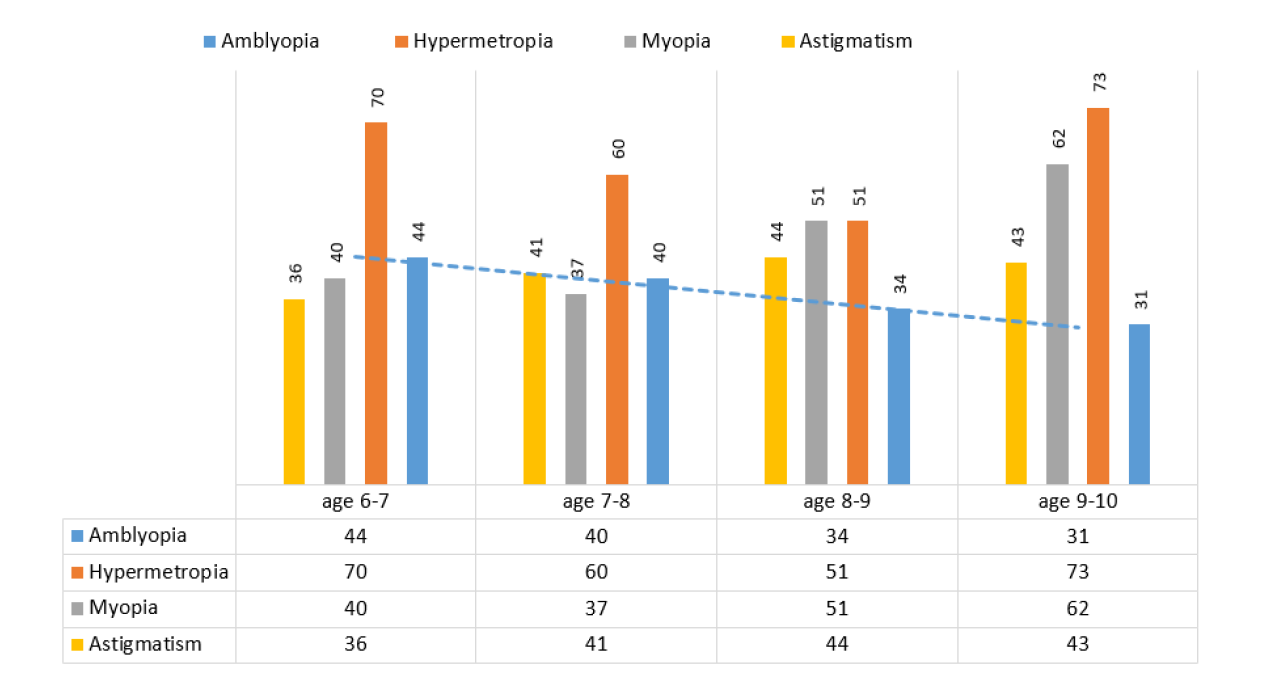
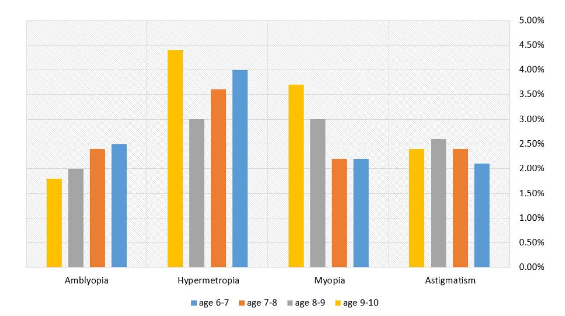
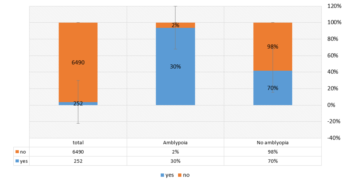
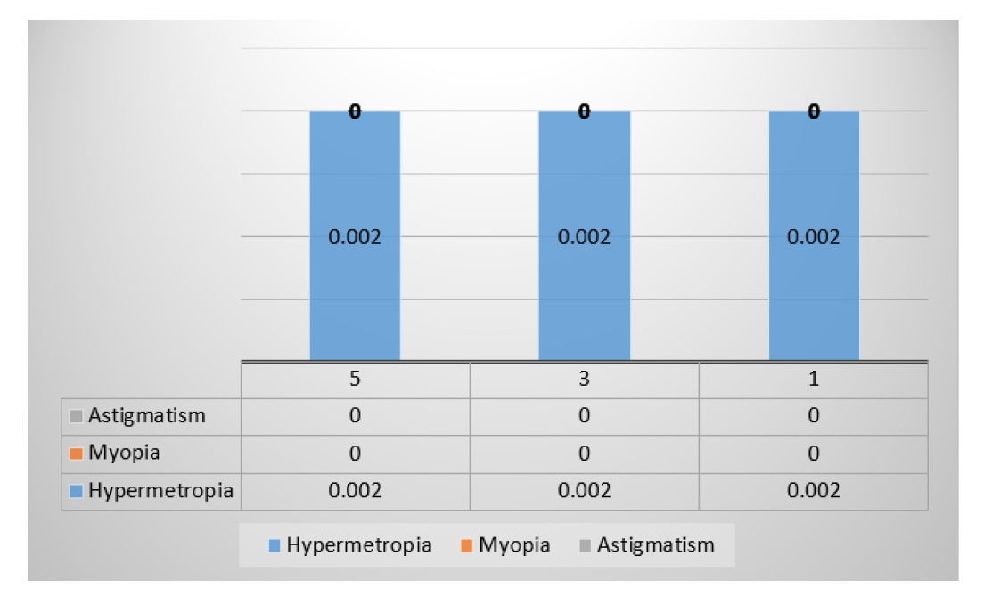
Amblyopia is a developmental defect of spatial visual processing that occurs in the central visual pathways of the brain which caused by form vision deprivation and/abnormal binocular interaction in the absence of visible pathological changes of the eye or visual pathway and it presents most dramatically as unilateral or (rarely) bilateral decrease of best-corrected visual acuity. But amblyopia is more than this where certain forms of amblyopia also present with diminished contrast sensitivity, Vernier acuity, grating acuity and spatial localization of objects.
Amblyopia could be either anisometropic (where it is caused by a difference in refractive error between both eyes, or even as small as 1.0 D) that results in abnormal binocular interaction from the superimposition of focused and unfocused images or from the superimposition of large and small images (aniseikonia). Strabismic amblyopia is another type that results from abnormal binocular interaction which occurs only if one eye is preferred for fixation, whereas stimulus-deprivation amblyopia results from form vision deprivation that caused by media opacity (as congenital cataract, corneal opacity from glaucoma, or dystrophy, lid masses, severe ptosis and persistent hyperplastic primary vitreous, from the point of pathogenesis, the cause of amblyopia is due to changes localized in the ventral visual pathway of the brain and external geniculate bodies. In some cases, the diagnosis of amblyopia presents certain difficulties as it is necessary to assess the morpho structural features of the retina, optic nerve and choroid and make sure that there is a functional inadequacy in the performance of the visual cortex with the introduction of the OCT in wide clinical practice it has become possible to study the morphometric features of the nerve fiber layer of the retina and optic nerve in children with amblyopia & hypermetropia including those with anisometropia, as well as determination of the thickness of the choroid in the macular region. The authors found that for those with amblyopia and hyperopic refraction, the peripapillary nerve fiber layer is thick more compared to what takes place in paired health out eyes [1]. Many researchers have obtained similar results [2–4]. The authors argue that the process may include postnatal reduction of ganglion cells depends on signals from the nature of focusing objects. They prefer lag that apoptosis of retinal ganglion cells in amblyopia is suppressed, this leads to an increase in the measured thickness of the layer of retinal nerve fibers in the amblyopic eyes. Amblyopia no such changes were found. The literature published forged and opposite results, proving that for children with amblyopia and hyperopic refractive the layer of nerve fibers in amblyopic eyes is thinner compared with paired, although the authors do not specify the form and the degree of amblyopia [5]. The result explains the reduction of ganglion cells, decrease shedding of nerve connections in the retina and thinning of the layer nerve fibers due to inadequate stimulation retina in the amblyopic eye.
Including the optic nerve OCT group. The area of the optic disc and the thickness of the layer of retinal nerve fibers in the heterogeneous strabismus is reliably less. The retinal neuron and the anterior region differences were also significantly different. Visual acuity Amblyopia does not correlate with the Ca zone, visual acuity is related to the area of the neural reticulum. In eyes with amblyopia, the retinal neurons were more common than eyes with medium and high amblyopia.
To some extent, however, these differences are statistically insufficient.
Patients, Sample Selection
Anisometropia with a slight difference in refraction between the eyes-one of the manifestations of asymmetry in the paired organ of vision - is widespread strange. According to the results of various studies, its frequency fluctuates, but within small limits. Robert W. Arnold has published the results of several studies in which the frequency of occurrence anisometropia more than 1.5 diopters as a risk factor for ornate amblyopia. So, according to Ottar et al., Ani-occurs in 0.9% of cases. Study MEPEDS showed that 1.6% of those surveyed have, as a result of the BPEDS study, the figure is 1.5%, VIPS-2.3% [6,7]. Donohue revealed anisometropia 1.0 diopters and more in 0.66% of the examined children of preschool age [8]. Believe that anisometropic hyperopia is the most common risk factor development of amblyopia [9,10] Anisometropia with a significant difference in refraction, especially concerning high ametropia of one eye should be considered a developmental abnormality. Cites a study in which studied the relationship of anisometropia with the general condition child. It is noted that in children with anisometropia, up to quite often there are other signs of a violation of symmetry in the body about the palpebral fissures, face, chin, skull bones. It was also revealed that anisometropia against the background of high and medium hyperopia degree is often combined with violations of the general health conditions, mainly with congenital pathology of the central nervous system. The authors expressed the assumption that the specified neurological pathological is due to a disorder of the central hemodynamic which is also one of the reasons for the slowdown, Eye growth and development of visual functions [11]. Note the following pattern: in children with congenital obstruction in the nasolacrimal duct One hundred anisometropic detection. Considering that 88-95% of Cases during the first year of a child's life from obstruction Quotas are allowed spontaneously, which is a theoretical possibility.
The intensity of this coincidence is 0.03%. The authors describe 5 cases when a child suffers from congenital Dacryocystitis-the size revealed an inequality of refraction with worse refraction on the affected side. For neonatal dacryocystitis patients Refraction against the background of ciliary palsy and observation of the refractive loop and optical functions [12] believe the anisometropia Up to 1.5 diopters, that is, does not lead to a decrease in vision, more than equal refraction is observed more often. To determine the frequency of anisotropy of refraction was performed by the authors Refractive adherence to 1,000 people on an unselected continent Ghent. Refractive contrast is detected at 54.8%, which is equal to refractive index -At 45.2%. In the predominant number of subjects with anisometropia, the difference in refraction between the two eyes was small -0.5-1.5 diopters and only 2.1% have 2.0 diopters and more [11].
The following classification of anisometropia:
Mild degree of anisometropia-the difference in refraction up to 1.5 diopters-binocular vision is preserved, the visual acuity of both eyes with correction is normal
Average degree-a difference of refraction 2.0-3.5 diopters-various degrees of impairment are noted binocular vision, on the eye with greater ame- tropia mild to moderate amblyopia
A high degree of anisometropia- the difference in ref-reactions 4.0 diopters and more-binocular vision present, there is a monocular nature of vision
In an eye with more ocular blurring in clinical practice, there are 3 types of presentation:
Deviation of the eye from one eye and ametropia from the other
Different degrees of ametropia of the same name on one and the second eye
Different types of ametropia in both eyes
With a high degree of anisometropia, especially with unilateral hypermetropia from birth, the child's binocular function is impossible as possible, it is absent. Take part in the horizon only an eye has a better view. Amblyopia is the worst. The eyes develop from the combination of two o'clock in the morning Generating Factors - Refractive and Sensory X. There is an active inhibition of fovea regions to eliminate perceptual interference caused by focused and non-focal overlap of the L-images. As a result, like a binocular Foveal inhibition of ocular anisotropic visual acuity at conditions of a less than monocular binocular [8]. In addition to the reduction in central visual acuity, there is a general decrease in contrast sensitivity, which also includes the periphery of the retina.
Disturbances of binocular vision, (due to the different size of the retinal images i.e. aniseikonia) is determined by the degree of refractive disparity and it is considered as another ambiguous factor, since retinal images of different sizes may represent an obstacle to fusion, the stability of the microscope vision with the asymmetry of refraction is determined not only by the anisometropia value and the percentage of aniseikonia, it is also individually determined by the individual ability to compensate for different sizes of objects due to cortical mechanisms to be compatible with binocular functions.
Clinically anisometropic amblyopia results from reduced vision in one or both eyes due to the lack of use of the eye during the period of visual learning. It is a disease that occurs in childhood and if it is not diagnosed and treated promptly, its complications will remain throughout adulthood. It is considered the most common cause of vision loss in children and young adults in developed countries and it affects 3-4% of children of primary school age.
After birth, the sense of vision is not yet complete, so we are not born with sight, but rather this ability develops little by little during the first years of life, the peak of its completion is at the age of 8 or 9 years (although the bulk of this visual learning is completed during the first four years of a child's life).
In many cases, amblyopia does not give symptoms, so the child does not know what the "correct vision" is and adjusts to the vision that he has. This occurs especially in cases where only one eye is affected because the injured can see well with a healthy eye Therefore, it is very important for children.
To be able to achieve optimal visual learning, it is necessary that both eyes receive a clear image of what we see, thus sending information to the brain and with it, we gradually acquire visual acuity. If the child suffers from an eye problem that makes the image that the brain receives of poor quality, at this stage of life, then what will happen is that the brain will "cancel" the development of this eye and the child will get used to the little he sees and his eye will become "lazy".
In most cases, the child has different refraction but continues his life normally. Only in the most obvious cases do we discover the child's "strange habits", such as head twisting, frequent occlusion of the eyelids, drooping upper eyelid, loss of mental focus.



 Download PDF
 Download XML