Congenital anophthalmia is a serious but rare eye deformities. It may be combined with other organ deformities often as part of a specific syndrome. We are reporting the case of a 07-month-old infant with congenital left anophthalmia associated with a cleft lip and palate: Anophtalmia Plus Syndrome (APS).
Keywords
Anophthalmia
Congenital
Cleft Lip and Palate
INTRODUCTION
Anophthalmia is the absence of the eyeball. The congenital form is due to the absence of formation or to the regression of the primary optic vesicle [1]. The involvement can be bilateral or unilateral. It can be isolated or associated with other deformities, which may or may not correspond to specific syndromes. The most frequent extraocular damage affects the craniofacial region, with abnormalities of the face, ears and neck, limbs and musculoskeletal systems [2]. The diagnosis can be clinical based on the anatomical absence of the eyeball. The imaging (ultrasound and or scenography) confirms the clinical observation and often highlights an outline of the eyeball. The management is disappointing, especially when it is associated with another facial anomaly involving the aesthetic prognosis. We are reporting the case of a 07-month-old infant received in consultation for not opening the palpebral fissure since birth.
CASE OBSERVATION
This is a 7-month-old female infant brought by her parents for lack of an eyelid opening since birth. It is the youngest of two siblings. There is no consanguinity between her parents. She had no specific history and is the result of a full-term pregnancy. The mother's infectious biological prenatal evaluation, particularly the rubella, toxoplasmic and syphilitic serologies turned into negative. Two obstetric ultrasounds were requested but not performed for lack of means according to the parents.
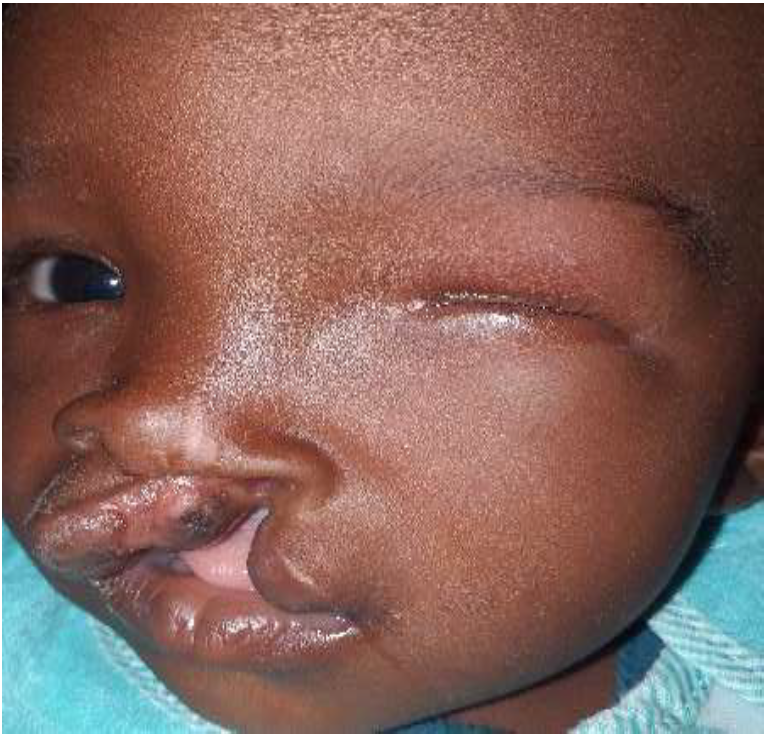
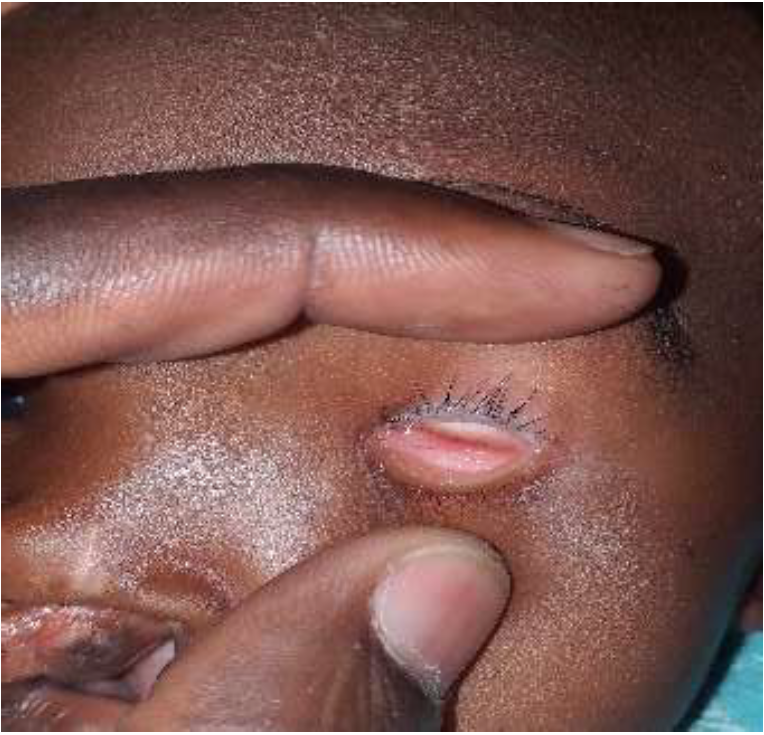
Upon examination, there was a left cleft lip and palate (Figure 1). On clinical ophthalmologic examination, the opening of the left eyelids revealed the absence of an eyeball (figure 2). The eyelid exam was normal. The contralateral eye and its appendages were unremarkable. A general paediatric examination ordered for other deformities was normal. An ocular ultrasound was requested but not performed by the parents.
The diagnosis of left anophthalmia associated with a cleft lip and palate (Anophthalmia Plus Syndrome) was thus made.
A maxillofacial surgery consultation has been requested and the child is awaiting a campaign of free cleft lip and palate surgery scheduled for soon. A proposal for a left ocular prosthesis was thus proposed to parents after surgical management of the cleft lip and palate.
DISCUSSION
Congenital anophthalmia is a rare deformity caused by the absence of any outline of the eyeball, due to an abnormality in embryological development. Its incidence in Great Britain has been estimated at 10 per 100,000 births [3], in France at 2.5 per 10,000 births [4]. Bilateral forms are very rare unlike our case which is unilateral. Like most birth defects, anophthalmia can also be associated with other conditions, often with specific syndromes. Kouassi et al. [5] reported a case of bilateral congenital anophthalmia in Patau syndrome.
Figure 1: cleft palate on examination
Ozcelik et al. reported a case of unilateral anophthalmia with cleft lip palate but associated with absence of the vomer bone and nystagmus of the contralateral eye [6]. Carrying out a paediatric evaluation to look for other malformed abnormality is therefore very useful in case of congenital anophthalmia.
The clinical diagnosis of anophthalmia is based on the absence of ocular structure (empty orbital cavity); which was the case in our child. Oculo-orbital imaging (ultrasound, scanner and nuclear magnetic resonance) provides more precision and makes it possible to distinguish two types of anophthalmia: clinical congenital anophthalmia which results in extreme microphthalmos and the presence of outline of the eyeball and optic nerve; congenital true anophthalmia expressed radiologically in absence of ocular outline and optic canal. Nowadays, with new imaging techniques, antenatal diagnosis is even possible. The aetiologies of congenital anophthalmia are variable. Chromosomal aberrations play an important role in most deformity syndromes. Among these chromosomal abnormalities, mention may be made of Patau's syndrome, deletions of chromosome 3 which may all be causes of poly-malformative syndromes including unilateral or bilateral anophthalmia. Genetic mutations in some genes have also been identified in carriers of anophthalmia: SOX2, PAX6, OTX2, STRA6, ALDH1A3 [7]. Performing a genetic evaluation is therefore necessary in the etiological research of anophthalmia but it was not possible in our context because of its cost and accessibility.
Mariman EMC considered that developmental environment influences on foetal development may be the cause of anophthalmia [8]. These environmental factors include exposure to x-rays during pregnancy, taking some drugs (thalidomide, nitrofen), embryo-fetopathy infections such as rubella [9].
Figure 2: anophthalmia after widening the eyelids
In some cases, etiological research finds no cause for anophthalmia.
The principle of treating congenital anophthalmia is based on improving the aesthetic appearance of the face, especially when it is associated with another face abnormality, like in our case with cleft lip and palate. In these cases, reconstructive surgery should be performed before any ocular prosthetic equipment is made. This is the approach that was offered to our patient. Because of the child's development, follow-up is necessary in order to adapt the prosthesis according to facial growth.
CONCLUSION
The association of anophthalmia with other facial deformities including cleft lip of the palate is relatively rare. Carrying out a comprehensive prenatal check-up often allows antenatal diagnosis and psychological preparation of the parents. The treatment is most often disappointing and just helps to reduce the aesthetic damage.
REFERENCE
Adenis, Jean-Pierre and Serge Morax. Pathologie Orbito-Palpébrale. Rapport de la Société Française d’Ophtalmologie, Masson, 1998, pp. 635–644.
Slavotinek, Anne M. “Eye development genes and known syndromes.” Molecular Genetics and Metabolism, vol. 104, 2011, pp. 448–456.
Busby, A. et al. “Compiling a national register of babies born with anophthalmia/microphthalmia in England 1988–94.” Archives of Disease in Childhood: Fetal and Neonatal Edition, vol. 79, 1998, pp. 168–173.
Stoll, C. et al. “Associated malformations among infants with anophthalmia and microphthalmia.” Birth Defects Research Part A: Clinical and Molecular Teratology, vol. 94, 2012, pp. 147–152.
Kouassi, F.X. et al. “Anophtalmies congénitales: à propos d’un cas de trisomie 13.” Journal Français d’Ophtalmologie, vol. 29, no. 4, 2006, e10.
Ozcelik, D. et al. “Anophthalmia, cleft lip/palate, absent vomer bone, nystagmus and mental-motor retardation: a new syndrome or Fryns ‘anophthalmia-plus’ syndrome?” Cleft Palate–Craniofacial Journal, vol. 45, no. 3, 2008, pp. 256–260.
Slavotinek, Anne. “Genetics of anophthalmia and microphthalmia. Part 2: Syndromes associated with anophthalmia–microphthalmia.” Human Genetics, vol. 138, 2019, pp. 831–846.
Mariman, E.C.M. “Clustering of anophthalmia and microphthalmia.” British Medical Journal, vol. 317, 1998, pp. 895–896.
Romero, C. et al. “Anophthalmia as an isolated congenital defect.” Archivos de la Sociedad Española de Oftalmología, vol. 77, 2002, pp. 571–574.
Recommended Articles
Research Article
Leigh Syndrome Should Not Be Diagnosed Exclusively Upon Cerebral MRI
Finsterer J
Published: 15/07/2020
Download PDF
Research Article
Vision for a Brighter Kangra: Unmasking the Truth about Pink Eye – A Comprehensive Study on Types, Symptoms, and Proactive Prevention in Himachal Pradesh’s Kangra District
Deepti ,
...
Garima
Published: 11/11/2023
Download PDF
Research Article
It Remains Unproven That the Variant M.8231C>A Causes Coronary Atherosclerosis
Finsterer J
Published: 15/07/2020
Download PDF
Research Article
Bioactive ingredients screening and antibacterial activity of Citrus aurantifolia leaf extract against some enteric isolates
Abdallah M.S,
Kabiru Sama’ila
Published: 10/08/2020
Download PDF
PO Box 101, Nakuru
Kenya.
Email: office@iarconsortium.org
Editorial Office:
J.L Bhavan, Near Radison Blu Hotel,
Jalukbari, Guwahati-India
J. P., T., J. M., T. & A., K. (2020). A Rare case of Anophtalmia Plus Syndrome (APS) or Fryns Anophtalmia. IAR Journal of Medical Sciences, 1(2), 1-3.
MLA
J. P., Théra, Tiama J. M. and Konipo A.. "A Rare case of Anophtalmia Plus Syndrome (APS) or Fryns Anophtalmia." IAR Journal of Medical Sciences 1.2 (2020): 1-3.
Chicago
J. P., Théra, Tiama J. M. and Konipo A.. "A Rare case of Anophtalmia Plus Syndrome (APS) or Fryns Anophtalmia." IAR Journal of Medical Sciences 1, no. 2 (2020): 1-3.
Harvard
J. P., T., J. M., T. and A., K. (2020) 'A Rare case of Anophtalmia Plus Syndrome (APS) or Fryns Anophtalmia' IAR Journal of Medical Sciences 1(2), pp. 1-3.
Vancouver
J. P. T, J. M. T, A. K. A Rare case of Anophtalmia Plus Syndrome (APS) or Fryns Anophtalmia. IAR Journal of Medical Sciences. 2020 Jul;1(2):1-3.



 Download PDF
 Download XML