The present study aimed to investigate the harmful effects of nicotine on serum alanine aminotransferase, aspartate aminotransferase and alkaline phosphatase activities and histological structure of the liver of male albino rats. And evaluate the protective effects of fenugreek seeds and curcumin on hepatotoxicity induced by nicotine in male albino rats. 30 male F-344/NHsd Fischer rats, weighing from 180 to 200g were used in the present study. The animals were divided into five groups (6 rats for each); Group I (control group), Group II (nicotine treated group), Group III (nicotine/fenugreek seeds co-administered), Group IV (nicotine/curcumin co-administered) and Group V (nicotine/curcuminand fenugreek seeds co-administered). At the end of the experimentation and 24 hours after the last dose, All animals were anaesthetized with ether and blood samples were collected by heart puncture. The blood samples were collected in clean dry tubes. and allowed to clot at room temperature for about 30 minutes and centrifuged at 3000 rpm for 15 minutes then, serum was separated and kept in a deep freezer at -20◦C until biochemical measurements were carried out. Animals were immediately dissected and small pieces of the liver were quickly removed and fixed in 10% formalin for histological examination. The results showed that the animals treated with nicotine for 4 weeks induced a significant increase in serum alanine aminotransferase, aspartate aminotransferase and alkaline phosphatase activities compared with the control group. Also, administration of nicotine to rats induced a severe structural change in the hepatic tissues. Co-administration of nicotine with fenugreek and/or curcumin caused improvement in serum liver function and histological structure of the liver when compared with nicotine group. It can be concluded that nicotine had a strong effect on the liver function and histological structure of the liver. The ingestion of fenugreek and/or curcumin prevent the hepatoxicity induced by nicotine. The current study suggests that fenugreek and curcumin may be useful in combating free radical-induced hepatotoxicity induced by nicotine.
Nicotine is an alkaloid found in the eggplants, green pepper, tomato, potato and tobacco [1]. It is one ofhundreds of substances contained in cigarette smoke [2]. It is a very toxic compound containing alkaloid and nitrogen [3]. It is responsible for addiction [4]. Nicotine accumulates markedly in saliva, gastric juice [5] and breast milk [6]. It crosses the placental barrier and accumulates in amniotic fluid and fetal serum [7].
Nicotine induces oxidative stress by production of generation of reactive oxygen [8]. It increases in blood pressure, pulse rate, blood sugar, plasma free fatty acids and blood catecholamines levels [9]. In addition, it disturbs the antioxidant defense mechanisms in animals fed a high fat diet. Stimulation of nicotinic receptors result in increased synthesis and release of epinephrine and norepinephrine. Chronic administration of nicotine activates tyrosine hydroxylase, that is the rate limiting enzyme in biosynthesis of catecholamine [10]. It is increased the chromosome aberration, induction of sister chromatide exchange and expression of heat shock proteins and suppressed apoptosis and inhibited cell proliferation [11]. Smoking induce a disturbance in various biological and metabolic processes in the body [2]. Nicotine can pass easily through the cell membrane and reacts with tubulin protein and causes cell division disorder in multiplying cells [12]. It increases the risk of coronary artery disease [13] and promotes tumor growth as well as atherosclerosis formation [14] and decreases fertility in males by inducing DNA damage and oxidative stress [15].
Natural antioxidants neutralize reactive species by reinforcing the endogenous antioxidants defenses and restore the optimal balance [16]. Curcumin as one of the naturally occurring dietary substances has been used since ancient times for promoting human health [17]. Curcumin is a main yellow pigments present in rhizomes of Curcuma longa. It is used as a spice and a coloring agent in foods [18]. Curcumin represents a class of antioxidant and anti-inflammatory that inhibit the of formation reactive oxygen species [19].
Trigonella foenumgraecum is an annual herb grown in Egypt, India and Middle East countries [20]. Fenugreek used with food as spice and in medicine. It used in diabetes mellitus due to the presence of different active constituents such as vitamins, alkaloids, flavonoids and amino acids [21]. The seeds of fenugreek used in beverages, nutrition, cosmetics, fragrances, medicine and in other industrial purposes [22]. Ground fenugreek seed is often mixed with breadstuffs and is an essential ingredient of curry powders [23].
In the folk medicine, herbs and Plant seeds are used for treatments of diseases due to its low side effects and their safetyness when compared with the chemical drugs [24]. Antioxidant potential of the fenugreek seeds and curcumin in the attenuation of nicotine induced oxidative stress need thorough investigation because these natural antioxidants has the potential for safe future use by humans. The evidence reporting the protective effect of fenugreek seeds and curcumin against nicotine induced hepato-toxicity are hardly found.
Objectives
The present study aimed to investigate the harmful effects of nicotine on serum alanine aminotransferase , aspartate aminotransferase and alkaline phosphatase activities and histological structure of the liver of male albino rats. And evaluate the protective effects of fenugreek seeds and curcumin on hepatotoxicity induced by nicotine in male albino rats.
Experimental Animal
Animals which were used in this study were 30 male F-344/NHsd Fischer rats, weighing from 180 to 200g. Animals purchased from Animal Welfare House of Libyan National Medical Research Centre, Zawia, Libya. Rats were kept under standard veterinary hygienic conditions for cleanliness and health care and normal conditions through the whole experimental periods. Rats were separated in plastic cages, 6 rats per cage and left one week of acclimation, before commencing the experiment. The rats were kept in a room under standard conditions of ventilation, temperature (25±4°C), humidity (65 ± 5 %) with light/dark cycle. A standard rodent pellet consisting of a mixture of protein, fat, fiber and ash were used to feed the rats. Food and water were supplied ad-libitum.
The Drug
Nicotine hydrogen tartrate salt (1-methyl-2-(3-pyridyl) pyrrolidine-bitartrate salt) will purchase from Sigma-Aldrich (St. Louis, MO, USA). Nicotine is a colorless organic Liquid. It was dissolved in physiological saline (0.9% sodium chloride) and was injected subcutaneously daily with 0.8 mg, nicotine/kg body weight for 30 days.
Curcumin and Fenugreek Seeds
Curcumin was given in diet as 20 g/kg diet daily for 30 days. Fenugreek seeds were finely grounded and added to the experimental diets as 7.5 g/kg diet daily for 30 days.
Experimental Design
After one week of acclimation, the animals were randomized and divided into five groups (6 male albino rats for each) as follow:
Group IV (Nicotine/Curcumin Co-Administered): The animals were injected subcutaneous daily with 0.8 mg, nicotine/kg body weigh concurrently with curcumin 20 g/kg diet daily for 30 days
Blood Sampling
At the end of the experimentation and 24 hours after the last dose, All animals were anaesthetized with ether and blood samples were collected by heart puncture. The blood samples were collected in clean dry tube and allowed to clot at room temperature for about 30 minutes and centrifuged at 3000 rpm for 15 minutes then, serum was separated and kept in a deep freezer at -20◦C until biochemical measurements were carried out.
Determination of Serum Aspartate Aminotransferase, Alanine Aminotransferase and Alkaline Phosphate Activities
Serum aspartate aminotransferase (AST) and alanine aminotransferase (ALT) activities were estimated by UV-method according to Bergmeyer and Horder [25]. Serum alkaline phosphatase (ALP) activity was determined according to Kind et al. [26].
Histological Preparation
At the end of the experimentation and 24 hours after the last dose, all animals were anaesthetized with ether. Animals were immediately dissected and small pieces of the liver were quickly removed and fixed in 10% formalin. After fixation, specimens were dehydrated in an ascending series of alcohol, then kept in terpineol for three days to ensure complete dehydration and clearing purposes. Cleared specimens were rinsed in three changes of xylol before embedding in paraffin wax (m.p.56-58C). Three sections of 5 microns thick were taken from each liver sample, each being at a distance of at least 500 microns from the proceeding one and mounted on clean slides without adhesive medium. For histological examination, sections were stained with Ehrlich's haematoxyline and eosin.
Statistical Analysis
Results were expressed as Mean±standard deviation, Data were analyzed by one way ANOVA. The difference between Means±SD was tested at p<0.05 using Duncan's multiple range test. In all statistical tests, the probability level of p<0.05 was considered significant.
Effect of Administration of Nicotine and Co-Administration of Nicotine with Fenugreek Seeds, Nicotine with Curcumin and Nicotine with Fenugreek Seeds and Curcumin on the Serum ALT, AST and ALP Activities in Male Rats
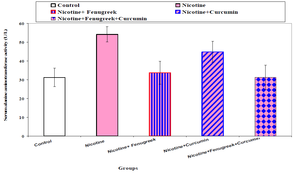
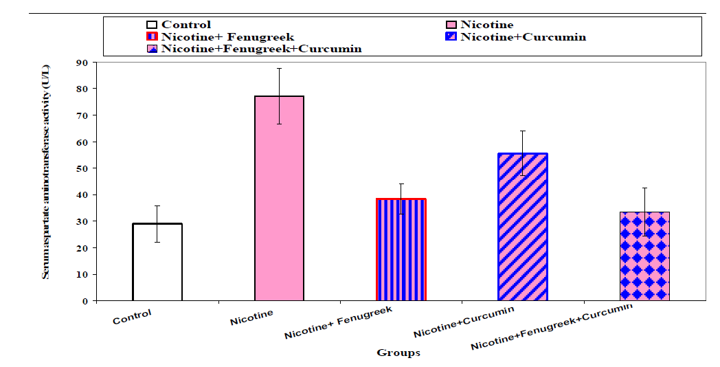
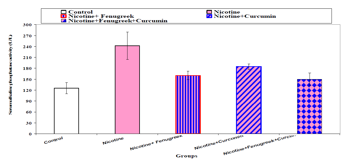
Serum ALT, AST and ALP activities of the different groups are shown in Table 1 and Figures (1-3). Male rats that received intraperitoneal injection of nicotine only (0.8 mg/kg body weight /day) for 30 consecutive days hadsignificantly (p<0.01), increased in serum ALT, AST and ALP activities when compared with the control group.
Co-administration of 0.8 mg, nicotine/kg body weight subcutaneously with fenugreek seeds 7.5 g/kg diet daily for 30 consecutive days resulted in a significant (p<0.01) increase in ALP activity as compared to the control group.
On the other hand, co-administration of fenugreek seeds with nicotine significantly (p<0.01) decreased serum ALT, AST and ALP activities when compared with nicotine group (Table1 and Figures 1-3).
Co-administration of 0.8 mg, nicotine/kg body weight subcutaneously with curcumin 20 g/kg diet daily for 30 consecutive days caused a significant (p<0.01) increase in serum ALT, AST and ALP activities as compared to the control group. Conversely, co-administration of curcumin with nicotine significantly (p<0.01) decreased serum ALT, AST and ALP activities when compared with nicotine group (Table1 and Figure 1-3).
On the other hand, the animals injected subcutaneous daily with 0.8 mg, nicotine/kg body weight concurrently with curcumin 20 g/kg diet and fenugreek seeds 7.5 g/kg diet daily for 30 consecutive days were showed a significant (p<0.01) decrease in serum ALT, AST and ALP activities when compared with nicotine group (Table.1 and Figure 1-3).
Histological Examinatons
Effect of Administration of Nicotine and Co-Administration of Nicotine with Fenugreek Seeds, Nicotine with Curcumin and Nicotine with Fenugreek Seeds and Curcumin on the Structural Changes in the Liver of Male Albino Rats
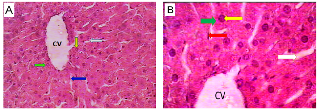
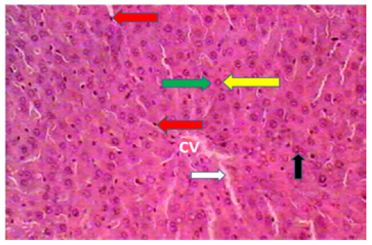
Liver Sections of Control Rats: Histologically, by light microscopic examination, the liver appeared with normal structure in the control group. As in other mammalian animals, the structural unit of the rat liver is the hepatic lobule, which is made up of radiating plates of cells forming a network around a central vein. The hepatic cells are polyhedral in shape with large, centrally located nuclei and granular cytoplasm. The sinusoids are narrow blood spaces with irregular boundaries composed of endothelial cells in addition to large kupffer cells, which are known to be actively phagocyte cells (Figure 4).
Table 1: Effect of Administration of Nicotine and Co-Administration of Nicotine with Fenugreek Seeds, Nicotine with Curcumin and Nicotine with Fenugreek Seeds and Curcumin on the Serum ALT, AST and ALP Activities in Male Rats
Groups Parameters | Control | Nicotine | Nicotine+Fenugreek | Nicotine+Curcumin | Nicotine+ Fenugreek+Curcumin |
| Mean±SD | Mean±SD | Mean±SD | Mean±SD | Mean±SD | |
ALT (U/L) | 31.17±4.88 | 54.17±4.07** | 33.67±6.12## | 44.83±5.56**## | 31.33±6.44## |
AST (U/L) | 29± 6.87 | 77± 10.43** | 38.33±5.72## | 55.50±8.41**## | 33.50±8.96## |
ALP (U/L) | 125.7±15.2 | 242±37.2** | 160.3±11.6**## | 184.8±7.4**## | 150±17.3## |
**: Significant at (p<0.01) when compared with control group, ##: Significant at (p<0.01) when compared with nicotine group

Figure 1: Effect of Administration of Nicotine and Co-Administration of Nicotine with Fenugreek Seeds, Nicotine it Curcumin and Nicotine with Fenugreek Seeds and Curcumin on the Serum ALT Activity in Male Rats

Figure 2: Effect of Administration of Nicotine and Co-Administration of Nicotine with Fenugreek Seeds, Nicotine with Curcumin and Nicotine with Fenugreek Seeds and Curcumin on the Serum AST Activity in Male Rats

Figure 3: Effect of Administration of Nicotine and Co-Administration of Nicotine with Fenugreek Seeds, Nicotine with Curcumin and Nicotine with Fenugreek Seeds and Curcumin the Serum ALP Activity in Male Rats

Figure 4: Light Micrograph of Section in the Liver of the Control Rat; Central Vein (CV); Hepatocyte (Green Arrow); Blood Sinusoid (White Arrow); Kupffer Cells (Blue Arrow); Nucleus (Yellow Arrow) (Haematoxylin and Eosin (A: ×400and B:×1000))
Liver Sections of Nicotine Treated Rats
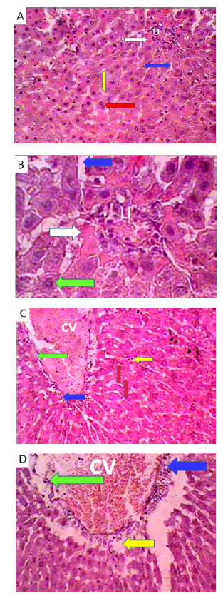
The liver sections in nicotine treated group show dilated congested central vein, damage in lining endothelium, leukocytic infiltration and inflammatory cells around portal blood vessels, dilation of blood sinusoids in zone2, 3, hepatocytes appear with vacuoles in cytoplasm, disorganized and focal necrosis associated with inflammatory cells (Figure. 5).

Figure 5: Light Micrographs of Sections in the Liver of the Rat Treated With Nicotine; A: Blood Sinusoid (Blue Arrow), Leukocytic Filtration (LI), Marked Vacuolar Degeneration Mainly Hydropic Degeneration (Yellow Arrow), Binucleated Hepatocyte (Red Arrow), Rbcs (White Arrow); B: Hepatocyte (Green Arrow); Blood Sinusoid (Blue Arrow), Leukocytic Filtration (LI), Rbcs (White Arrow); C: Dilated Congested Central Vein (CV); Marked Vacuolar Degeneration Mainly Hydropic Degeneration (Red Arrow), Necrosis and Leukocytic Infiltration (Blue Arrow); Damage In Lining Endothelium (Green Arrow); Dilation of Blood Sinusoids (Yellow Arrow); D: Dilated Congested Central Vein (CV); Leukocytic Infiltration (Blue Arrow); Damage in Lining Endothelium (Green Arrow); Necrosis (Yellow Arrow). (H and E, (A: ×400and B:×1000))

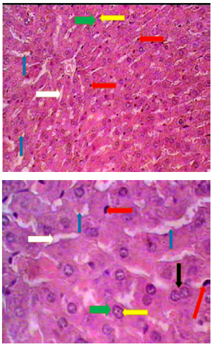
Figure 6: Light Micrograph of Section in the Liver of the Nicotine and Fenugreek Treated Rat, Congested Central Vein (CV); Hepatocyte (Green Arrow); Dilated Blood Sinusoid (White Arrow), Activated Kupffer Cells (Blue Arrow); Nucleus (Yellow Arrow) (H and E (A: ×400and B:×1000))

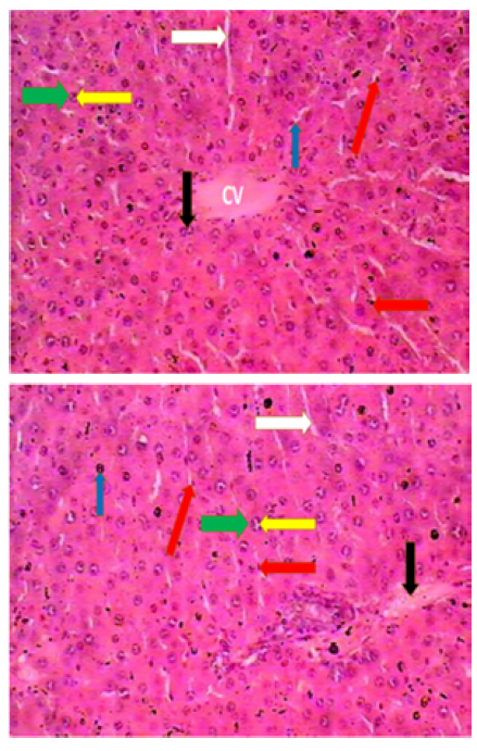
Figure 7: Light Micrograph of Section in the Liver of the Nicotine and Curcumin Treated Rat; Dilated Congested Central Vein (CV); Hepatocyte (Green Arrow); Leukocytic Infiltration In Portal Area (Black Arrow); Dilated Blood Sinusoid (White Arrow); Activated Kupffer Cells (Blue Arrow); Nucleus (Yellow Arrow) (H and E (A: ×400and B:×1000))
Liver Sections of Nicotine and Fenugreek Treated Rats
Giving the animal fenugreek with nicotine for the same period caused improvement in the histological structure of the liver tissues. The structure of the hepatic lobule appears normal. Some liver sections show congested central veins, activated kupffer cells, dilated blood sinusoids (Figure 6).

Figure 8: Light Micrograph of Section in the Liver of the Nicotine, Fenugreek, and Curcumin Treated Rat; Central Vein (CV); Dilated Blood Sinusoid (White Arrow); Hepatocyte (Green Arrow); (Yellow Arrow ); (Red Arrow), (H And E (A: ×400and B:×1000)).
Liver Sections of Nicotine and Curcumin Treated Rats
Treatment of rats with nicotine and curcumin for 4 weeks caused improvement in the histological structure of the liver tissues. The structure of the hepatic lobule disorganized. Some liver sections show dilated congested central veins, activated kupffer cells, leukocytic infiltration in portal area and congested portal vein dilated blood sinusoids (Figure7).
Liver Sections of Nicotine, Fenugreek and Curcumin Treated Rats
Co-administration of nicotine with fenugreek and curcumin to rats for 4 weeks caused improvement in the histological structure of the liver tissues. The structure of the hepatic lobules appeared normal (Figure 8).
Liver is a principle organ of detoxification and the major site of intense metabolism in generally, thus undergoing to various disorders because of exposure to the toxins [27, 28]. Its injury may be leads to intense metabolic disorders. Liver injury induced by chemicals has been recognized as one of the most toxicological problems [28,29]. Nicotine is metabolized in liver to a number of metabolites. About 80% of nicotine is converted to cotinine in human body [28,30].
Van der Vaart et al. [31] reported that smoking exposure causes a reduction in antioxidants and increases of the production of reactive oxygen metabolites. Nicotine causes oxidative damage to liver, lung, kidney and heart. Nicotine induces a production of free radicals which react with biomembranes causing a destruction of polyunsaturated fatty acids and forming cytotoxic aldehydes by lipid peroxidation [32,33]. Nicotine produces a damage to endothelium and plays a major role in the development of numerous human diseases [34].
In the hepatotoxicity studies, serum ALT and AST activities are known as toxicity markers and an increase in the activities of these enzymes is termed as the early recognition of toxic hepatitis [28,35]. Alkaline phosphatase is found in a high concentrations in the bones and liver. It has been reported that serum alkaline phosphatase activity is increased in hepatobiliary diseases [36] and during alcoholic hepatitis [37].
The present study showed that intraperitoneal injection of rats with nicotine for 30 consecutive days caused a significant increases in the serum AST, ALT and ALP activities when compared with the control group. Similar results were reported by previous studies [28,38-42]. Abushofa et al. [28]reported that subcutaneous injection of male F-344/NHsd Fischer rats with 4 mg of nicotine/kg body weight /day for 6 weeks caused a significant increase in serum ALT, AST and ALP activities compared with the control group. Fahim et al., [43] found that intraperitoneal injection of mice with 1 mg of nicotine/Kg of body weight for 3 weeks induced an increase in hepatic ALT and AST levels. Also, Mahmoud and Amer, [44] recorded that a significant elevations in the activities of ALT, AST and alkaline phosphatase in liver homogenate of nicotine treated rats compared with control group. The results obtained in this study confirm the finding of Sharif et al. [40] found that subcutaneous injections of mice with 1 mg/kg body weight of nicotine in the scruff of the neck daily for 6 weeks caused a significant increase in the serum alanine amino transferase, aspartate aminotransferase and alkaline phosphatase activities compared with the control group. It is concluded that nicotine administration in mice resulted in deleterious effects on ALT, AST and ALP. Balakrishnan and Menon, [38] indicated that nicotine administration can significantly increase marker enzymes in liver.
Cell membrane damage can trigger the release of hepatic enzymes into the blood circulation [45]. The increases in serum alanine aminotransferase activity observed in the current study may reflect hepatotoxic potency of nicotine exposure on the liver. This may be attributed to the state of hypoxia of the parenchyma for contracting fibrous tissue and the increased permeability of hepatic cell membrane due to nicotine treatment which release ALT enzyme into the circulation. The increased level of ALT is marked as liver parenchymal cell destruction induced by nicotine treatment [44]. Cellular damage by nicotine may be leads to accumulation of lipids and glucose in the hepatic cells, so the histological section revealed liver injury [46]. Nicotine can induce the production of free radicals and consequently oxidative stress, which is one of the most important causes of liver cells damage [47]. Also, nicotine can cause lipid peroxidation by affecting the membrane of hepatocytes, which, in turn, causes a change in membrane permeability and lipid degeneration and accumulation in the liver cells. Free radicals seem to change the enzymatic activity and necrosis by attacking polyunsaturated fatty acids and alkylating groups of proteins and other cellular macromolecules [41, 48].
In the current study, the animals injected subcutaneous daily with 0.8 mg, nicotine/kg body weight concurrently with curcumin 20 g/kg diet daily for 30 consecutive days were showed a significant (p<0.01) decrease in serum ALT, AST and ALP activities when compared with nicotine group. These results similar to the results of Al Anany et al. [33] who found that treatment of adult male albino rats with curcumin with nicotine decreased plasma ALT, AST and ALP activities compared with nicotine group. Authors suggested that curcumin exerts protective effects by improving the antioxidant system, inhibiting the oxidative stress in liver tissues induced by nicotine. Curcumin could exert antioxidative effects either directly as a chemical antioxidant due to its ability to scavenge reactive oxygen and nitrogen free radicals or by modulating cellular defenses which themselves exert antioxidant effects [33, 49]. The obtained results may be due to the antioxidant effects of curcumin and/or quercetin [33, 50].
The present study showed that co-administration of 0.8 mg, nicotine/kg body weight subcutaneously with fenugreek seeds 7.5 g/kg diet daily for 30 consecutive days resulted in a significant (P<0.01) decrease in serum ALT, AST and ALP activities when compared with nicotine group. These results were in agreement with the study done by Das, [51] who reported that administration of extract of fenugreek seeds in CCl4 treated rats caused reduction in Serum ALT, AST, ALP levels. A water extract of Fenugreek seeds concurrently during 60 days of alcohol ingestion was associated with a reduction in the rise of oxidation and liver enzymes noted in the serum of rats given ethanol alone, suggesting protective effects [52]. Kumar and Bhandari, [53] demonstrated that the activities of serum ALT and AST were increased in rats treated with monosodium glutamate. Administration of aqueous Trigonella foenum-graecum seeds significantly reduced the elevated ALT and AST levels, which could be attributed to the protective effect on hepatic tissues.
The present study showed that the animals injected subcutaneous daily with 0.8 mg, nicotine/kg body weight concurrently with curcumin 20 g/kg diet and fenugreek seeds 7.5 g/kg diet daily for 30 consecutive days were caused a significant improvement in all hematological parameters and serum levels of ALT, AST, ALP, urea, creatinine and uric acid and the structural changes in the liver and kidney when compared with nicotine group. These parameters and histological structure of the liver and kidneys were nearly similar to that in the control groups. These results may be due to the additive antioxidant effect of fenugreek and curcumin together. Al Anany et al.[33] reported that treatment of adult male albino rats with curcumin or quercetin alone or in combination improved all parameters deteriorated by nicotine, i.e. the elevation in plasma ALT, AST and ALP levels. Authors suggested that curcumin and/or quercetin exerts protective effects by improving the antioxidant system, inhibiting the oxidative stress in liver tissues induced by nicotine. Combined therapy with both curcumin and quercetin was much better than each one alone. Because, previous studies reported that natural antioxidants strengthen the endogenous antioxidants defenses from reactive oxygen species and restore the optimal balance by neutralizing reactive species [54,55]. Curcumin has anti-inflammatory and antioxidant properties with a potent ability to inhibit reactive oxygen species formation [56]. Curcumin represents a class of anti-inflammatory and anti-oxidant reported to be a potent inhibitor of reactive oxygen species formation [19]. Fenugreek had a different active constituents such as flavonoids, alkaloids, vitamins and amino acids [21]. The ameliorative effect of fenugreek and curcumin against nicotine induced hematotoxicity and hepatorenal toxicity may be due to decrease nitric oxide production, uremic toxin and increasing radical-scavenging enzyme activity through scavenging reactive oxygen and nitrogen species and chelating redex-active transition metal ions.
In the current study, the liver sections in nicotine treated group show dilated congested central vein, damage in lining endothelium, leukocytic infiltration and inflammatory cells around portal blood vessels, dilation of blood sinusoids, hepatocytes appear with vacuoles in cytoplasm, disorganized and focal necrosis. These results are coinciding with these recorded by the study of Abushofa et al. [28] who reported that the histopathological examination of the hepatic tissues of rats treated with nicotine showed congestion of central and portal veins, congestion of blood sinusoids, hepatocytes degeneration. Also, Gawish et al. [57] found the same effect of nicotine on the hepatic tissues.
Hepatocellular necrosis is probably due to attach of a cell membrane by the hepatotoxin or interaction with some specific components of the metabolic pathways leading to the alteration of their structure and function [28, 58]. Oxidative stress by nicotine occurs when there are excess free radicals or low antioxidant defense and result in chemical alteration of biomolecules causing structural and functional modification [28,59]. Hence, logically there is association between nicotine and liver cell inflammation and activation of Kuppfer cells which may be attributed to proinflammatory cytokines [28, 60].
The current study showed that treatment of male rats with nicotine concurrent with fenugreek seeds powder caused improvement in histological structure of the liver tissues. This result similar to the result of Das, [51] and Botsoglou et al. [61] who reported that the livers of rats treated with extract of fenugreek seeds showed a significant attenuation from CCl4-induced liver damage as evident from normal hepatocytes with well-defined nuclei. The improvement of histological changes in the liver are well correlating with the biochemical estimations. These results suggest that the extract of fenugreek seeds has potential clinical applications for treating liver disorders.
It can be concluded that nicotine had a strong effect on the liver function and histological structure of the liver. The ingestion of fenugreek and/or curcumin prevent the hepatotoxicity induced by nicotine. The current study suggests that fenugreek and curcumin may be useful in combating free radical-induced hepatotoxicity induced by nicotine.
Siegmund, B. et al. “Determination of the nicotine content of various edible nightshades (Solanaceae) and their products and estimation of the associated dietary nicotine intake.” Journal of Agricultural and Food Chemistry, vol. 47, 1999, pp. 3113–3120.
Abdel-Aziz, H.O. “Morphological evaluation on the protective effect of curcumin on nicotine induced histological changes of the adrenal cortex in mice.” Egyptian Journal of Histology, vol. 33, no. 3, 2010, pp. 552–559.
Jana, K. et al. “Nicotine diminishes testicular gametogenesis, steroidogenesis and steroidogenic acute regulatory protein expression in adult albino rats: Possible influence on pituitary gonadotropins and alteration of testicular antioxidant status.” Toxicological Sciences, vol. 116, no. 2, 2010, pp. 647–659.
Benowitz, N.L. et al. “Prevalence of smoking assessed biochemically in an urban public hospital: A rationale for routine cotinine screening.” American Journal of Epidemiology, vol. 170, no. 7, 2009, pp. 885–891.
Lindell, G. et al. “Transdermally administered nicotine accumulates in gastric juice.” European Journal of Clinical Pharmacology, vol. 51, 1996, pp. 315–318.
Dahlstrom, A. et al. “Nicotine and cotinine concentrations in the nursing mother and her infant.” Acta Paediatrica Scandinavica, vol. 79, 1990, pp. 142–147.
Dempsey, D.A. and N.L. Benowitz. “Risks and benefits of nicotine to aid smoking cessation in pregnancy.” Drug Safety, vol. 24, 2001, pp. 277–322.
Yildiz, D. et al. “Nicotine enantiomers and oxidative stress.” Toxicology, vol. 130, 1998, pp. 155–165.
Waldum, H.L. et al. “Long term effects of inhaled nicotine.” Life Sciences, vol. 16, 1996, pp. 1339–1346.
Hiremagular, B. et al. “Nicotine increases expression of tyrosine hydroxylase gene.” Journal of Biological Chemistry, vol. 268, 1993, pp. 23704–23711.
Yamashita, H. and S. Nakamura. “Nicotine rescues PC12 cells from death induced by nerve growth factor deprivation.” Neuroscience Letters, vol. 213, 1996, pp. 145–147.
Gorrod, J.W. “The mammalian metabolism of nicotine: An overview.” Nicotine and Related Alkaloids: Absorption, Distribution, Metabolism and Excretion, edited by J.W.Gorrod and J.Wahren, Chapman & Hall, 1993, pp. 31–44.
Swislocki, A.L. et al. “Smokeless nicotine administration is associated with hypertension but not with a deterioration in glucose tolerance in rats.” Metabolism, vol. 46, 1997, pp. 1008–1012.
Heeschen, C. et al. “Nicotine stimulates angiogenesis and promotes tumor growth and atherosclerosis.” Nature Medicine, vol. 7, 2001, pp. 833–839.
Jalili, C. et al. “Protective effect of curcumin against nicotine-induced damage on reproductive parameters in male mice.” International Journal of Morphology, vol. 32, no. 3, 2014, pp. 844–849.
Ho, C. et al. “Phytochemicals in teas and rosemary and their cancer preventive properties.” Food Phytochemicals for Cancer Prevention, American Chemical Society, 1994, pp. 2–19.
Joe, B. et al. “Biological properties of curcumin: Cellular and molecular mechanisms of action.” Critical Reviews in Food Science and Nutrition, vol. 44, 2004, pp. 97–111.
Tirkey, N. et al. “Curcumin attenuates cyclosporine induced renal dysfunction and oxidative stress in rat kidneys.” Journal of Biosciences, vol. 22, no. 2, 2005, pp. 233–246.
Venkatesan, N. et al. “Curcumin prevents adriamycin nephrotoxicity in rats.” British Journal of Pharmacology, vol. 12, 2000, pp. 231–234.
Flammang, A.M. et al. “Genotoxicity testing of fenugreek extract.” Food and Chemical Toxicology, vol. 42, 2004, pp. 205–208.
Basch, E. et al. “Therapeutic application of fenugreek.” Alternative Medicine Review, vol. 8, no. 1, 2003, pp. 20–27.
Djeridane, A. et al. “Antioxidant activity of some Algerian medicinal plants extracts containing phenolic compounds.” Food Chemistry, vol. 97, 2006, pp. 654–660.
Blank, I. et al. “The principal flavor components of fenugreek (Trigonella foenum-graecum L.).” Spices: Flavor Chemistry and Antioxidant Properties, edited by S.J.Risch and C.T.Ho, American Chemical Society, 1997, pp. 12–28.
Alhawari, S. “Medical plants as food and medicine.” Saudi Arabian Journal, vol. 21, 1986, pp. 70–71.
Bergmeyer, H. and C. Horder. “ALT kit.” Journal of Clinical Chemistry and Clinical Biochemistry, vol. 18, 1980, pp. 521–534.
Kind, P.R.N. et al. Practical clinical biochemistry. Heinmann, 1980, pp. 899–900.
Aebi, H. “Catalase in vitro.” Methods in Enzymology, vol. 105, 1984, pp. 121–126.
Kakkar, P. et al. “A modified spectrophotometric assay of superoxide dismutase.” Indian Journal of Biochemistry and Biophysics, vol. 21, 1984, pp. 130–132.
Ellman, G.L. “Tissue sulfhydryl groups.” Archives of Biochemistry and Biophysics, vol. 82, 1959, pp. 70–77.
Lowry, O.H. et al. “Protein measurement with the Folin phenol reagent.” Journal of Biological Chemistry, vol. 193, 1951, pp. 265–275.
Sedlak, J. and R.H. Lindsay. “Estimation of total, protein-bound and nonprotein sulfhydryl groups in tissue with Ellman’s reagent.” Analytical Biochemistry, vol. 25, 1968, pp. 192–205.
Ohkawa, H. et al. “Assay for lipid peroxides in animal tissues by thiobarbituric acid reaction.” Analytical Biochemistry, vol. 95, 1979, pp. 351–358.
Snedecor, G.W. and W.G. Cochran. Statistical methods. 8th ed., Iowa State University Press, 1989.
Benowitz, N.L. “Pharmacology of nicotine: Addiction and therapeutics.” Annual Review of Pharmacology and Toxicology, vol. 36, 1996, pp. 597–613.
Benowitz, N.L. and P. Jacob. “Metabolism of nicotine to cotinine studied by a dual stable isotope method.” Clinical Pharmacology and Therapeutics, vol. 56, 1994, pp. 483–493.
Sener, G. et al. “Protective effects of melatonin, vitamin E and N-acetylcysteine against nicotine-induced oxidative damage in rat liver.” Life Sciences, vol. 69, 2001, pp. 183–194.
Pari, L. and P. Murugan. “Protective role of tetrahydrocurcumin against erythromycin estolate-induced hepatotoxicity.” Pharmacological Research, vol. 49, 2004, pp. 481–486.
Manju, V. and N. Nalini. “Chemopreventive efficacy of curcumin on 1,2-dimethylhydrazine induced colon carcinogenesis.” Food and Chemical Toxicology, vol. 43, 2005, pp. 165–172.
Hensley, K. et al. “Reactive oxygen species and cell signaling: A review.” Free Radical Biology and Medicine, vol. 28, 2000, pp. 1456–1462.
Valko, M. et al. “Free radicals and antioxidants in normal physiological functions and human disease.” The International Journal of Biochemistry and Cell Biology, vol. 39, 2007, pp. 44–84.
Ames, B.N. et al. “Oxidants, antioxidants and the degenerative diseases of aging.” Proceedings of the National Academy of Sciences of the United States of America, vol. 90, 1993, pp. 7915–7922.
Halliwell, B. and J.M.C. Gutteridge. Free radicals in biology and medicine. 4th ed., Oxford University Press, 2007.
Pryor, W.A. and K. Stone. “Oxidants in cigarette smoke: Radicals, hydrogen peroxide, peroxynitrate and peroxynitrite.” Annals of the New York Academy of Sciences, vol. 686, 1993, pp. 12–27.
Das, S. et al. “Curcumin ameliorates nicotine-induced oxidative stress and mitochondrial dysfunction in rat brain.” Neurochemical Research, vol. 36, 2011, pp. 1283–1293.
Kalpana, C. and V.P. Menon. “Curcumin ameliorates oxidative stress during nicotine-induced lung toxicity.” Indian Journal of Experimental Biology, vol. 42, 2004, pp. 798–802.
El-Beshbishy, H.A. “Hepatoprotective effect of curcumin in rats.” Pharmacological Research, vol. 51, 2005, pp. 111–117.
Maheshwari, R.K. et al. “Multiple biological activities of curcumin: A short review.” Life Sciences, vol. 78, 2006, pp. 2081–2087.
Aggarwal, B.B. and K.B. Harikumar. “Potential therapeutic effects of curcumin.” International Journal of Biochemistry and Cell Biology, vol. 41, 2009, pp. 40–59.
Sharma, R.A. et al. “Pharmacodynamic and pharmacokinetic study of oral curcuma extract.” Clinical Cancer Research, vol. 7, 2001, pp. 1894–1900.
Gupta, S.C. et al. “Multitargeting by curcumin as revealed by molecular interaction studies.” Natural Product Reports, vol. 28, 2011, pp. 1937–1955.
Reddy, A.C. and B.R. Lokesh. “Effect of curcumin on iron-induced hepatic toxicity.” Biochemical Pharmacology, vol. 44, 1992, pp. 1177–1182.
Naidu, P.S. and M. Kulkarni. “Protective effect of curcumin on nicotine-induced toxicity.” Indian Journal of Pharmacology, vol. 36, 2004, pp. 85–88.
Trivedi, A.H. et al. “Protective effect of curcumin on nicotine-induced hepatotoxicity.” Journal of Environmental Biology, vol. 30, 2009, pp. 363–367.
Yadav, R. et al. “Effect of curcumin on oxidative stress in nicotine-treated rats.” Indian Journal of Physiology and Pharmacology, vol. 54, 2010, pp. 162–170.
Khan, N. et al. “Curcumin and health: Perspectives.” Clinical Nutrition, vol. 27, 2008, pp. 1–12.
Sreejayan and M.N.A. Rao. “Nitric oxide scavenging by curcuminoids.” Journal of Pharmacy and Pharmacology, vol. 49, 1997, pp. 105–107.
Jurenka, J.S. “Anti-inflammatory properties of curcumin.” Alternative Medicine Review, vol. 14, 2009, pp. 141–153.
Shishodia, S. et al. “Curcumin: Getting back to the roots.” Annals of the New York Academy of Sciences, vol. 1056, 2005, pp. 206–217.
Anand, P. et al. “Bioavailability of curcumin: Problems and promises.” Molecular Pharmaceutics, vol. 4, 2007, pp. 807–818.
Chainani-Wu, N. “Safety and anti-inflammatory activity of curcumin.” Journal of Alternative and Complementary Medicine, vol. 9, 2003, pp. 161–168.
Gupta, B. et al. “Therapeutic roles of curcumin: Lessons learned from clinical trials.” AAPS Journal, vol. 15, 2013, pp. 195–218.