Acute mesenteric ischemia (AMI) is typically defined as a group of diseases characterized by an interruption of the blood supply to varying portions of the small intestine, leading to ischemia and secondary inflammatory changes. If untreated, this process will result in life threatening intestinal necrosis. The incidence is low, estimated at 0.09–0.2% of all acute surgical admissions. Early diagnosis and timely surgical intervention are the cornerstones of modern treatment and are essential to reduce the high mortality associated with this entity. The advent of endovascular approaches may provide new options. We herein report a case of adult male patient with total occlusion of superior and inferior mesenteric artery along with critical Celiac artery stenosis in patient presented with weight loss, abdominal pain and gastrointestinal bleeding and celiac artery stenting done successfully.
Keywords
Acute Mesenteric Ischemia (AMI)
Celiac Artery Stenting
INTRODUCTION
Acute mesenteric ischemia (AMI) may be defined as a sudden interruption of the blood supply to a segment of the small intestine, leading to ischemia, cellular damage, intestinal necrosis, and eventually patient death if untreated [1]. AMI may be non-occlusive (NOMI) or occlusive, with the primary etiology further defined as mesenteric arterial embolism (50%), mesenteric arterial thrombosis (15–25%), or mesenteric venous thrombosis (5–15%) [2,3]. The overall incidence is low (0.09 to 0.2% of all acute admissions to emergency departments), representing an uncommon cause of abdominal pain [2]. Prompt diagnosis and intervention are essential to reduce the high mortality rates . There is currently no level 1 evidence to guide the evaluation and treatment of suspected AMI, with no clearly defined treatment guidelines.
Case
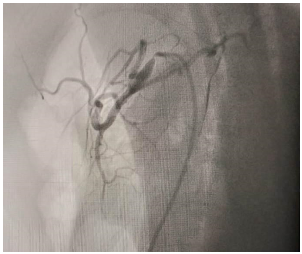
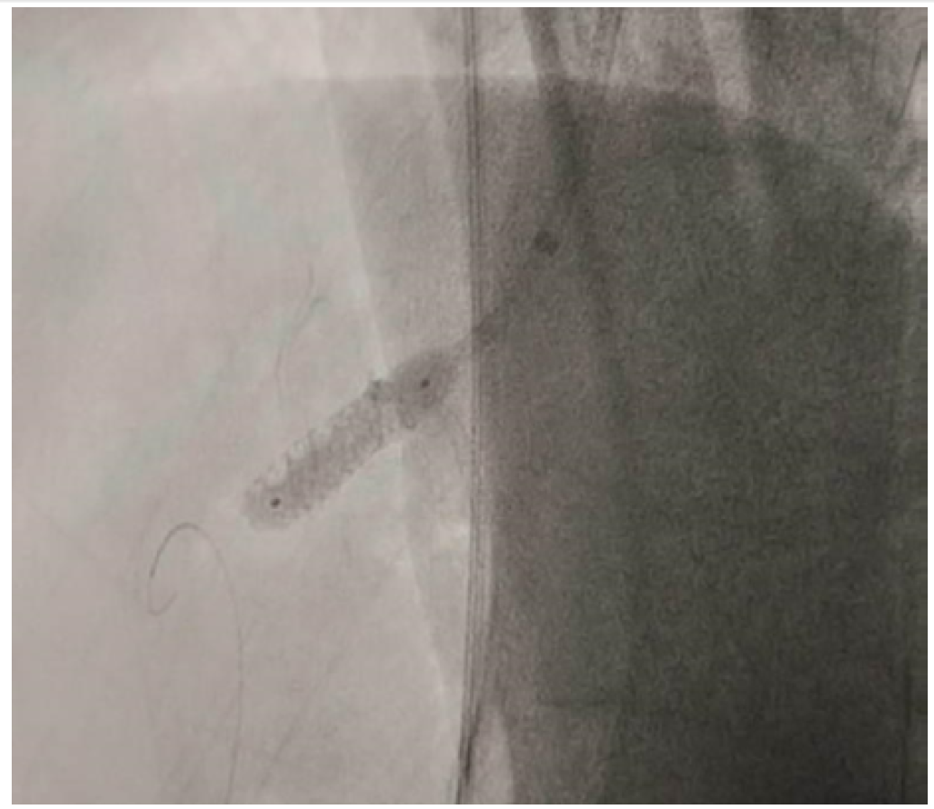
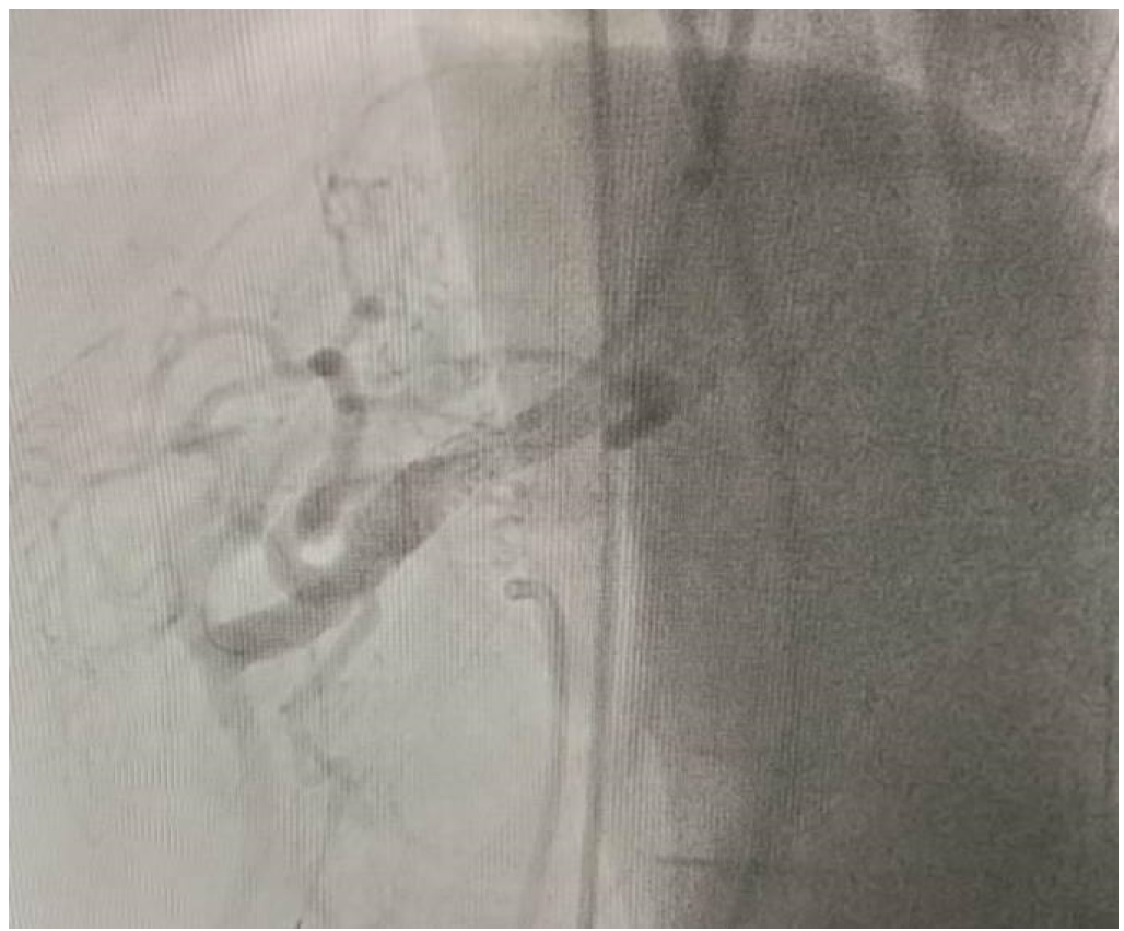
A Adult male aged 52 year smoker nonalcoholic without any family history of atherosclerotic cardiovascular disease presented in emergency with pain abdomen for for one day with 1 year history of weight loss , poor appetite, postprandial angina. 3- to 4 hour after admission patient started having bleeding per rectum. Patient developed a fever of 100 degrees f., the patient had a bp of 90/60 and pulse rate of 100/min at admission. On abdominal examination no distension, no guarding ,rigidity, tenderness with decreased bowel sounds.Patients Hb was 10g%, TLC count 13800/ mm3 ,RFT ,LFT, were normal. serum amylase was 399. Proctoscopy was normal. Usg abdomen was normal. CECT abdomen reported total cut off of IMA, SMA and near total occlusion of celiac artery. Patients were managed with fluid resuscitation antibiotics. In view of borderline hemodynamics and ongoing lower gi bleed, the patient was considered for endovascular revascularization. Selective angiography Figure 1 demonstrates ostial stenosis of 99% with thrombus . Lesion was crossed with fielder wire predilated with 3*15 nc balloon stent Herculink elite 7/18 deployed in ostial proximal celiac artery Figure 2. Figure 3 demonstrates good ostial coverage with good flow in the celiac trunk. Patient improved clinically with reduction in pain in abdomen and gi bleed and started orally on the second day.patient was kept on close clinical follow up .patient had significant weight gain from 46 kg at admission to 52 kg at 6 months.
Figure 1: Preprocedural Angiogram Showing Severe Stenosis of Ostium of Celiac Artery with Some Thrombus
Figure 3: Angiographic Image Showing Complete Ostial Coverage and Normal Flow to Celiac Artery
DISCUSSION
Roughly, 50% of all cases of AMI are due to acute mesenteric embolism .Thrombosis (approximately 25% of cases) is usually associated with pre-existing chronic atherosclerotic disease leading to stenosis. Many of these patients have a history consistent with chronic mesenteric ischemia (CMI). The key to early diagnosis is a high level of clinical suspicion. The clinical scenario of a patient complaining of excruciating abdominal pain with an unrevealing abdominal exam is classic for early AMI [4]. 95% of patients presented with abdominal pain, 44% with nausea, 35% with vomiting, 35% with diarrhea, and 16% with blood per rectum [5]. Approximately one-third of patients present with the triad of abdominal pain, fever, and hemoccult-positive stools. There are no laboratory studies that are sufficiently accurate to identify the presence or absence of ischemic or necrotic bowel, although elevated l-lactate, and D-dimer may assist.Computed tomography angiography (CTA) should be performed as soon as possible for any patient with suspicion for AMI [6]. When the diagnosis of AMI is made, fluid resuscitation should commence immediately to enhance visceral perfusion. Electrolyte abnormalities should be corrected, and nasogastric decompression initiated [7]. Broad-spectrum antibiotics should be administered immediately[8]. Unless contraindicated, patients should be anticoagulated with intravenous unfractionated heparin. Prompt laparotomy should be done for patients with overt peritonitis. Endovascular revascularization procedures may have a role with partial arterial occlusion [8].
CONCLUSION
AMI is a true surgical emergency. First and foremost, important evidence is a high index of suspicion. This clinical scenario should prompt imaging (CTA) in order to establish the diagnosis. In parallel with rapid resuscitation and after careful assessment, the patient should be explored to assess bowel viability, re-establish vascular flow, and resect non-viable bowel. There are no randomized controlled trials comparing laparotomy versus endovascular treatment as a first line strategy for the management AMI. Endovascular therapy can be considered for patients with low likelihood of bowel infarction.
REFERENCE
Patel, A. et al. “Pathophysiology of mesenteric ischemia.” Surgical Clinics of North America vol. 72, 1992, pp. 31–41.
Acosta, S., and Bjorck, M. “Acute thrombo-embolic occlusion of the superior mesenteric artery: a prospective study in a well-defined population.” European Journal of Vascular and Endovascular Surgery vol. 26, 2003, pp. 179–183.
Acosta, S. “Mesenteric ischemia.” Current Opinion in Critical Care vol. 21, 2015, pp. 171–178.
Carver, T.W. et al. “Mesenteric ischemia.” Critical Care Clinics vol. 32, 2016, pp. 155–171.
Park, W.M. et al. “Contemporary management of acute mesenteric ischemia: factors associated with survival.” Journal of Vascular Surgery vol. 35, 2002, pp. 445–452.
Evennett, N.J. et al. “Systematic review and pooled estimates for the diagnostic accuracy of serological markers for intestinal ischemia.” World Journal of Surgery vol. 33, 2009, pp. 1374–1383.
Oldenburg, W.A. et al. “Acute mesenteric ischemia: a clinical review.” Archives of Internal Medicine vol. 164, 2004, pp. 1054–1062.
Arthurs, Z.M. et al. “A comparison of endovascular revascularization with traditional therapy for the treatment of acute mesenteric ischemia.” Journal of Vascular Surgery vol. 53, 2011, pp. 698–705.
None
None
Recommended Articles
Research Article
Knowledge of the Patient's Family about the Level of Emergency Based on Triage with the Satisfaction of the Patient's Family in the Emergency Department of the Sumedang Area General Hospital
Roganda Situmorang,
...
Raida Rihadatul Aisyi
Published: 30/03/2024
Download PDF
Research Article
The Influence of Murottal Al Qur'an Surat Ar Rahman Irama Hijaz Therapy on the Pain Scale of Post-Explorative Laparatomy Patients in the Icu Room of Bandung City Hospital
Andri Nurmansyah,
...
Fifi Siti Fauziah Yani
Published: 30/03/2024
Download PDF
Research Article
The role of IFN-g gene polymorphism (+874 T/A) as predisposing factor for pulmonary tuberculosis
Download PDF
Research Article
Caesarean Scar Pregnancy
Komal Sharma,
...
Jyotshna Sharma
Published: 02/02/2023
Download PDF
PO Box 101, Nakuru
Kenya.
Email: office@iarconsortium.org
Editorial Office:
J.L Bhavan, Near Radison Blu Hotel,
Jalukbari, Guwahati-India
Sharma, A. & sharma, C. (2024). A Case of Celiac Artery Stenosis and Stenting in Patient with Critical Mesenteric vascular Disease and Symptomatic Intestinal Ischemia. Scientific Research Journal of Clinical and Medical Sciences, 4(1), 1-3.
MLA
Sharma, Ambudhar and Charu sharma. "A Case of Celiac Artery Stenosis and Stenting in Patient with Critical Mesenteric vascular Disease and Symptomatic Intestinal Ischemia." Scientific Research Journal of Clinical and Medical Sciences 4.1 (2024): 1-3.
Chicago
Sharma, Ambudhar and Charu sharma. "A Case of Celiac Artery Stenosis and Stenting in Patient with Critical Mesenteric vascular Disease and Symptomatic Intestinal Ischemia." Scientific Research Journal of Clinical and Medical Sciences 4, no. 1 (2024): 1-3.
Harvard
Sharma, A. and sharma, C. (2024) 'A Case of Celiac Artery Stenosis and Stenting in Patient with Critical Mesenteric vascular Disease and Symptomatic Intestinal Ischemia' Scientific Research Journal of Clinical and Medical Sciences 4(1), pp. 1-3.
Vancouver
Sharma A, sharma C. A Case of Celiac Artery Stenosis and Stenting in Patient with Critical Mesenteric vascular Disease and Symptomatic Intestinal Ischemia. Scientific Research Journal of Clinical and Medical Sciences. 2024 Jan;4(1):1-3.


 Download PDF
 Download XML