The present study was performed to evaluate the ameliorative effect of celastrus paniculatus against CCl4 induced hepatotoxicity in rats. In the present study, Celastrus paniculatus was selected for hepatoprotective activity owing to its traditional use. The liver function is assessed by determining the activities of SGOT, SGPT, SALP, i.e. enzymes present in the cytoplasm. In case of hepatotoxicity, the enzymes move into the bloodstream and their amount confirms the liver damage extent. The plant material was extracted by continuous hot percolation Soxhlation and the percentage yield calculated by the following formula was found to be 2.41 % (by ethanol). Phytochemical screening of ehanolic extract of Celastrus paniculatus showed the presence of carbohydrate, alkaloid, protein, flavonoids, tannin and phenolic compound, saponin, triterpenoids and steroids and absence of glycoside. In case of acute oral toxicity study when animals were treated with 5, 50, 300 and 2000 mg/kg b.w. of dose for 72 hours there was no mortality and any behavioural changes, so the dose will be choosen as 200 mg/kg b.w. on the basis of LD50. Administration of ethanolic extract showed a substantial decrease in SGPT, SGOT, SALP level (p <0.05) at a dose of 200mg/kg. Similarly, treatment with hepatoprotective drug such as silymarin showed a significant decrease in SGPT, SGOT and SALP levels. Therefore, it was reported that Celastrus paniculatus extract treatment normalize few morphological features of the liver in rats with CCl4- induced hepatotoxicity.
Keywords
Ethanolic Extract of Celastrus Paniculatus
Soxhlation
Hepatotoxicity
Silymarin
CCl4 Induced Hepatotoxicity
INTRODUCTION
Hepatotoxicity means damage to the liver caused by drugs and other factors resulting in problems in its functioning. Chemicals or drugs that because hepatotoxicity are called hepatotoxins. The liver is the largest solid organ in the upper abdomen that aids in digestion and removes waste products and worn out cells from the blood. Itis considered to be one of the most vital organs that functions as centre of metabolism of nutrients such as carbohydrates, proteins and lipids and excretion of waste metabolites. Additionally, it is also handling the metabolism and excretion of drugs and other xenobiotics from the body thereby providing protection against foreign substances by detoxifying and eliminating those (Figure 1) [1-6].
The seeds of Celastrus Paniculatus are acrid, bitter, thermogenic, emollient, stimulant, intellect promoting, digestive, laxative, emetic, expectorant, appetizer, aphrodisiac, cardiotonic, antiinflammatory, diuretic, emmenagogue, diaphoretic, febrifuge and tonic, abdominal disorders, leprosy, pruritus, skin diseases, paralysis, cephalalgia, arthralgia, asthma, leucoderma, cardiac debility, inflammation, nephropathy, amenorrhoea. The seed oil is useful in abdominal disorders, beri-beri and sores (Table 1-4) [7,8].
Figure 1: Picture of Celastrus Paniculatus (Malkangani) Seeds
Table 1: Botanical Description of Celastrus Paniculatus
Height
10m
Leaves
Simple, broad, oval withtoothed margins
Flowers
Borne in large panicles at the endof branches
Pod
Round
Seeds
Oval –shaped, 6 seeds per pod
Stem
Upto 10 cm in diameter and 6m long
Bark
Rough, pale brown exfoliating bark
Roots
Strong root system
Table 2: Taxonomical Classification of Celastrus Paniculatus
Kingdom
Plantae
Order
Celastrales
Family
Celastraceae
Genus
Celastrus
Species
C. paniculatus
Table 3: Vernacular Names of Celastrus Paniculatus
Language
Plant name
Hindi
Malkangani
English
Staff tree
Kannada
Kariganne
Tamil
Valuluvai
Telugu
Malkangani
Sanskrit
Jyotishmati
Table 4: Chemical Constituents of Celastrus Paniculatus
Central Nervous System: The crude CP seed oil administered orally, intramuscularly (i.m.) and intraperitoneal (i.p.) in a dose of 1 g/kg produced sedation in rats. The oil administered orally (100 mg/kg) as an emulsion showed no sedative effect in rabbits. The same emulsion (1 g/kg i.p.) to mice produced mild sedation. Administration of oil (1 g/kg i.m.) to mice showed a significant reduction of movement. CP seed oil given as emulsion showed tranquilizing effect on adrenaline and amphetamine-induced excitement in mice [9,10].
Cardiovascular Activity
The crude CP seed oil administered as emulsion (50-100 mg/kg) produced a gradual fall in cardiac output, bradycardia and marked increase in pulse pressure on isolated heart lung preparation in cat. A similar action with 1 g of emulsified oil was also observed in dogs. The aqueous extract of CP seed showed 50% Angiotensin Converting Enzyme (ACE) inhibition; ethanol extract showed mild activity while the acetone extract was devoid of it [11,12].
Antifertility Activity
The seed oil when given in a dose of 0.2 mL/animal/48 h to adult albino rats for 30 days showed antispermatogenic effects as evidenced by vacuolization of seminiferous tubules, germ cell depletion and exfoliation culminating into an arrest in spermatogenesis. The shrunken tubules revealed only sertoli cells and spermatogonia in the final stage of impairment of spermatogenesis. The livers revealed focal necrosis in animals receiving 0.2 mL (i.p.) seed oil for 30 days, but 45 days’ post treatment these lesions were absent. These results indicate that CP oil may have useful antifertility effects and that the degenerative changes seen in the liver are reversible with time [13].
MATERIALS AND METHODS
Selection of Plant and Authentication
Seeds of Celastrus paniculatus collected from the cultivated farm and the open field of Kolhapur, Maharastra and authenticated by botanist from the Department of Botany, Safia Science College, Bhopal. Four hundred gram of the powder prepared from shade dried seeds was subjected to Soxhlet extraction for 16 h using Petroleum ether (40 - 60ºC) as nonpolar solvent at first. Exhausted plant material (marc) was dried and then extracted with ethanol. Colourless solvent was collected from siphon tube and evaporated for residue. Absence of residue confirmed the completion of extraction. Obtained extracts were evaporated using rotary vacuum evaporator (Bucchi type) at 40°C. The percentage yield of the extract was 2.41%, based on the starting quantity. The doses of 200 mg/kg body weight (b.w.) were prepared by suspending the dried extract in water and administering it to the rats by the per os (p.o.) route.
Extraction of Plant
In the present study, extraction was performed using continuous hot percolation ‘Soxhlation’. Dried seeds of Celastrus paniculatus willd. were placed in thimble of Soxhlet apparatus. Soxhlation was performed at 60°C using Petroleum ether (40 - 60ºC) as non-polar solvent at first. Exhausted plant material (marc) was dried and then extracted with ethanol. For confirmation of exhausted plant marc (i.e. completion of extraction), colourless solvent was collected from siphon tube and evaporated for residue. Absence of residue confirmed the completion of extraction. Obtained extracts were evaporated using rotary vacuum evaporator (Bucchi type) at 40°C.
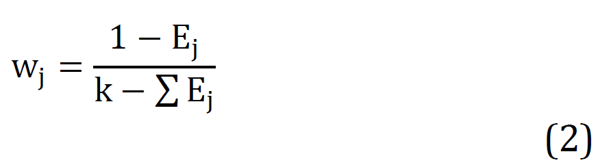
Dried extract was weighed and percentage yield for each extract was determined using the following formula:
Preparation of Ethanolic Extracts
About 400 grams of the powder prepared from shade dried seeds of Celastrus Paniculatus was subjected to Soxhlet extraction for 16 h using Petroleum ether (40 - 60ºC) as non-polar solvent at first. Exhausted plant material (marc) was dried and then extracted with ethanol. Colorless solvent was collected from siphon tube and evaporated for residue. Absence of residue confirmed the completion of extraction. Obtained extracts were evaporated using rotary vacuum evaporator (Bucchi type) at 40°C.
Phytochemical Screening
Qualitative photochemical investigation. The phytochemical investigation was carried out by procedure given in Kokate et al.
Tests for Carbohydrates
Molish Test: About 2 mL of aqueous extract was treated with 2 drops of alcoholic α-naphthol solution in a test tube and then 1 mL of concentrated sulphuric acid was added carefully along the sides of the test tube. Formation of violet ring at the junction indicates the presence of carbohydrates
Benedict’s Test: Equal volume of Benedict’s reagent and extract were mixed in a test tube and heated in the water bath for 5-10 minutes. Solution appears green, yellow or red depending on the amount of reducing sugar present in the test solution which indicated the presence of reducing sugar
Tests for Protein and Amino acids
Biuret’s Test: The extract was treated with 1 mL of 10% sodium hydroxide solution in a test tube and heated. A drop of 0.7% copper sulphate solution was added to the above mixture. The formation of violet or pink colour indicates the presence of proteins
Tests for Glycosides
Borntrager’s Test: To 3 mL of test solution, dilute sulphuric acid was added, boiled for 5 minutes and filtered. To the cold filtrate, equal volume of benzene or chloroform was added and shake it welled. The organic solvent layer was separated and ammonia was added to it. Formation of pink to red colour in ammonical layer indicates presence of anthraquinone glycosides.
Tests for Alkaloids
To the extract, dilute hydrochloric acid was added, shake it well and filtered. With the filtrate, the following tests were performed.
Mayer’s Test: To 2-3 mL of filtrate, few drops of Mayer’s reagent were added along sides of tube. Formation of white or creamy precipitate indicates the presence of alkaloids
Hager’s Test: To 1-2 mL of filtrate, few drops of Hager’s reagent were added in a test tube. Formation of yellow colour precipitate indicates the presence of alkaloids
Wagner’s Test: To 1-2 mL of filtrate, few drops of Wagner’s reagent were added in a test tube. Formation of reddish brown precipitate indicates the presence of alkaloids. Tests for Saponins: Froth Test: The extract was diluted with distilled water and shaken in graduated cylinder for 15 minutes. The formation of layer of foam indicates the presence of saponins
Tests for Flavonoids
Lead Acetate Test: The extract was treated with few drops of lead acetate solution. Formation of yellow precipitate may indicate the presence of flavonoids
Alkaline Reagent Test: The extract was treated with few drops of sodium hydroxide separately in a test tube. Formation of intense yellow colour, which becomes colour less on addition of few drops of dilute acid, indicate presence of flavonoids
Tests for Triterpenoids and Steroids
Salkowski’s Test: The extract was treated with chloroform and filtered. The filtrate was added with few drops of concentrated sulphuric acid, shaken and allowed to stand. If the lower layers turn red, sterol are present. Presence of golden yellow layer at bottom indicates the presence of triterpenes.
Tests for Tannin and Phenolic Compounds
Ferric Chloride Test: Some amount of extract was dissolved in distilled water. To this solution 2 mL of 5% ferric chloride solution was added. Formation of blue, green or violet colour indicates presence of phenolic compounds
Dilute Iodine Solution Test: To 2-3 mL of extract, few drops of dilute iodine solution were added. Formation of transient red colour indicates presence of phenolic compounds
Preparation of Ethanolic Extracts
About 400 grams of the powder prepared from shade dried seeds of Celastrus Paniculatus was subjected to Soxhlet extraction for 16 h using Petroleum ether (40 - 60ºC) as non-polar solvent at first. Exhausted plant material (marc) was dried and then extracted with ethanol. Colourless solvent was collected from siphon tube and evaporated for residue. Absence of residue confirmed the completion of extraction. Obtained extracts were evaporated using rotary vacuum evaporator (Bucchi type) at 40°C.
Pharmacological Screening Model Design
CCl4 induced toxicity model.
CCl4- induced Hepatotoxicity in Rats
CCl4-induced hepatotoxicity was done by intraperitoneally injecting 1 mL/kg b. w. CCl4 dissolved in liquid paraffin in ratio 1:1 after animals are being denied food for 18 hours. The rats were randomLy divided into four groups of six rats each.
Group I: Normal control and were administered 1 mL/kg normal saline
Group II: CCl4 intoxicated (0.7mL/kg by Intraperitonial injection)
Group III: Standard drug treated, Silymarin, 100 mg/kg, orally
Group IV: CCl4 - induced hepatotoxic rats treated with 100 mg/kg b.w Ethonolic extract of Celastrus Paniculatus
Extracts and Standards Used
Extracts Used: Ethanolic extract of dried seeds of Celastrus Paniculatus
Standard Drug: Silymarin 100mg/kg
Treatment of Animals
Healthy male Albino Wistar rat of age 8-10 weeks and weight 150 to 200gm were selected after physical and veterinary examination. All experiments involving animals comply with the ethical standard of animal handling and are approved by Institutional Animal Ethics Committee (IAEC). All the selected animals were kept under acclimatization on the same day. The animals were acclimatized one week prior to dosing. Identification of animals by cage number and marking on animal. The rats were housed in separate standard polypropylene cages with stainless steel top grill. Clean autoclaved paddy husk was used as bedding. The paddy husk was changed at least thrice in a week. The animals were kept in a clean environment with 12-hour light and 12-hour dark cycles. The air was conditioned at 22±2°C and the relative humidity was maintained between 30-70% with 100% exhaust. Standard rat pellet feed was provided ad libitum throughout the study, except overnight fasting prior to blood collection and was offered the feed immediately after completion of blood collection of all the animals. Drinking water was provided ad libitum in polypropylene bottles with a stainless steel sipper tube throughout study period.
RESULTS
Plant Extraction
The plant material was extracted by continuous hot percolation Soxhlation and the percentage yield calculated by the following formula was found to be 2.41 % by ethanol.
Acute Oral Toxicity (OECD)
The acute oral toxicity study was carried out according to OECD 423 guidelines. Four ranges of dose were used for toxicity studies, i.e 5mg/Kg, 50 mg/Kg, 300 mg/Kg, 2000 mg/Kg. animals were observed individually for next 4 hours after dosing for the presence of mortality during this period and 72 hours after sample administration.
Histology
Hepatotoxicity in rat as Shown in Figure 2.
Figure 2: Hepatotoxicity in Rat
DISCUSSION AND CONCLUSION
According to the World Health Organization (WHO), herbal medicines are being used by about 60% of world population primarily in developing countries for primary health care. The objective of the biological screening of plants is to find the sources of the biologically active chemical compounds which can be developed as a drug or to discover the lead molecules which can be modified through chemical procedures into useful drugs.It has been suggested that aqueous and ethanolic extracts from plants are potential source of antiviral, antitumoral and antimicrobial agents [14].
Historically, plants have provided a good source of anti-infective agents; emetin, quinine and berberin, which remain highly effective in the fight against microbial infections. Phytomedicines derived from plants have shown great promise in the treatment of intractable infectious diseases [15].
CCl4 is commonly used hepatotoxin for the experimental study of liver disorder. CCl4 induced liver cell injury involves CCl4 biotransformation caused by cytochrome P450 leading to the production of trichloromethyl free radical which causes peroxidative degradation in adipose tissue causing hepatocyte fatty infiltration. Lipid peroxidation is caused by trichlo- romethyl free radicals in the presence of oxygen produced by metabolic leakage from mitochondria. All these changes inhibit the damage of hepatic tissue and loss of integrity of cell membrane. CCl4 increases SGPT, SGOT and induced hepatotoxicity.
In the present study, Celastrus paniculatus was selected for hepatoprotective activity owing to its traditional use. The liver function is assessed by determining the activities of SGOT, SGPT, SALP, i.e. enzymes present in the cytoplasm. In the case of hepatotoxicity, the enzymes move into the bloodstream and their amount confirms the liver damage extent.
The plant material was extracted by continuous hot percolation Soxhlation and the percentage yield calculated by the following formula was found to be 2.41 % (by ethanol). On phytochemical screening of ehanolic extract of Celastrus paniculatus showed the presence of carbohydrate, alkaloid, protein, absence of glycoside. In case of acute oral toxicity study when animals were treated with 5, 50, 300 and 2000 mg/kg b.w. of dose for 72 hours. There was no mortality and any behavioural changes so they decided dose will be choosen as, 200 mg/kgb.w. On the basis of LD50.
Phytochemical tests of ethanolic extract of leaves of Celastrus paniculatus indicated the existence of saponins, flavonoids and phenolic compounds. Administration of ethanolic extract showed a substantial decrease in SGPT, SGOT, SALP level (p<0.05) at a dose of 200mg/kg. Similarly, treatment with hepatoprotective drug such as silymarin showed a significant decrease in SGPT, SGOT and SALP levels. Therefore, it was reported that Celastrus paniculatus extract treatments normalize few morphological features of the liver in rats with CCl4 induced hepatitis.
REFERENCES
Firenzuoli, F. and L. Gori. “Herbal medicine today: Clinical and research issues.” Evidence-Based Complementary and Alternative Medicine, vol. 4, suppl. 1, 2007, pp. 37–40.
Kampa, M. and E. Castanas. “Human health effects of air pollution.” Environmental Pollution, vol. 151, no. 2, 2008, pp. 362–367.
Nicholson, J.K. et al. “Metabonomics: A platform for studying drug toxicity and gene function.” Nature Reviews Drug Discovery, vol. 1, no. 2, 2002, p. 153.
Kumar, A. “A review on hepatoprotective herbal drugs.” International Journal of Research in Pharmaceutical Chemistry, vol. 2, no. 1, 2012, pp. 96–102.
Akaerue, B.I. and G.I. Onwuka. “Evaluation of the yield, protein content and functional properties of Mungbean (Vigna radiata L. Wilczek) Protein Isolates as Affected by Processing.” Pakistan Journal of Nutrition, vol. 9, no. 8, 2010, pp. 728–735.
Lee, W.M. “Drug-induced hepatotoxicity.” The New England Journal of Medicine, vol. 333, no. 17, 1995, pp. 1118–1127.
Watanabe, S. and M.J. Phillips. “Acute phalloidin toxicity in living hepatocytes: Evidence for a possible disturbance in membrane flow and for multiple functions for actin in the liver cell.” The American Journal of Pathology, vol. 122, no. 1, 1986, pp. 101–111.
Trauner, M. et al. “Molecular pathogenesis of cholestasis.” The New England Journal of Medicine, vol. 339, no. 17, 1998, pp. 1217–1227.
Cullen, J.M. “Mechanistic classification of liver injury.” Toxicologic Pathology, vol. 33, no. 1, 2005, pp. 6–8.
Faubion, W.A. et al. “Toxic bile salts induce rodent hepatocyte apoptosis via direct activation of fas.” The Journal of Clinical Investigation, vol. 103, no. 1, 1999, pp. 137–145.
Pessayre, D. et al. “Hepatotoxicity due to mitochondrial dysfunction.” Cell Biology and Toxicology, vol. 15, 1999, pp. 367–373.
Arora, N. and S.Z. Goldhaber. “Anticoagulants and transaminase elevation.” Circulation, vol. 113, 2006, pp. 698–702.
Shimizu, S. et al. “Metabolism-dependent hepatotoxicity of amodiaquine in glutathione-depleted mice.” Archives of Toxicology, vol. 83, 2009, pp. 701–707.
Kaplowitz, N. “Idiosyncratic drug hepatotoxicity.” Nature Reviews Drug Discovery, vol. 4, 2005, pp. 489–499.
Stedman, C. “Herbal hepatotoxicity.” Seminars in Liver Disease, vol. 22, 2002, pp. 195–206.
Recommended Articles
Research Article
Oral Manifestations of Myasthenia Gravis, Rheumatoid Arthritis, Pernicious Anemia, Graft Vs Host Disease, Linear IgA Disease and Chronic Bullous Disease of Childhood
Download PDF
Research Article
Tlics Score & Denis Scale and Their Association with Various Variables among Patients Undergoing Operative Treatment for the Thoraco-Lumbar Fracture at Tertiary Care Hospital
Bahrat Bhushan,
...
Vineet Aggarwal
Published: 10/04/2022
Download PDF
Research Article
Socio-Demographic Characteristics, Classification, Management, Complication and Implant Status of Patients Undergoing Operative Treatment for the Thoraco-Lumbar Fracture at Tertiary Care Hospital.
Bahrat Bhushan,
...
Vineet Aggarwal
Published: 10/04/2022
Download PDF
Mini Review Article
The Significant and Profound Impacts of the Studies on the Rate of Diffusion-Controlled Reactions of Enzyme
Kuo- Chen Chou
Published: 10/10/2020
Download PDF
PO Box 101, Nakuru
Kenya.
Email: office@iarconsortium.org
Editorial Office:
J.L Bhavan, Near Radison Blu Hotel,
Jalukbari, Guwahati-India
Shantaram, C. P., Bargode, R. S., Jain, S. & Shukla, K. (2021). Reformative Effect of Jyotishmati (C. Paniculatus) In Chemical Induced Hepatotoxicity in Experimental Animals. IAR-Medical Series, 2(2), 1-5.
MLA
Shantaram, Chaudhari P., et al. "Reformative Effect of Jyotishmati (C. Paniculatus) In Chemical Induced Hepatotoxicity in Experimental Animals." IAR-Medical Series 2.2 (2021): 1-5.
Chicago
Shantaram, Chaudhari P., Ranjeet S. Bargode, Sourabh Jain and Karunakar Shukla. "Reformative Effect of Jyotishmati (C. Paniculatus) In Chemical Induced Hepatotoxicity in Experimental Animals." IAR-Medical Series 2, no. 2 (2021): 1-5.
Harvard
Shantaram, C. P., Bargode, R. S., Jain, S. and Shukla, K. (2021) 'Reformative Effect of Jyotishmati (C. Paniculatus) In Chemical Induced Hepatotoxicity in Experimental Animals' IAR-Medical Series 2(2), pp. 1-5.
Vancouver
Shantaram CP, Bargode RS, Jain S, Shukla K. Reformative Effect of Jyotishmati (C. Paniculatus) In Chemical Induced Hepatotoxicity in Experimental Animals. IAR-Medical Series. 2021 Jul;2(2):1-5.


 Download PDF
 Download XML