Background: In the majority of general surgical units around the world, Secondary Peritonitis is the common surgical emergency and has high risk of morbidity and mortality. The present study was done to evaluate the surgical management among Secondary Peritonitis cases in a tertiary care Hospital. Material and Methods: This cross-sectional study was conducted by Department of Surgery, IGMC, Shimla over a period of twelve months from January 2021 – December 2021 among patients with a diagnosis of secondary peritonitis after fulfilling the inclusion and exclusion criteria. Relevant information was collected and further analysed by using IBM SPSS Statistics. Results: A total of 250 individuals with a diagnosis of perforation peritonitis were assessed in the current study. Males made up 205 (82%) of the total while females made up 45 (18%). The patients' average age was 47.28 17.34 years, ranging from 18 to 87. In the study, 99 (39.6%) of the total patients were under the age of 40, while 151 (60.4%) were beyond the age of 40. Of the total, 32 (12.80%) passed away while 218 (87.20%) were discharged after receiving treatment. In the present study, most of patients 89(35.6%) of secondary peritonitis were managed by Cellan jones repair followed by Loop ileostomy in 43(17.2%) patients End ileostomy in 27 (10.8%) patients and rest by other procedures. Conclusion: The study concluded that most of patients of secondary peritonitis were managed by Cellan jones repair followed by Loop ileostomy and End ileostomy.
In the majority of general surgical units around the world, Secondary Peritonitis is the common surgical emergency and has high risk of morbidity and mortality. The most typical type of peritonitis that develops after an intra-peritoneal source, typically from the puncture of hollow viscera, is secondary peritonitis. A serious and potentially fatal situation is acute widespread peritonitis brought on by an underlying hollow viscous perforation [1,2].
Abdominal surgical infections have a complex nature that makes it challenging to correctly define the illness, evaluate its severity, and track the effectiveness of treatment. The outcome is influenced by the physiologic compromise it causes, as well as the anatomic source of the infection, to a greater extent [3].
Despite spectacular advances in understanding pathogenesis of disease, in diagnostic modalities discovery of broad-spectrum antibiotics, invention of modern advance equipments like ventilators, advancement of knowledge in surgical and anesthesiology field, peritonitis still poses major problem for surgeons as far as morbidity and mortality is concerned [1-3].
The aim of this study is to evaluate the surgical management among Secondary Peritonitis casesin a tertiary care Hospital.
Aims and Objectives
To evaluate the surgical management among Secondary Peritonitis cases in a tertiary care Hospital.
This study was conducted in the Department of Surgery IGMC, Shimla over a period of twelve months from January 2021 – December 2021.
Inclusion Criteria
All adult patients (> 18 years of age) presenting with the clinical diagnosis of perforation peritonitis of either sex
All Patients willing to participate in the study
Exclusion Criteria
Patients operated elsewhere before presentation
Patients unwilling to participate in the study
Patients on drugs which alter the level of arterial lactate
Study Methodology
The proposed study was a longitudinal, prospective study
All patients presenting to the Department of Surgery, IGMC, Shimla with a diagnosis of secondary peritonitis and fulfilling the inclusion and exclusion criteria were recruited
The diagnosis of secondary peritonitis was established based on clinical examination, investigations and operative findings
Informed and written consent was taken from the patient / relative
Resuscitation, preoperative and postoperative treatment was performed according to the established protocol for perforation peritonitis
Relevant information was collected
Statistical Analysis
Data compiled in the excel sheet and further analysed by using IBM SPSS Statistics 28 and result displayed in number and percentage. To access the relationship between parameters under study outcome various hypothesis was designed and results were compiled in results and observation section using t-test and Chi-square test.
In this retrospective study total 250 patients with diagnosis of perforation peritonitis were evaluated. Among the total Males were 205 (82%) and Females were 45 (18%). Mean age of the patients was 47.28±17.34 years with range from 18 to 87 years. Among the total, 99(39.6%) patients were age <40 years while 151(60.4%) patients were aged > 40 years (Table 1).
Table 1: Age and gender Distribution of Study Participants
Age (Years) | ≤ 40 years | > 40 years | Total |
Male | 93 (93.93%) | 112 (74.17%) | 205 (82%) |
Female | 16 (6.06%) | 39 (25.82%) | 45(18%) |
Total | 99 (39.6%) | 151 (60.4%) | 100 % |
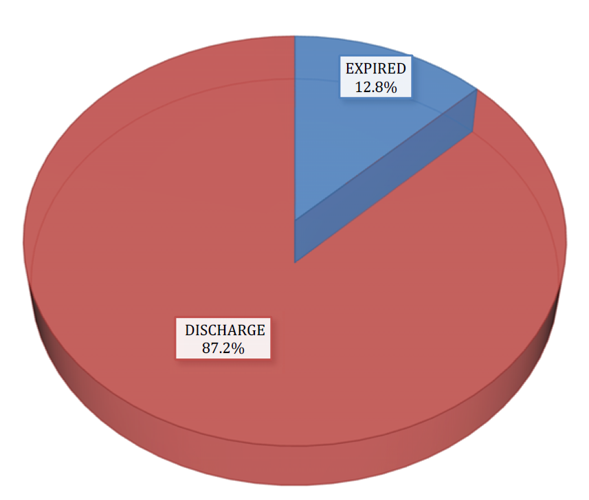
Among the total, 32(12.80%) were expired while 218(87.20%) were discharged after treatment (Figure 1).

Figure 1: Distribution of Outcome of the Study Participants
In the present study, most of patients 89(35.6%) of secondary peritonitis were managed by Cellan jones repair followed by Loop ileostomy in 43(17.2%) patients End ileostomy in 27 (10.8%) patients and rest by other procedures. Table 2 shows the distribution of procedure of the study participants and percentage of study participants.
Table 2: Distribution of Procedure of the Study Participants
Procedure | Number of Patients | Percentage of Study Participants |
Cellan jones repair | 89 | 35.6 |
Loop ileostomy | 43 | 17.2 |
End ileostomy | 27 | 10.8 |
Cellan jones repair.F | 19 | 7.6 |
Primary repair of any perforation,Loop ileostomy | 16 | 6.4 |
Primary repair of any perforation | 11 | 4.4 |
Appendectomy | 6 | 2.4 |
End ileostomy,Limited ileocaecal resection | 5 | 2 |
Primary repair of any perforation,End ileostomy | 3 | 1.2 |
Primary repair of any perforation,Colostomy end | 3 | 1.2 |
Cholecystectomy | 3 | 1.2 |
Resection and anastomosis | 2 | 0.8 |
Resection and anastomosis.F | 2 | 0.8 |
Tube repair.F | 2 | 0.8 |
Jejunostomy | 2 | 0.8 |
Loop colostomy | 2 | 0.8 |
Limited ileocaecal resection,End ileostomy | 2 | 0.8 |
Right hemicolectomy,End ileostomy | 2 | 0.8 |
Primary repair of any perforation,Appendectomy | 1 | 0.4 |
Cellan jones repair, End ileostomy | 1 | 0.4 |
Cellan jones repair, T. | 1 | 0.4 |
Tube repair | 1 | 0.4 |
Total gastrectomy with oesophagojejunostomy, rouex en y jj with fj.F | 1 | 0.4 |
Rectus abdominal repair | 1 | 0.4 |
Gastrojejunostomy | 1 | 0.4 |
End ileostomy,Right hemicolectomy | 1 | 0.4 |
Limited ileocaecal resection | 1 | 0.4 |
Gastrectomy with loop gastrojejunostomy | 1 | 0.4 |
Gastrectomy with loop gastrojejunostomy,Loop ileostomy | 1 | 0.4 |
Total | 250 | 100 |
In our study, most of patients 89(35.6%) of secondary peritonitis were managed by Cellan jones repair followed by Loop ileostomy in 43(17.2%) patients End ileostomy in 27 (10.8%) patients and rest by other procedures. Gupta et al. [4] performed Cellan- jones repair in 119 patients out of 162 patients, jejunal patch repair in 5 patients, pyloroplasty in 3 patients, antrectomy and Bilroth II in 1 patient. Leeman et al.[5] also reported Cellan jones repair as the most common surgery performed for perforated peptic ulcer disease. For ileal perforations most common procedure done in our study was stoma formation in almost 90 percent case, primary repair of the ileal perforation was done in 10% patients when the intraperitoneal contamination was minimal and period of presentation was less than 24 hours, similar in cases of traumatic perforations. In patients with late presentation resulting in contamination of peritoneal cavity, low BMI and malnutrition, ileostomy is more preferred surgical procedure when compared with primary repair [6]. Other procedures limited ileo-caecal resection with ileostomy, appendectomy, colostomy, right hemicolectomy, and cholecystectomy was performed as per indication.
The present study concluded that most of patients of secondary peritonitis were males and less than 40 years old. Most of them were managed by Cellan jones repair followed by Loop ileostomy and End ileostomy.
Bhaskar, A. and L. Alishala. “Acute peritonitis: A clinical study.” International Journal of Surgical Sciences, vol. 4, no. 1, 2020, pp. 85–90.
Ahuja, A. and R. Pal. “Prognostic scoring indicator in evaluation of clinical outcome in intestinal perforations.” Journal of Clinical and Diagnostic Research, vol. 7, no. 9, 2013, pp. 1953–1955.
Naveen, P. and P. K. Dhannur. “Modified APACHE II scoring and mannheim’s peritonitis index in predicting the outcome of patients with peritonitis secondary to hollow viscus perforation.” International Journal of Surgical Sciences, vol. 3, no. 3, 2019, pp. 403–407.
Gupta, S. et al. “The management of large perforations of duodenal ulcers.” BMC Surgery, vol. 5, 2005, pp. 15.
Leeman, M. F. et al. “The management of perforated gastric ulcers.” International Journal of Surgery, vol. 11, 2013, pp. 322–324.
Verma, H. et al. “Surgical audit of patients with ileal perforations requiring ileostomy in a tertiary care hospital in India.” Surgery Research and Practice, 2015, pp. 1–5.