Aim: The purpose of the current study is to show how acute regional contractility impairment, as determined by 3D echocardiography, can be used to predict LV remodelling in individuals who have experienced an Acute Myocardial Infarction (AMI). Methods: We enrolled 48 AMI patients in the trial who underwent primary PCI and afterwards received the best available medical care. Regional contractility in the immediate post-infarction period, as measured by 3D echo parameters at baseline: Regional Index of Contraction Amplitude (RICA) and the index of contraction amplitude, were correlated with the amplitude of ventricular remodelling at 6 months post-infarction in all of these cases (ICA). A rise in LV end-diastolic global volume of more than >15% from baseline was considered Positive Remodelling (PR). Results: Patients with Positive Remodeling (PR) presented at baseline a significantly lower ejection fraction (44.75% versus 49.95%, p = 0.009), associated with a higher end-systolic volume (80.34 ml vs. 70.63 mL, p = 0.02) and lower values for index of contraction amplitude-ICA (3.05 vs. 3.53, p = 0.01) and for regional index of contraction amplitude-RICA (1.38 vs. 2.78, p<0.0001), in comparison with the patients who did not experience ventricular re-modeling. RICA achieved the best statistical significance for predicting the development of LV remodeling during the evolution of the disease. For RICA, the ROC curve using logistic analysis showed an Area Under the Curve (AUC) of 0.88, highly significant (p = 0.0001). Conclusion: Impairment of regional contractility is associated with development of LV re-modeling to a more significant extent than the global impairment of ventricular contractility.
Keywords
Remodeling
3D Echocardiography
Acute Myocardial Infarction
Ventricular Dysfunction
INTRODUCTION
Loss of myocardial tissue after an Acute Myocardial Infarction (AMI) is one of the factors that leads to detrimental ventricular remodelling and the development of heart failure, with the degree of ventricular remodelling serving as one of the most crucial indicators of worse outcomes in the post-infarction period [1,2]. The severity of co-existing lesions in other coronary arteries, the pre-existing left ventricular dysfunction and dilatation, the size of the infarct, treatment administered during the acute phase or during the post-acute phase and many other factors affect the amplitude of the left ventricular remodelling process after an acute myocardial infarction [3-5]. The development of ventricular remodelling is also strongly influenced by wall stress, which is a key factor in oxygen consumption. It has been demonstrated that other factors, such as infarct size, have less of an impact on the stress concentration around an infarcted region than does the ionotropic state of the non-infarct border zone [6]. Modern echocardiographic techniques enable the accurate quantification of wall motion based on the semi-automated calculation of endocardial systolic excursion or wall thickening, making it possible to assess regional contractility. The new 3D echocardiographic techniques, which provide a more objective and reliable basis for evaluation of ventricular volumes and regional wall motion using quantitative assessment of volume and contractility for the heart, have removed many limitations of 2D echocardiography. These limitations included the high degree of interobserver variability and the impossibility of the Simpson technique, which is based on a geometrical assumption, in patients with re-modelled ventricles [7,8]. Therefore in research, we wanted to show the importance of regional contractility assessed with 3D echocardiography following the initial myocardial infarction in the region surrounding the infarcted myocardium in predicting the onset and progression of left ventricular remodelling following a re-vascularized myocardial infarction.
MATERIALS AND METHODS
Patients: The study included a total of 48 patients with acute myocardial infarction who underwent primary PCI within the first 12 hours after the onset of symptoms and then received the best available medical care (ACE inhibitors, beta blockers, aspirin, clopidogrel and statins). Every patient had their demographic information, medical history and risk factors (such as age, gender, smoking status, diabetes, hyperlipidemia, obesity and hypertension) documented.
Analysis of 3D Echocardiography
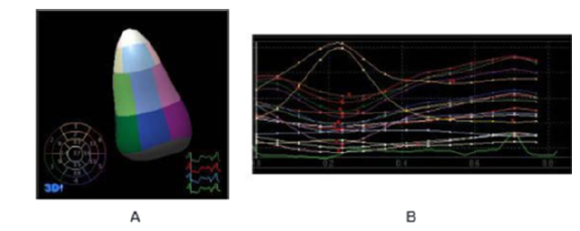
Using Philips Sonos 7500 equipment, all echocardiographic exams were performed at the baseline (days 2±3 post infarction) and during follow-up at 3 months after the infarction. For data processing, measurements and interpretation, all captured images were uploaded to the QLab workstation. Delineation of the endocardial border and the determination of ventricular segments were the first steps in a 3D echocardiographic examination. The American Heart Association advised that the left ventricle be divided into 17 segments. Consequently, 816 ventricular segments from the 48 individuals were examined and a contractility curve was obtained for each segment (Figure 1).
Figure 1: D Echocardiography-Segmentation of the Left Ventricle (a) and Wall Motion Analysis (b) using ontractility Curves for each of the 17 Segments
The greatest contraction amplitude (endocardial excursion), maximum and minimum volume during the cardiac cycle and segmental ejection fraction were all calculated for each segment. We got the global left ventricular end-diastolic and end-systolic volume as well as the 3D-determined ejection fraction after summing these data from all 17 segments. Using 3D echocardiography, serial changes in the global LV volume were measured at baseline and three months later to evaluate remodelling.
Definitions
The greatest contraction amplitude for all segments added together and divided by the total number of segments was called the Index of Contraction Amplitude (ICA). The maximal contraction amplitude for the segments irrigated by the infarct-related artery divided by the total number of these segments was defined as the Regional Index of Contraction Amplitude (RICA). An increase in LV end-diastolic global volume of more than >15% from baseline was considered Positive Remodelling (PR).We looked at the relationship between these parameters from 3D echocardiography and the degree of remodelling after myocardial infarction, which was measured by serial changes in global LV volume using 3D echocardiography at baseline and 3 months later.
Statistical Analysis
The statistical programme JMP 10 was used to conduct the statistical analysis (SAS Institute Inc., Cary, North Carolina). In order to compare the baseline characteristics of patients between the PR and non-PR populations, we employed Fisher's exact test (or Student's t-test for age). The Mann-Whitney test was used to establish the statistical significance of continuous values, which are represented as the mean and standard deviation. Percentages are used to express categorical variables. A p value of <0.05 or lower was regarded as statistically significant and all p values were two-sided. We used logistic regression analysis to examine the accuracy of the variables in predicting the onset of ventricular remodelling for individuals with p<0.05.
RESULTS
Twenty-seven patients experienced positive remodeling (PR group) following the infarction and 21 patients showed no signs of remodeling (non-PR group). The clinical baseline characteristics of the study population showed no significant differences between the PR and non-PR group in respect to age (p = 0.5), gender (p = 0.2), the presence of diabetes (p = 0.7), hypertension (p = 0.7), hyperlipidemia (p = 0.4), obesity (p = 0.4), smoking status (p = 0.2) or comorbidities (p = 0.3), as presented in Table 1.
Table 1: Comparison of 3D Echo Characteristics at Baseline in Patients with PR versus Non-PR
Parameters
Group 1-positive remodeling
Group 2-no remodeling
p value
LV end-diastolic volume (mL)
Mean±SD
95% confidence interval
146.34±28.92
134.66–158.03
140.82±20.20
131.86–149.78
0.5
LV end-systolic volume (mL)
Mean±SD
95% confidence interval
80.34±14.58
74.45–86.23
70.63±13.32
64.72–76.54
0.02
Ejection fraction (%)
Mean±SD
95% confidence interval
44.75±4.3
43.01–46.49
49.95±5.43
47.54–52.36
0.009
ICA
Mean±SD
95% confidence interval
3.05±0.60
2.80–3.30
3.53±0.47
3.32–3.74
0.01
RICA <0.0001
Mean±SD
95% confidence interval
1.38±0.45
1.20–1.57
2.78±0.88
2.39–3.17
<0.0001
3D Echo Analysis of Global and Regional Contractility at Baseline
The 3D echo analysis of left ventricular global and regional contractility demonstrated that patients who developed post infarction LV remodeling tend to have at lower ejection fraction (44.75% vs. 49.95%, p = 0.009), larger endsystolic volumes (80.34 ml vs. 70.63 ml, p = 0.02), lower index of global contraction amplitude (3.05 vs. 3.53, p = 0.01) and lower index of regional contraction amplitude (1.38 vs. 2.78, p<0.0001) at baseline than patients who did not develop left ventricular remodeling (Figure 2).
Figure 2:3D Echocardiographic Characteristics at Baseline in the PR vs. Non-PR Group
However, the highest statistical significance to differentiate between the two groups was reached for the index of regional contraction amplitude (Table 2).
3D Echo Analysis of LV Remodeling
Despite successful first PCI, a sizable portion of patients experienced favourable remodelling. By using 3D echo to evaluate the remodeled ventricles, it was discovered that the ventricular cavity had enlarged at the site of the infarction (Figure 3).
Figure 3: Remodeled Left Ventricle following an AMI-3D Echo Analysis Indicating a Pronounced Dilatation in the Apical Area
The amplitude of LV remodelling had the highest correlation with the RICA, a parameter expressing the regional contractility in the infarction border zone, according to a linear regression analysis of 3D echocardiography-derived parameters in relation to the extent of remodelling at 3 months post infarction (Figure 4).
Figure 4:Correlation between Remodeling Index and 3D Echo Derived Parameters of Ventricular Contraction and Volume
3D Echo based Predictors of LV Remodeling
RICA was shown to be a good indicator for anticipating the onset of LV remodelling (area under the curve 0.88), according to a logistic analysis based on receiver-operator characteristics (ROC) curves of the 3D echo generated parameters that substantially correlated with LV remodelling (Figure 5).
Figure 5: Liniar Regression Analysis for Echo based Predictors of LV Remodeling
The development of LV remodelling in the post-infarction era could be predicted with high sensitivity (92%), 90% positive predictive value and a cut-off point of 1.8 regional index of contraction amplitude.
Compared to the other measures examined, the accuracy of RICA in predicting LV remodelling was much higher (AUC = 71 for ICA, 78 for EF, 55 for EDV and 68 for ESV). However, only 26% of people were able to predict ventricular remodelling using this measure (Table 2).
Table 2:Sensitivity, Specificity and Predictive Values for Parameters Expressing Ventricular Function
Parameters
AUC
Cut-off value
Specificity (%)
Sensitivity (%)
PPV (%)
NPV (%)
RICA
0.88
1.80
26.00
92.00
90.00
79.31
ICA
0.71
3.20
73.00
65.00
73.91
64.00
FE
0.78
46%
87.00
61.00
84.21
65.52
VED
0.55
167.00
95.50
30.00
88.89
53.85
VES
0.68
79.00
78.00
65.00
73.91
64.00
DISCUSSION
When done within the advised period, successful revascularization of acute myocardial infarction should restore the function of the injured myocardium. This improvement in circulation ought to halt the ventricular remodelling process, which has a detrimental effect on patients' prognoses [9,10]. What fraction of patients is more likely to respond to this medication, however, is not yet known. A precise difference between prospective "responders" or "non-responders" to revascularization is not conceivable given the existing state of knowledge.
In this study we demonstrated that there is a strong correlation between alterations in regional contractility of the border zone areas adjacent to infarction, immediately after infarction and the development of left ventricular remodeling. In our experience, the impairment of regional contractility was associated with the development of LV remodeling to a more significant extent than the global impairment of ventricular contractility.
Continuity of contraction in the vicinity of the myocardial boundary infarction is influenced by a number of factors, including the degree of ischaemia in this region, the structure of the coronary circulation, the micro-circulation and the collaterals [11]. All These causes of regional contractility include a variety of variations or changes in the acute phase on a scale infarctions of the myocardium [12,13]. Consequently, given the context evaluation of an acute coronary event should consider not only the extent of the damage, as well as the state of the border zone as a significant factor influencing ventricular function, regional or worldwide. Despite the recent recognition of the significance of regional contractility in predicting subsequent cardiac events following an acute coronary event, no study has examined the impact of changes in regional contractility on the onset and progression of ventricular remodelling. In this investigation, regional wall motion quantification using 3D echocardiography yielded new characteristics that might be used to predict the onset of left ventricular remodelling in patients with acute myocardial infarction receiving primary PCI. A new metric created by 3D echocardiography that is simple to measure and provides an objective framework for evaluating absolute regional contractility is the index of regional contraction amplitude.
Despite successful repermeabilization of the occluded coronary artery, a low index of regional myocardial contraction in the infarct region had the strongest correlation with the presence and extent of the LV remodelling process in our study, outperforming more traditional parameters like ventricular volumes or ejection fraction at baseline. This finding sheds new light on the need for more thorough revascularization following acute myocardial infarction because partial revascularization, which leaves a coronary artery stenosis in a neighbouring, non-infarcted vessel untreated, may result in decreased regional contractility, which may then hasten or accelerate the remodelling process. Another open question raised by these findings is whether this index of regional contraction could be used to predict the regression of remodelling following reopening of chronic total occlusions. It has been established that regional contractility plays a significant role in the beginning and continuation of the ventricular remodelling process. However, a more complicated strategy would be required in this situation because the preservation of myocardial viability is also necessary for the reversal of remodelling and regional contractility.
CONCLUSION
In conclusion, using a 3D based objective assessment of ventricular volumes and quantification of regional wall motion, our study showed that impairment of regional contractility in the border area of the infarction was associated with development of LV remodeling to a more significant extent than the ventricular volumes or the global impairment of ventricular contractility. This underlines the role of regional contractility immediately after the infarction in predicting future evolution of these patients.
Conflicts of Interest
The authors declare that there are no conflicts of interest regarding the publication of this paper.
REFERENCES
Wenk, J.F. et al. “First evidence of depressed contractility in the border zone of a human myocardial infarction.” Annals of Thoracic Surgery, vol. 93, 2012, pp. 1188–1193.
Lipiecki, J. et al. “Different patterns of left ventricular enlargement and long-term prognosis after reperfused acute myocardial infarction.” Archives of Cardiovascular Diseases, vol. 102, 2009, pp. 599–605.
Ghanem, A. et al. “Quantitation of myocardial borderzone using reconstructive 3-d echocardiography after chronic infarction in rats: Incremental value of low-dose dobutamine.” Ultrasound in Medicine and Biology, vol. 34, 2008, pp. 559–566.
Barletta, G. et al. “Left Ventricular shape and function in primary coronary angioplasty.” International Journal of Cardiology, vol. 125, 2008, pp. 364–375.
Carluccio, E. et al. “Patients with hibernating myocardium show altered left ventricular volumes and shape, which revert after revascularization: Evidence that dyssynergy might directly induce cardiac remodeling.” Journal of the American College of Cardiology, vol. 47, 2006, pp. 969–977.
Silva, J.C. et al. “Late coronary artery recanalization effects on left ventricular remodelling and contractility by magnetic resonance imaging.” European Heart Journal, vol. 26, 2005, pp. 36–43.
Danchin, N. et al. “Effect of late revascularization of the responsible artery after infarction on left ventricular function and remodeling.” Archives des Maladies du Cœur et des Vaisseaux, vol. 90, 1997, pp. 47–51.
Sutton, M.J. and N. Sharpe. “Left ventricular remodeling after myocardial infarction: Pathophysiology and therapy.” Circulation, vol. 101, 2000, pp. 2981–2988.
Bolognese, L. et al. “Left Ventricular remodeling after primary coronary angioplasty: Patterns of left ventricular dilation and long-term prognostic implications.” Circulation, vol. 106, 2002, pp. 2351–2357.
Nijland, F. et al. “Myocardial viability: Impact on left ventricular dilatation after acute myocardial infarction.” Heart, vol. 87, 2002, pp. 17–22.
Jenkins, C. et al. “Reproducibility and accuracy of echocardiographic measurements of left ventricular parameters using real-time three-dimensional echocardiography.” Journal of the American College of Cardiology, vol. 44, 2004, pp. 878–886.
Mannaerts, H. et al. “Early identification of left ventricular remodelling after myocardial infarction, assessed by transthoracic 3d echocardiography.” European Heart Journal, vol. 25, 2004, pp. 680–687.
Udelson, J.E. “The Occluded Artery Trial (OAT) Viability Ancillary Study (OATNUC): Influence of infarct zone viability on left ventricular remodeling after percutaneous coronary intervention vs. optimal medical therapy alone.” American Heart Journal, vol. 161, 2011, pp. 611–621.
Recommended Articles
Research Article
Vision for a Brighter Kangra: Unmasking the Truth about Pink Eye – A Comprehensive Study on Types, Symptoms, and Proactive Prevention in Himachal Pradesh’s Kangra District
Deepti ,
...
Garima
Published: 11/11/2023
Download PDF
Research Article
It Remains Unproven That the Variant M.8231C>A Causes Coronary Atherosclerosis
Finsterer J
Published: 15/07/2020
Download PDF
Research Article
Leigh Syndrome Should Not Be Diagnosed Exclusively Upon Cerebral MRI
Finsterer J
Published: 15/07/2020
Download PDF
Research Article
Bioactive ingredients screening and antibacterial activity of Citrus aurantifolia leaf extract against some enteric isolates
Abdallah M.S,
Kabiru Sama’ila
Published: 10/08/2020
Download PDF
PO Box 101, Nakuru
Kenya.
Email: office@iarconsortium.org
Editorial Office:
J.L Bhavan, Near Radison Blu Hotel,
Jalukbari, Guwahati-India
Dahal, T., Chaure, A. & Tamang, A. (2022). Link between Left Cavity Remodeling and Acute Alteration of Regional Contractility after Re-Vascularized Critical Myocardial Infarction. IAR Journal of Medical Sciences, 3(2), 1-5.
MLA
Dahal, Tshetiz, Ashish Chaure and Anil Tamang. "Link between Left Cavity Remodeling and Acute Alteration of Regional Contractility after Re-Vascularized Critical Myocardial Infarction." IAR Journal of Medical Sciences 3.2 (2022): 1-5.
Chicago
Dahal, Tshetiz, Ashish Chaure and Anil Tamang. "Link between Left Cavity Remodeling and Acute Alteration of Regional Contractility after Re-Vascularized Critical Myocardial Infarction." IAR Journal of Medical Sciences 3, no. 2 (2022): 1-5.
Harvard
Dahal, T., Chaure, A. and Tamang, A. (2022) 'Link between Left Cavity Remodeling and Acute Alteration of Regional Contractility after Re-Vascularized Critical Myocardial Infarction' IAR Journal of Medical Sciences 3(2), pp. 1-5.
Vancouver
Dahal T, Chaure A, Tamang A. Link between Left Cavity Remodeling and Acute Alteration of Regional Contractility after Re-Vascularized Critical Myocardial Infarction. IAR Journal of Medical Sciences. 2022 Jul;3(2):1-5.



 Download PDF
 Download XML