Contents
Download PDF
pdf Download XML
101 Views
30 Downloads
Share this article
Research Article | Volume 3 Issue 2 (Jul-Dec, 2022) | Pages 1 - 8
Clinical Presentations of Idiopathic Thrombocytopenic Purpura
 ,
1
Al-Batool Teaching Hospital, Diyala, Iraq
Under a Creative Commons license
Open Access
Received
April 3, 2022
Revised
May 9, 2022
Accepted
June 19, 2022
Published
July 10, 2022
Abstract

Background: ITP is the most common cause of thrombocytopenia in children and is a result of immunologically mediated increased platelet destruction, which is usually acute and self-limiting but may be a recurrent or chronic disorder. Aim of study: Assessment of clinical manifestation of ITP in children. Patient and Methods: A prospective study was performed on 60 patients diagnosed as idiopathic thrombocytopenic purpura admitted to central child teaching hospital from 1st of January to 3o September 2011. Diagnosis was based on clinical and laboratory tests including bone marrow aspiration. Results: The age ranged from 6 months to 13 years and majority of cases were between 1-5 years, 68.3% were males and 31.7% were females, male to female ratio 2.1: 1History of upper respiratory tract infection preceded the onset of illness in 71.7% of patients. Where the Duration between Preceding URTI and ITP Up to 1 week in 30.2% and >1 week in 79.8% Concerning the distribution of cases according to the season of the year: One case (1.7%) found during January, four cases (6.7%) during February, six cases (10.0%) during March, five cases (8.3%) during April, ten cases (16.7%) during May, eight cases (13.3%) during June, 14 cases (23.3%) during July , five cases (8.3%) during August and seven cases (11.7%) during September. This distribution was significant (p<0.05). The commonest presentation was cutaneous bleeding in 60%, Mucous Membrane bleeding 1.7%, both 38.3%Splenomegaly with bleeding 6.7%, Hepatomegaly with bleeding 8.3%, Lymphadeno-pathy with bleeding 5%. In 69% of cases the initial platelet count was less than 20.000/mm³ 37.9% had anemia. Bone marrow examination done in 56 case and all were normal. In this study, one case die due to intracranial hemo– rrhage we didn’t include the management because the patients receive different types of treatments (mostly by steroid, some by IVIG and anti-D), most of patients examined by bone marrow aspiration which was normal and also we didn’t include the follow up because of most of the patients didn’t come for follow up. Conclusion: The disease is self-limited most cases at age of 1-5 years, male affected more than female, most cases preceded by upper respiratory tract infections , most cases presented by skin bleeding.

Keywords
INTRODUCTION

Immune (Idiopathic) Thrombocytopenic Purpura: Immune Thrombocytopenic Purpura (ITP) is a syndrome characterized by:


  • Thrombocytopenia (platelet count less than 150,000/mm3)

  • Shortened platelet survival

  • Presence of antiplatelet antibody in the plasma

  • Increased megakaryocytes in the bone marrow


This condition may be acute, chronic, or recurrent. In the acute form, the platelet count returns to normal (>150,000/mm3) within 6 months after diagnosis. In the chronic form, the platelet count remains low beyond 6 months. In the recurrent form, the platelet count decreases after having returned to normal levels. In adults, the chronic form is more common, whereas in children, the acute form is more common [1]. In acute ITP, it is associated with purpura, petechiae, mucocutaneous bleeding and occasionally hemorrhage into internal tissues [2]. In 70–80% of children who present with acute ITP, spontaneous resolution occurs within 6 mo. Approximately 20% of children who present with acute ITP go on to have chronic ITP [2]. 


Etiology and Pathogenesis

The most frequent cause of ITP is immune mediated platelet destruction due to auto antibodiesA recent history of viral illness is described in 50 _ 65% of cases of childhood ITP. The reason why some children respond to a common infection with an autoimmune disease remain unknown [1,2]. Most common infectious viruses have been described in association with ITP, including rubella, variclla, measles, parvovirus, influenza, EBV and HIV [3]. Other infections that might cause ITP are; Helicobacter pylori. Cytomegalovirus and hepatitis C virus [4,5]. Acute ITP had been also observed following vaccination in children notably; Measles-Mumps-Rubella vaccine (MMR), Pneumococcus, Haemophilus influenzae b, hepatitis B vaccine and varicella zoster vaccine [6,7]. After binding of the antibody to the platelet surface, circulating antibody-coated platelets are recognized by the Fc receptor on the splenic macrophages, ingested and destroyed [2]. In the circulation platelet survival is markedly decreased, splenic sequestration accounts for the shortened survival in most patients, but the liver and even the reticuloendothelial cells of the bone marrow, can play a major role in the sequestration of antibody-coated platelets, especially in patients with very low platelet counts or continued thrombocytopenia after splenectomy. The spleen has also been implicated as a site of antibody formation [8].


Incidence

The true incidence of ITP is unknown because the disease is often transient. The estimated incidence is about 1 in 10,000 children per year [1,9].


Clinical Manifestations

The classic presentation of ITP is that of a previously healthy 1–4 yr old child who has sudden onset of generalized petechiae and purpura. There is a history of a preceding viral infection 1–4 wk before the onset of thrombocytopenia. Often there is bleeding from the gums and mucous membranes, particularly with profound thrombocytopenia (platelet count <10×109/L) [2,10,11]. Bleeding from gastrointestinal tract, or kidneys is not uncommon. Menorrhagia may occur and may be severe. Hematemesis and melena are infrequent [1]. Findings on physical examination are normal, other than the finding of petechiae and purpura. Splenomegaly is rare, as is lymphadenopathy or pallor. An easy to use classification system has been proposed from the U.K. to characterize the severity of bleeding in ITP on the basis of symptoms and signs, but not platelet count:


  • No Symptoms

  • Mild Symptoms: Bruising and petechiae, occasional minor epistaxis, very little interference with daily living

  • Moderate: More severe skin and mucosal lesions, more troublesome epistaxis and menorrhagia

  • Severe: Bleeding episodes-menorrhagia, epistaxis, melena-requiring transfusion or hospitalization, symptoms interfering seriously with the quality of life


The presence of abnormal findings, such as hepatosplenomegaly or remarkable lymphadenopathy, suggests other diagnoses (leukemia) [2,10,12]. Fewer than 1% of patients have intracranial hemorrhage.


Laboratory Findings

Platelet Count


  • Always less than 150,000/mm3

  • Often less than 20,000/mm3 in patients with severe generalized hemorrhagic manifestations.

  • MPV increased (on automated counting equipment); normal MPV,8.9±1.5\m3


Blood Smear


  • Blood smear normal, apart from thrombocytopenia (if active infection, increased neutrophils, lymphocytes, or atypical mononuclear cells may be present)

  • Anemia present only in proportion to amount of blood loss


Bone Marrow


  • Increased megakaryocytes, often immature and with absence of budding

  • Normal erythroid and myeloid cells

  • Increased eosinophils occasionally seen

  • Erythroid hyperplasia if significant blood loss.

  • (Marrow findings are diagnostic of ITP only when consistent with the clinical state and presence of a low platelet count and when other causes of secondary thrombocytopenia are excluded. The main purpose of performing a bone marrow examination is to exclude other hematologic disorders, e.g., (leukemia.)


Coagulation Profile


  • Bleeding time-usually abnormal

  • Prothrombin Time (PT), Activated Partial Thromboplastin Time (APTT) and fibrinogen level-normal [1]


Other laboratory tests should be done as indicated by the history and physical examination. In adolescents with new-onset ITP, an antinuclear antibody test should be done to evaluate for SLE. A Coombs test should be done if there is unexplained anemia to rule out Evans syndrome (autoimmune hemolytic anemia and thrombocytopenia) or before instituting therapy with IV anti-D [2,11]. 


Differential Diagnosis

The well-appearing child with moderate to severe thrombocytopenia, an otherwise normal complete Blood Cell Count (CBC) and normal findings on physical examination has a limited differential diagnosis [2].


ITP must be differentiated from:


  • Exposure to some medications may give clinical picture similar to ITP (e.g., heparin, quinine, sulfa, rifampin, carbamazepin, valproic acid and H1 blocker)

  • Familial non immune thrombocytopenia: 

  • Wiskott-Aldrich Syndrome: X-linked recessive immunodeficiency characterized by thrombocytopenia , eczema and recurrent infections 

  • Bernard Soulier Syndrome: There is a deficiency in glycoprotein Ib lead to defect in adhesion. Platelet are giant and few and do not aggregate with ristocetin 

  • A megakaryocytic thrombocytopenia 


These disorders are suspected when the patient fail to respond to therapy and by finding on bone marrow aspiration:


  • Disorders that associated with ITP, like HIV infection, SLE, humoral immunodeficiency, especially when there is atypical presentation

  • Other cytopenias; aplastic anemia, leukemia, myelodysplasia, Evans syndrome [12]


Treatment

Acute ITP in childhood is usually a self-limiting condition. Medical treatment does not seem to alter the natural history of the disease. Treatment may be required to minimize the danger of life-threatening intracranial hemorrhage, a more rapid rise in platelet count to the theoretically safe level of >20×109/L, although there are no data indicating that early therapy prevents intracranial hemorrhage. Antiplatelet antibodies bind to transfused platelets as well as they do to autologous platelets. Thus, platelet transfusion in ITP is usually contraindicated unless life-threatening bleeding is present [2,10]. Treatment is indicated for children with platelet counts less than 20,000/mm3 and significant mucous membrane bleeding and those with platelet counts less than 10,000/mm3 and minor purpura [1].


General Measures

The general management of children with ITP includes: 


  • Avoiding aspirin and other drugs that can impair platelet function

  • Avoiding intramuscular injection

  • If the platelet count is very low, children should avoid stimulus which carries significant risks of severe trauma [13]


Initial approaches to the management of ITP include the following:


No Therapy

Other than education and counseling of the family and patient for patients with minimal, mild and moderate symptoms, as defined earlier. This approach emphasizes the usually benign nature of ITP and avoids the therapeutic roller coaster that ensues once interventional therapy is begun. This approach is far less costly and side effects are minimal [2].


Intravenous Immunoglobulin (IVIG)

IVIG at a dose of 0.8–1.0 g/kg/day for 1–2 days induces a rapid rise in platelet count (usually>20×109/L) in 95% of patients within 48 hr. IVIG appears to induce a response by down regulating Fc-mediated phagocytosis of antibody-coated platelets. IVIG therapy is both expensive and time-consuming to administer. Additionally, after infusion, there is a high frequency of headaches and vomiting, suggestive of IVIG-induced aseptic meningitis [2].


IVGG Toxicity


  • Anaphylaxis in IgA-deficient patients because of preexisting IgA antibodies that react with small amounts of IgA present in commercially available gamma globulin

  • Post infusion headache in 20% of patients; transient and possibly severe (in severe cases, administer postinfusion dexamethasone 0.15–0.3 mg/kg IV). Severe headache in ITP may suggest the presence of intracranial hemorrhage and if clinically indicated may require a CT scan

  • Fever and chills in 1–3% of patients; prophylactic acetaminophen (10–15 mg/kg, 4 hourly, as required) and diphenhydramine (1 mg/kg, 6–8 hourly, as required) to reduce the incidence and severity

  • Coombs-positive hemolytic anemia because of the presence of blood group antibodies (anti-A, anti-B and anti-D) present in IVGG

  • Hepatitis C Virus (HCV) infection reported in patients receiving IVGG; however, no reports of HIV infection with any licensed preparation in the United States has been described [1]


Intravenous Anti-D Therapy

For Rh positive patients, IV anti-D at a dose of 50–75μg/kg causes a rise in platelet count to>20×109/L in 80–90% of patients within 48–72 hr. When given to Rh positive individuals, IV anti-D induces mild hemolytic anemia. RBC-antibody complexes bind to macrophage Fc receptors and interfere with platelet destruction, thereby causing a rise in platelet count. IV anti-D is ineffective in Rh negative patients.


Adverse Drug Reactions


  • Fever

  • Chills

  • Headache


Drop in hemoglobin and hematocrit attributed to anti-D-related hemolysis. These symptoms are directly attributable to anti-D-related hemolysis and a positive Coombs reaction [1,2,13,14].


Prednisone

Corticosteroid therapy has been used for many years to treat acute and chronic ITP in adults and children. Doses of prednisone of 1–4 mg/kg/24 hr appear to induce a more rapid rise in platelet count than in untreated patients with ITP. Whether bone marrow examination should be performed to rule out other causes of thrombocytopenia, especially acute lymphoblastic leukemia, before institution of prednisone therapy in acute ITP is controversial [2].


The mechanism of action of steroids in ITP is multifactorial; by inhibiting phagocytosis of antibody coated platelets in the spleen and prolonging platelet survival, by improving capillary resistance and inhibiting platelet antibody production. A course of prednisone, 2 mg/kg/day (maximum 60 mg/day), is given in divided doses. Prednisone is reduced in stepwise fashion at 5 to 7 days intervals irrespective of the platelet count and is stopped at the end of 21-28 days, regardless of the response. A shorter course of prednisone at 4 mg/kg/day for 4 days has also been used with success [4]. In severe cases, methylprednisolone (Solu-medrol) 30 mg/kg/day, (maximum1g/day) for 3 days produces a more rapid response than steroids in conventional doses [1]. Prolonged use of steroids in ITP is undesirable. Large doses or prolonged steroid usage may perpetuate the thrombocytopenia and depress platelet production. It also leads to side effects including weight gain, cushingoid facies, fluid retention, acne, hyperglycemia, hypertension, mood swings, pseudotumor cerebri, cataracts, growth retardation and avascular necrosis [1]. Each of these medications may be used to treat exacerbations of ITP, which commonly occur several wk after an initial course of therapy. In the special case of intracranial hemorrhage, multiple modalities should be used, including platelet transfusion, IVIG, high-dose corticosteroids and prompt surgical consultation, with plans for emergency splenectomy [2].


Splenectomy

Splenectomy should be reserved for patients with acute life threatening bleeding and those whose symptoms lasted more than one year (chronic ITP) and older than 4 years. Its effective in controlling thrombocytopenia in 64 – 88% of cases [2]. Splenectomy is associated with lifelong risk of overwhelming sepsis by encapsulated organisms. Before splenectomy corticosteroids, IVIG, or anti-D given to raise platelet count [1]. Presplenectomy immunization with meningococcal, pneumococcal and haemophilus influenza type b vaccines and subsequent penicillin prophylaxis are necessary for all age groups [4]. When possible, surgery should be performed using laparoscopic technique [15].


Other Modalities of Treatment

Are uncommonly used and tried only after failure of response to immunoglobulin, steroids, or splenectomy, these include [1,8,16]: 


  • Immunosuppressive therapy like: danazol, vincristine, vinblastine, cyclosporine, cyclophosphamide and azathioprine

  • Rituximab

  • Plasmapheresis

  • Platelet transfusion


Management of Life Threatening Hemorrhage: 


  • Platelet transfusion

  • Methylprednisolone 500 mg/m² IV per day for 3 days

  • Intravenous immunoglobulin 2g/kg

  • Emergency splenectomy


These measures can be used singly or in combination, depending on the severity and response to treatment. Patient’s refractory to these measures may benefit from vincristine sulfate (1.5 mg/m² IV maximum 2 mg) and plsmapheresis [1].


 Prognosis


  • Excellent; 50% recover usually within 1 month and 70–80% recover within 6 months

  • Spontaneous remission after 1 year is uncommon, although may occur even after several years

  • Age older than 10 years, insidious onset and female gender are associated with the development of chronic ITP

  • Of all chronic patients, 50–60% eventually stabilize without any therapy and without need for splenectomy [1]


Aims of the Study

Assessment of clinical manifestation of ITP in children.


To enlighten the possible risk factors of acute ITP regarding to age, sex, residence, history of antecedent event and date variation.

MATERIALS AND METHODS

To Study the Laboratory Finding at Diagnosis: A prospective study was performed on 60 patients diagnosed as acute idiopathic thrombocytopenic purpura, who were admitted to central child teaching hospital from 1st of January to 3o September 2011. Data were collected according to preceded questionnaire from information regarding age, sex, date of admission, Residence, Rural or Urban, Antecedent event (the events like upper respiratory tract infections, fever, etc.), Onset of event, Duration between antecedent event and generalized petechiae and purpura, site of bleeding, associated clinical features, lab tests which include CBC, blood smear, bone marrow exam. 


The criteria of inclusion for the study group were:


  • Signs and symptoms of ITP

  • The necessary laboratory investigation


Stastical Analysis

Statistical Package for Social Sciences version 18 (SPSS 18) was used for data input and analysis. Discrete variables presented as numbers and percentages. Continuous variables presented as mean and Standard Deviation (SD). Chi square test for independence used to test the significance of association between two discrete variables. T test used to test the significance of difference in mean between two independent samples. Chi square test for goodness of fit used to test the significance of observed distribution. Findings with P value less than 0.05 were considered significant.


Note

P value is the probability of error/chance (sampling error, error because of sampling). The finding is not considered significant if the probability of error more than 5% (p>0.05), here you cannot say there is no relation but say there is no significant relation because still the probability of trueness is more than 90% in some instances but the probability of chance is high (>5%).

RESULTS

Age

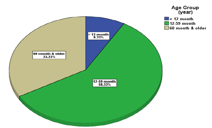
The sample is consisted of 60 child aged 1 to 156 months with mean age 49.2±38.4 month. Dominant age group was 12-59 months (1-5 years) (p<0.05, Table 1 and Figure 1).


Table 1: Demographic Characteristics of Study Sample

Variables

N (%)

Chi-squarep value

60 (100.0)

Age (month); mean±SD

49.2±8.4

---

---

Age Group

<12 month

5 (8.3)

22.500

0.006

12-59 month

35 (58.3)

≥60 month

20 (33.3)

Gender

Male

41 (68.3)

8.067

0.005

Female

19 (31.7)

Residence

Urban

34 (56.7)

1.067

0.302

Rural

26 (43.3)

N; number, %; percent, SD; Standard Deviation


Figure 1: Distribution of Study Sample According to Age


Sex Differences

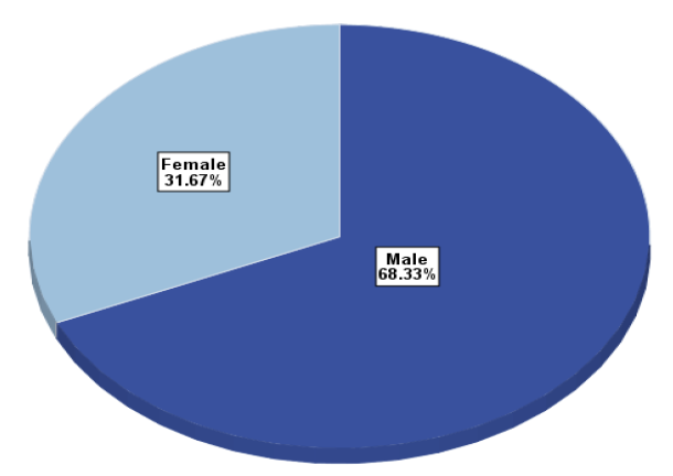
As shown in Table 1, Males were 41 (68.3%) and females 19 (31.7%) and males significantly constituted about two thirds of cases (p<0.05, Table 1 and Figure 2).


Figure 2: Distribution of Study Sample According to Gender


Residence (Rural or Urban)

Thirty four patients constitute (56.7%) of patients were from urban areas and 26 patients constitute (43.3%) from rural areas as shown in (Table 1). while the observed distribution regarding residence was not significant (p>0.05, Table 1).


Date of Admission

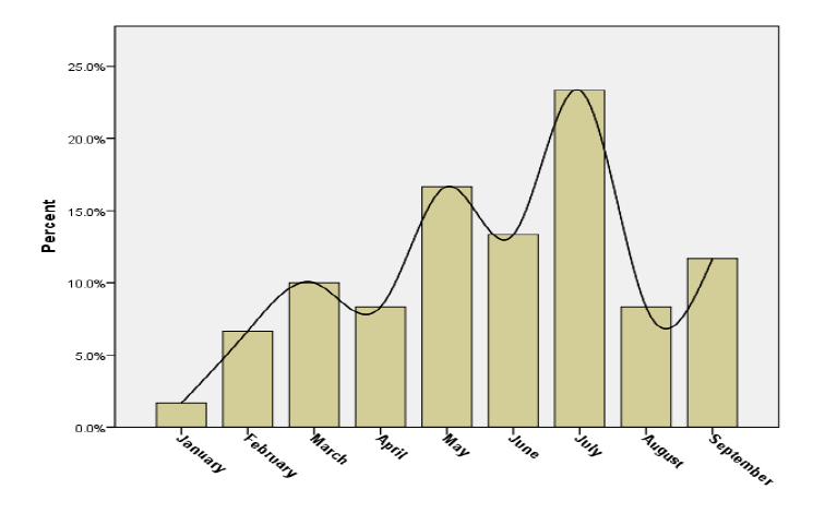
Concerning the distribution of cases according to the month of the year: One case (1.7%) found during January, four cases (6.7%) during February, six cases (10.0%) during March, five cases (8.3%) during April, ten cases (16.7%) during May, eight cases (13.3%) during June, 14 cases (23.3%) during July, five cases (8.3%) during August and seven cases (11. 7%) during September. This distribution was significant (p<0.05, Table 2 and Figure 3). 


Table 2: Distribution of ITP Cases According to the Month of the Presentation

Month of the year

N (%)

Chi-square

p value

January

1 (1.7)


February

4 (6.7)


March

6 (10.0)


April

5 (8.3)

16.800

0.032

May

10 (16.7)


June

8 (13.3)


July

14 (23.3)


August

5 (8.3)


September

7 (11.7)


Total

60(100.0)


Figure 3: Distribution of Study Sample According to Month of the Presentation


The preceding upper respiratory tract infection: 


  • It was significant to find 43 cases (71.7%) preceded with upper respiratory tract infection (p<0.05, Table 3)

  • Duration between upper respiratory tract infections and appearance of ITP lesions was 3 days in minimum, maximum 23 days and in average was 11.6±4.8 days (Table 3)


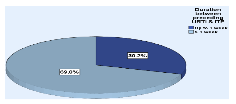
Those preceded with URTIs of more than one week were the majority; 30 cases (79.8%). This distribution was significant (p<0.05, Table 3 and Figure 4).


Figure 4: Distribution of Study Sample According to Duration between Preceding URTI and Onset of ITP


Table 3: Relation between ITP and Preceding URTI

VariablesN (%)Chi-squarep value

60 (100.0)

Preceding URTI

43 (71.7)

9.600

0.002

Duration between Preceding URTI and ITP (days); mean ± SD

11.6±4.8

---

---

Duration between Preceding URTI and ITP 


Up to 1 week

13 (30.2)


>1 week

30 (79.8)

6.721

0.010

Total

43 (100.0)


N; Number, %; Percent, SD; Standard Deviation; URTI; Upper Respiratory Tract Infection


Table 4: Clinical Findings for the Study Sample

FindingsN (%) 60 (100.0)Chi-squarep value
Presenting Site of ITP
Skin36 (60.0)  
Mucous Membrane1 (1.7)31.3000.034
Both23 (38.3)  
Splenomegaly with bleeding 4 (6.7)45.0670.005
Hepatomegaly with bleeding 5 (8.3)41.6670.004
Lymphadenopathy with bleeding 3 (5.0)48.6000.004

N; Number, %; Percent


Clinical Manifestations

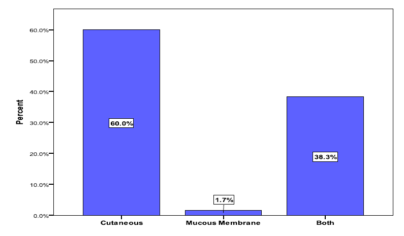
Table 4 and Figure 5, show the clinical features of cases presented in our study , presenting site of bleeding is skin 60%, skin and mucuos memberane 38.3%, mucuos memberane only 1.7% Splenomegaly with bleeding 6.7%, Hepatomegaly with bleeding 8.3%, Lymphadenopathy with bleeding 5%.


Figure 5: Distribution of Study Sample According to Site of Presenting ITP Lesions


Lab Findings

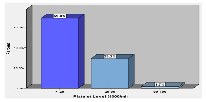
Table 5 and Figure 6, show the initial lab finding.

There is one case dead due to intracranial hemorrhage noticed during this study 

Bone Marrow Examination

Sixty fife patients had bone marrow exam. , All of which were normal. For patients didn’t examine the bone marrow due to family rejection.


Figure 6: Distribution of Study Sample According to Platelet Level


Table 5: Laboratory Findings for the Study Sample

FindingsN (%)Chi-squarep value
60 (100.0)
PCV%; mean±SD32.5 ± 5.4------
Presence of Anemia   
Yes22(37.9)  
No36(62.1)3.3790.066
 Total                                     60(100.0)  
Platelets (109/ml) ; mean±SD13.7 ± 11.6------
Platelet Level (109/ml)   
< 2040(69.0)  
20-5017(29.3)39.7590.005
50-1501(1.7)  
Total60(100.0)  

N; Number, %; Percent, SD; Standard Deviation; URTI; Upper Respiratory Tract Infection

DISCUSSION

In our study we want to clarify the most common presenting features of ITP in attempt to identify the risk factors of this disease, but it is a descriptive study, so we didn’t include the management because the patients receive different types of treatments (mostly by steroid, some by IVIG and anti-D), most of patients examined by bone marrow aspiration which was normal and also we didn’t include the follow up because it was poor. In this study the distribution of cases according to the age and sex of the patients; Males were 41 (68%) and females 19 (31.7%). And males significantly constituted about two thirds of cases. Which compatible with [17] and in [1], but incompatible with [18-20]. 


In our study the dominant age group was 12- 59 month (66.6%), this result was consistent with many studies like [18,20-23]. History of preceding infection usually viral upper respiratory infection is noted in 71.7% that’s similar to a finding by [17,18,20,21]. The distribution of cases according to the duration of the disease between the antecedent event and generalized petechiae and purpura in which the highest duration noticed was more than 1 wk in which 79.8% of cases, while the least patient recorded of duration less than 1 wk 30.2%, which is against what reported by [17,24]. The distribution of cases according to the month of the year: One case (1.7%) found during January, four cases (6.7%) during February, six cases (10.0%) during March, five cases (8.3%) during April, ten cases (16.7%) during May, eight cases (13.3%) during June, 14 cases (23.3%) during July, five cases (8.3%) during August and seven cases (11.7%) during September. The largest number in July and May, may be due to increased cases of viral infections. The distribution of cases according to the residence: Where 34 (56.7%) four patients constitute (56.7%) of patients were from urban areas and 26 patients constitute (43.3%) from rural areas. While the observed distribution regarding residence was not significant (p>0.05). The presenting signs and symptoms were skin bleeding (petechiae and purpura) 60% which is the highest, Mucous Membrane (epistaxis and gum bleeding) 1.7%, both 38.3%, the least presentations were Splenomegaly with bleeding 6.7%, Hepatomegaly with bleeding8.3%, Lymphadenopathy with bleeding 5%, these abnormal findings may be due to the causative agent like viral disease. All of these were consistent with [17,18,20,21]. Majority of our cases had initial platelet count <20.000/mm³ (69% of cases) from 20-50 (29.3%) more than 50 (1.7%), a finding that's similar to [18,20,21]. where's anemia present in 37.9% which may be due to bleeding or nutritional anemia.

CONCLUSION
  • The majority of cases of ITP and children had mild bleeding symptoms and self-limited

  • The most affected patients were between 1-5 yr age group

  • Male affected more than female

  • Most of cases preceded by upper respiratory tract infection or by fever

  • The most presenting sign was skin bleeding

  • Bone marrow examination was done routinely in most cases and the results were normal


Recommendations


  • The disease should be suspected in the risky age group of 1-5 yr, who presented with petechia or mucous membrane bleeding

  • The disease is a self-limited and should wait to know the platelets count and the full blood count to decide the treatment


Routine bone marrow examination is not necessary in the typical cases but should be done in all atypical cases.

REFERENCES
  1. Lanzkowsky, P., editor. Manual of Pediatric Hematology and Oncology. Elsevier, 2005, pp. 250–263.

  2. Kliegman, R.M. et al. “Acquired Platelet Disorders-ITP.” Nelson Textbook of Pediatrics, W.B. Saunders, 2007, pp. 2082–2084.

  3. Imbach, P. et al. “Abnormalities of platelet number or function.” Current Diagnosis and Treatment: Pediatrics, edited by W.H.Williams et al., 19th ed., McGraw-Hill, 2008, pp. 831–833.

  4. Cines, D.B. and V.S. Blanchette. “Immune thrombocytopenic purpura.” New England Journal of Medicine, vol. 346, no. 13, 2002, pp. 995–1008.

  5. Cines, D.B. et al. “The ITP syndrome: Pathogenic and clinical diversity.” Blood, vol. 113, no. 26, 2009, pp. 6511–6521.

  6. France, E.K. et al.“Risk of immune thrombocytopenic purpura after measles-mumps-rubella immunization in children.” Pediatrics, vol. 121, no. 3, 2008, pp. e687–e692.

  7. Jadavji, T.A.J. et al. “Thrombocytopenia after immunization of Canadian children, 1992 to 2001.” The Pediatric Infectious Disease Journal, vol. 22, no. 2, 2003, pp. 119–122.

  8. Levine, S.P. “Thrombocytopenia caused by immunologic platelet destruction.” Wintrobe’s Clinical Hematology, edited by J.P. Greer et al., 11th ed., Lippincott Williams and Wilkins, 2003, vol. 2, p. 59.

  9. Craig, J.I.O. et al. “Immune thrombocytopenic purpura.” Principles and Practice of Medicine, 20th ed., Churchill Livingstone, 2006, pp. 1056–1057.

  10. Jonas, M.M. et al.“The liver and bile duct.” Rudolph’s Pediatrics, 21st Edn., McGraw-Hill, 2003, pp. 1133–1150.

  11. Paul, S.J. “Autoimmune thrombocytopenic purpura of childhood.” Nelson Essentials of Pediatrics, R.E. Behrman and R.M. Kliegman (Eds.), 4th Edn., W.B. Saunders, 2002, p. 637.

  12. Bussel, J. and D. Cines. “Immune Thrombocytopenic Purpura.” Hematology: Basic Principles and Practice, R. Hoffman et al. (Eds.), Churchill Livingstone, 2000, pp. 2102–2104.

  13. Tarantino, M.D. “Acute Immune ITP in Children.” Blood Reviews, vol. 16, no. 1, 2002, pp. 19–21.

  14. Gaines, A.R. “Acute Onset Hemoglobinemia and/or Hemoglobinuria Following RhD Immune Globulin Administration in Immune Thrombocytopenic Purpura Patients.” Blood, vol. 95, 2000, pp. 2523–2529.

  15. Kahn, M.J. and K.R. McCrae. “Splenectomy in immune thrombocytopenic purpura: Recent controversies and long-term outcomes.” Current Hematology Reports, vol. 3, no. 5, 2004, pp. 317–323.

  16. Bennet, C.M. et al. “Prospective study of rituximab in childhood and adolescent chronic immune thrombocytopenic purpura.” Blood, vol. 107, 2006, p. 2639.

  17. Khashan, A. Idiopathic Thrombocytopenic Purpura in Children: Clinical Presentation and Complications. Dissertation, Iraqi Committee for Medical Specialization, 2008.

  18. Nariman, F.I. Idiopathic Thrombocytopenic Purpura: Clinical Manifestations, Outcome, Complications and Response to Treatment. Dissertation, Iraqi Committee for Medical Specialization, 1998.

  19. Sawsan, S.A. “Childhood ITP: A retrospective analysis of clinical features and response to treatment.” Journal of the Faculty of Medicine Baghdad, vol. 48, no. 3, 2006, pp. 258–261.

  20. Hussein, K.M. Idiopathic Thrombocytopenic Purpura: Clinical Manifestations and Treatment in Children. Dissertation, Iraqi Committee for Medical Specialization, 2010.

  21. Al-Nadawi, M.N. et al. “ITP: Clinical manifestation and treatment.” Journal of the Faculty of Medicine, vol. 44, no. 1, 2002, pp. 71–76.

  22. Zeller, B. et al. “ITP in childhood in Norway: A prospective population-based registration.” Pediatric Hematology and Oncology, vol. 17, no. 7, 2000, pp. 551–558.

  23. Jacobs, P. et al. “Intravenous immunoglobulin has no advantage over corticosteroid as primary therapy for ITP.” Seminars in Thrombosis and Hemostasis, vol. 24, no. 6, 2003, pp. 605–617.

  24. Erduran, E. et al. “A randomized and comparative study of IVIG and mega-dose methylprednisolone treatment in children with acute ITP.” Turkish Journal of Pediatrics, vol. 45, no. 4, 2003, pp. 295–300.

Recommended Articles
Research Article
Vision for a Brighter Kangra: Unmasking the Truth about Pink Eye – A Comprehensive Study on Types, Symptoms, and Proactive Prevention in Himachal Pradesh’s Kangra District
...
Published: 11/11/2023
Download PDF
Research Article
It Remains Unproven That the Variant M.8231C>A Causes Coronary Atherosclerosis
Published: 15/07/2020
Download PDF
Research Article
Leigh Syndrome Should Not Be Diagnosed Exclusively Upon Cerebral MRI
Published: 15/07/2020
Download PDF
Research Article
Release Kinetic Study of Matrix Type Transdermal Patch Using an Analgesic Drug
...
Published: 10/08/2020
Download PDF
Chat on WhatsApp
Flowbite Logo
PO Box 101, Nakuru
Kenya.
Email: office@iarconsortium.org

Editorial Office:
J.L Bhavan, Near Radison Blu Hotel,
Jalukbari, Guwahati-India
Useful Links
Order Hard Copy
Privacy policy
Terms and Conditions
Refund Policy
Shipping Policy
Others
About Us
Team Members
Contact Us
Online Payments
Join as Editor
Join as Reviewer
Subscribe to our Newsletter
+91 60029-93949
Follow us
MOST SEARCHED KEYWORDS
Copyright © iARCON International LLP . All Rights Reserved.