Breast cancer is the most common carcinoma in women in the western world. Breast cancer is the leading cause of death in those aged 44-50 years. This cross-sectional study was performed to evaluate the role of mammography in the detection of occult malignant breast lesions in high-risk women. The screened women were clinically asymptomatic for breast cancer. The study population was 238 high-risk women in Mosul city; over 13 months. The age range was (35-75) years; the mean age was (42.7 years) in (62.2%) of the total. Ten groups of high-risk women were identified:-Those with a previous history of modified radical mastectomy 89 (37.4)%, mammography was positive in eight cases (8.99%). Of those who have a positive family history of breast cancer in first-degree relatives 68 (28.7%), mammographically positive cases were six (8.8)%. For those with a positive family history of malignancy elsewhere in the body other than the breast 35 (14.8)%, mammography was positive in three (8.6%). Of those with conservative surgery for stage one breast cancer 19(7.98%) (4) cases (21.1%) three were mammographically positive (15.8%) in the ipsilateral breast and one woman in her contralateral breast 5.3%. Nulliparous women about forty years old were 9 (3.8%) and only two were mammographically positive (22.2%). Patients who were subjected to surgery for ovarian tumors 8 (3.4%). Two were mammographically positive (25%). Patients with operations for benign ipsilateral breast lesions were five (2.1%), only one was mammographically positive (20%).Two obese ladies with bulky fatty breasts (0.84%), one of them was mammographically positive (50%). Patients with a history of hysterectomy for fibroids were 2(0.84%), only one was mammographically positive (50%).One woman 74 years old (0.42%) was mammographically positive (100%). Ultrasonography and mammography were performed for 387 breasts. The size of tumors detected was in the range of 4-10 millimeters with an average of 4.6 millmeters. In conclusion, mammography is an X-ray picture of the breast and doctors use it for early screening programs for early detection of breast cancer, especially in high-risk women, sometimes two to three years before it can be felt.
Breast health care has become an international priority, the complexity of the diagnosis and treatment of breast diseases and in particular breast cancer, make breast care a rapidly growing subspecialty area of medicine [1]. Breast cancer grow at different rates. Abreast cancer isn’t the same in every woman, properly the whole process of going from a normal breast cell, going through several changes, DNA mutations, that actually lead to the cancer , takes many years may be even as long as five to ten years [2]. Screening refers to the application of a test to people who are as yet asymptomatic for the purpose of classifying them as to their likelihood of having a particular disease [3]. The importance of early diagnosis is emphasized by the fact that breast cancer has the highest morbidity and mortality preceding cervical carcinoma amongst women [4]. The classic feature of breast cancer on mammography are tissue asymmetry, mass effect, irregular shape, speculated margins, microcalcification, skin thickening and nipple inversion. One or more of these feature may be present, the most reliable being a combination of mass effect with localized microcalcification. While mammography is the most effective tool for screening for breast cancer who do not show any sign of the disease, its sensitivity can be reduced in women with dense breast tissue which is common in young women mammography is considered the Gold Standered in breast cancer detection [5]. Preoperative mammography is not only of diagnostic importance but also detects potential multi-centricity and acts as abase-line for radiological evaluation of the breast in the years after intial diagnosis [6]. With regular mammograms, women may reduce their risk of dying from breast cancer by 60%. The amount of radiation the women get from a four view mammogram is about the same as a two view chest x-ray. As far as a mammogram every year, that is an insignificant amount of radiation [7]. Screening mammography is used to detect unexpected cancer of breast in women with no symptoms, mammography more accurate than clinical examination in discovering occult breast cancer up to two years before they can be detected with a self-examination [8].
Study population; Women with high-risk factors who attend the breast clinic in AL-Kansas teaching hospital total number of 238 women with the age range of 35-75 years over thirteen month. The study design; was a cross-sectional community-based study, all the studied women were subjected to detailed history by special questionnaire paper, also subjected to mammographic study unilateral or bilateral, all the attendants were checked by ultrasound, all positive cases are checked by fine-needle aspiration cytology and histopathology and those who need surgical intervention were scheduled for operations.
Methods of Evaluation
Base line assessment of women (personal, obstetric, family, past medical and surgical history)
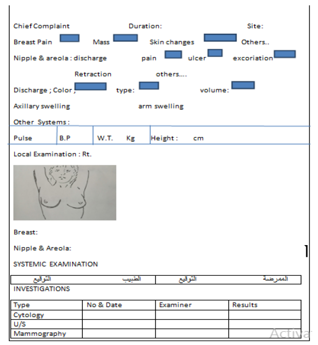
Perfect physical examination (Appendix I)
Precise evaluation by mammography this technique uses X-ray equipment by which compression of breast between it is kind of a clear plexiglass plates which can raise and lowers and that to lessen the thickness of the tissue through which the radiation must pass, two-view mammography as two pictures of each breast a Mediolateral Oblique (MLO) view and a Craniocaudal (CC) view, film screen mammography delivers an average glandular dose of radiation less than 100mrad (0.1cGyo.). The procedure probably takes about half an hour in the office. The actual imaging takes less time, around five to seven minutes.
Ultrasonography, Doppler ultrasound
Fine needle aspiration cytology
Excisional biopsy and histopathological examination for the specimen
Follow up by scheduled short-term (4monthly)screening mammography in still negative cases during the first exposure
The result of the study was238 women who enrolled in the screening program, the age range was were 35-75 years old and ten groups of high-risk women were identified (Table 1).
Table 1: The Total and Subgroups of High–Risk Women
Subgroup | Number | % | Mammographically+ve | % |
Modified Radical Mastectomy | 89 | 37.4 | 8 | 8.99 |
+ve Family History of Breast Cancer | 68 | 28.7 | 6 | 8.8 |
+ve Family History of other Cancers | 35 | 14.8 | 3 | 8.6 |
+ve Breast Cancers S-1 | 19 | 7.98 | 4 | 21.1 |
Nulliparous | 9 | 3.8 | 2 | 22.2 |
Surgery of ovarian tumars | 8 | 3.4 | 2 | 25 |
Benign breast lesion | 5 | 2.1 | 1 | 20 |
Obesity | 2 | 0.84 | 1 | 50 |
Hysterectomy | 2 | 0.84 | 1 | 50 |
Old age | 1 | 0.42 | 1 | 100 |
Total | 238 | 100% | 29 |
|
All women were clinically negative for breast lesions, occult breast lesions were detected mammographically in 32 women (13.5%) (Table 2).
Table 2: The Total and Subgroups of High-Risk Women
Results | Physiological examination | Mammograpy | Ultrasound | ||
True +ve | 0 | 29 | 12.2 % | 29 | 12.2 % |
True –ve | 238 | 209 | 87.8 % | 209 | 87.8 % |
False +ve | 0 | 3 | 9.4 % | 0 | 0 % |
False –ve | 29 | 0 | 0 % | 0 | 0 % |
Sensitivity |
|
| 100 % |
|
|
Specificity |
|
| 98.6 % |
| -- |
Malignancy was confirmed by ultrasound and Fine-Needle Aspiration Cytology (F.N.A.C) and tissue biopsy was positive in 29 cases (True positive) i.e. sensitivity of 100%. Three women were mammographically positive, but negative by both ultrasound and (F.N.A.C) and (false positive) in three cases i.e. specificity of 98.6%. The size of the tumor detected was in the range of 4-10 millimeters with an average of 4.6 millimeters.
The results showed that the age group most affected was those with a mean of 42.7 years age (62.2%), which indicates that breast cancer usually appears after the age of forty years [1,2]. Women who experienced previous modified radical mastectomy (37.4%) of whom (8.99%) were clinically negative for breast cancer but mammographically positive this clarifies that breast cancer can present as synchronous, metachronous, or as a systemic disease from the onset of the pathological process [1-3]. In the second group whom first degree relatives were diagnosed and treated for breast cancer (28.7%), out of them (8.8%) were clinically negative and positive by mammograph, this finding reflects the genetic and familial predisposition for breast cancer disease [3,8].
About 5% of breast cancer patients have inherited their disease because of a mutation in genes encoding either the BRCA-1 or BRCA-2 proteins [9]. The third group was women with a positive family history of cancer in organs apart from the breast (14.8%), of them (8.9%) were mammographically positive, the highest figure was 23 women comes from families have colonic cancer (65.7%) [10]. In the fourth group of women were lumpectomy was done for stage one cancer of the breast in about three years (7.98%) out of them (21.1%) were mammographically positive, this reflects the multifocality nature of the disease process and our results are earlier than what was in other studies, in which it appears about 8-10 years after lumpectomy which might be due to reluctance of most of our patients to be exposed to post-operative deep X-ray therapy in certain cases of lumpectomy, however the rate of cancer recurrence in ipsilateral breast after lumpectomy was higher in women who not receiving post lumpectomy radiation therapy [11]. The fifth group represents nine nulliparous ladies (3.8%), (22.2%) of them were mammographically positive; and this fact was strongly stressed by others due to the absence of the protective factor of pregnancy [12]. The mammograph was positive in two women (25%) out of eight (3.4%) who had abdominal surgery for the removal of ovarian tumors one of them was 53 years old her operation done six years ago and the other 58 years old her operation done nine years ago [13]. Surgery for ipsilateral benign breast lesions represents (2.1%) of the total, fifty six years old lady (20%) was positive for a malignant lesion in her breast. The relationship between benign breast disease and the risk of cancer is bedeviled by ascribing cancer risk to fibroadenomas and fibrocystic change. There is no increased cancer risk for these two entities [14.15]. Obesity, the average weight was 85 kg, the height of was 147 centimeters was seen in two women (0.84%); one of them (50%) was found to be mammographically positive despite the absence of any other risk factor apart from obesity, obese women are at increased risk of developing breast cancer. The theoretical basis for this association is that fatty tissue raises estrogen levels and increases esrogen activity in the setting of obesity [12,15]. Hysterectomy was done for two ladies for fibromyomas (0.84%), fifty two years old lady was positive by mammogram (50%) [16]. Seventy-four years old lady with bulky breasts for her built (0.42%) was mammographically positive (100%) [8]. Mammography was 100% sensitive in our study I.e. the false-negative result was zero, which is higher than what is present in the literature which is about (80-90)%; and this comes from the high precision of the criteria used in the selection of study population [6,10]. The specificity of mammography in our results was (98.6%) because three false-positive cases (9.4%) were diagnosed by mammography and proved to be negative by ultrasound and F.N.A.C. because one of our aims is to reduce the needless disfigurement and trauma to the breast caused by surgery in clinically negative women [10]. The true negative cases were 209 women (87.82%) were so by clinical examination, mammography and ultrasound study; all these were done as a baseline assessment and repeated after four months as a form of short term follow up of the study population; in order not to miss any possible false-negative case or any case of interval breast cancer [17].

The purpose of screening is to detect cancer at the earliest stage and improve the outcome of patient by treating the cancer as soon as possible. At the present time, self-exams and mammograms represent the best methods for early detection 0f breast cancer. Breast cancer is a common disease and the most common type of cancer in women, represents the second most common cause of cancer-related death in women. There is widespread disagreement over the age at which a woman should first get tested, the best time to start mammography screening should be based on the family history of breast cancer and to start mammography screening ten years before the age at which the youngest primary relative was diagnosed with breast cancer. The doctors and the general public are continuing to understand more about the management of this disease. The rate of receiving a mammogram was below the recommended level because of the low educational level of most women. Some women feel because they don’t have a family history of breast cancer they don’t need to be screened. Unfortunately, there are women at above-average risk that frequently don’t recognize their risk. Rates of receiving a mammogram were below the recommended levels, because of low educational level of most women. In addition to the psychologic consequences, many women undergo procedures that may, in fact, be unnecessary. Mammography is very reproducible. The radiation dose from a single mammogram is approximately 0.1msev.
Recommendation
To provide health care professionals as devoted employees and medical staff in the community to increase awareness and appropriate breast health education
Screening mammography for women aged 40-49 every 1-2 years and annually after age 50
Support and encouragement for the high-risk families
The best time to have a mammogram is about five to seven days after the menstrual period
Women with a strong personal or family history of breast cancer should be screened in their 40s
To provide facilities to identify women who are BRCA-1 or BRCA-2 carriers genetic counseling, that may get benefit from doing mammograms at an earlier time
Strongly encourage all women to do their self-exams.
To provide mobile mammography units for screening purposes
Doherty, G.M. et al. The Washington manual of surgery. In: Lippincott Williams and Wilkins, Philadelphia, 1999. “Breast Surgery,” J.H. Yim et al. (Eds.), pp. 471–472.
Strombeck, J.O. and F.E. Roseta. Surgery of the breast. In: “Diagnosis and Treatment of Breast Disease,” H.J. Frischbier (Ed.), Thieme Inc., New York, 1999, pp. 8–20.
Wright, C. and C.B. Mueller. “Screening mammography and public health policy: The need for perspective.” Lancet, vol. 348, 2000, pp. 29–32.
Smart, C. “The role of mammography in the prevention of mortality from breast cancer.” Cancer Prevention, 1999.
Way, Lawrence W. and D.M. Lawrence. Current surgical diagnosis and treatment. In: Appleton and Lange, 1999. “Breast,” by A.E. Guiliano (Eds.), pp. 293–296.
Moskowitz, M. “Breast cancer: Age-specific growth rates and screening strategies.” Radiology, 1986.
Mettler, F.A. et al. “Benefits versus risks from mammography: A critical assessment.” Cancer, vol. 77, 1996, p. 903.
Schwartz, S.I. and G.T. Shire. Principles of Surgery. In: “Breast,” K.I. Bland and Michael, P. (Eds.), McGraw-Hill, New York, 2014. pp. 551–554.
Struewing, J.P. et al. “The risk of cancer associated with specific mutations of BRCA1 and BRCA2 among Ashkenazi Jews.” New England Journal of Medicine, vol. 336, 1997, pp. 1401–1408.
Doherty, G.M. et al. The Washington Manual of Surgery. In: “Breast Surgery,” JH. Yim (Eds.), Lippincott Williams and Wilkins, Philadelphia, 1999. pp. 469–470.
Schwartz, S.I. and G.T. Shire. Principles of Surgery. In: “Breast,” K. Bland and Michael P. (Eds.), McGraw-Hill, New York, 2014. p. 538.
Gail, M.H. et al.“Projecting individualized probabilities of developing breast cancer for females.” Journal of the National Cancer Institute, vol. 81, 1999, pp. 1879–1886.
Claus, E.B. et al.“Autosomal dominant inheritance of early-onset breast cancer.” Cancer, vol. 37, 1994, p. 643.
Obwegeser-Berghammer, P. et al. “99m-Tc-tetrofosmin scintigraphy for the evaluation of suspicious palpable and non-palpable breast lesions.” Breast Cancer Research and Treatment, vol. 62, no. 3, August 2000, pp. 253–258.
Crobtree, T.D. and R.G. Sawyer. Board Review Series in General Surgery. In: “Mammography,” L.H. Brincherhoff and Craig, L. (Eds.), Lippincott Williams and Wilkins, 2014. pp. 431–432.
American Cancer Society. Breast cancer facts. American Cancer Society, Atlanta, 1995.
Burke, W. et al. “Recommendations for follow-up care of individuals with an inherited predisposition to cancer.” JAMA, vol. 277, 1997, pp. 997-1003.