Evaluation of Visual Analogue Scale, Oswestry Disability Macnab Index and Criteria Post Action Percutaneous Laser Disc Decompression (Pldd) on Types of Lumbal Disc Herniation in Rsud Dr. Moewardi Surakarta
Bagus Iman Brilianto
1
,
Bintang Soetjahjo
1
,
Rieva Ermawan
1
,
Romaniyanto
1
1
Faculty of Medicine Sebelas Maret, University, Surakarta, Indonesia
Percutaneous Laser Disc Decompression (PLDD) is a minimally invasive procedure for disc herniation that is known to be faster and effective.This retrospective study is analytic observational. The study was conducted at the Regional General Hospital dr. Moewardi Surakarta. Subjects were patients who had PLDD from January 2020 to December 2020 with total sample of 30 respondents. Evaluation of the Visual Analogue Scale (VAS) and The Oswestry Disability Index (ODI) was performed before the PLDD action was carried out, 1 month and 3 months after. The Macnab criteria was evaluated 3 months after the PLDD action.There was a significant difference in VAS between before and after PLDD, 1 month and 3 months. There was a significant difference in the ODI score before and after 1 month and 3 months, but not in the ODI value after 1 month and 3 months. Twenty patients (66.7%) showed good satisfaction following PLDD with Macnab criteria. The significant improvement in VAS of patients with lumbar disc herniation after PLDD were directly as a result of decreasing intradiscal pressure due to vaporization by laser energy, especially in the herniated area and tissue pressure. As the pain subsided, ODI scores in this patients would improve.PLDD in patients with intact lumbar disc herniation led to a significant improvement in VAS and ODI scores. PLDD can be used as an option in the management of patients with low back pain due to an intact lumbar disc herniation.
Keywords
Lumbar Disc Herniation
PLDD
VAS
ODI Score
INTRODUCTION
Lumbar disc herniation is one of the main causes of low back pain. This complaint can arise at a young age to old age, with various basic causes [1]. Complaints of low back pain can appear in a mild spectrum, to complaints that interfere with daily activities, can even lead to paralysis in patients if not treated properly. Percutaneous Laser Disc Decompression (PLDD) is a minimally invasive procedure developed as a therapy for low back pain caused by lumbar disc herniation. This method has advantages, namely a faster procedure, more minimally invasive because it does not damage a lot of tissue such as a discectomy procedure and is quite effective in dealing with complaints of low back pain due to lumbar disc herniation [2].PLDD is performed under local anesthesia via a fiber laser that is inserted percutaneously into the nucleus pulposus. Irradiation is applied through the fibers to vaporize the contents of the nucleus pulposus. Discogenic pain can be reduced with PLDD which causes disc size to decrease due to water loss. In vitro studies confirm that a small decrease in intervertebral disc volume can lead to a significant decrease in intradiscal pressure [3].
MATERIALS AND METHODS
This study is an analytic observational with a retrospective review. The study was conducted at the Regional General Hospital Dr. Moewardi Surakarta by collecting data on patients who had PLDD from January 2020 to December 2020. The inclusion criteria were patients with low back pain in the lumbar region with and without complaints of radicular leg pain, low back pain patients with lumbar disc herniation, HNP was established based on MRI and those who showed no improvement after 3 months of conservative therapy. The exclusion criteria were patients with back pain other than in the lumbar area, patients with complaints of low back pain with causes other than lumbar disc herniation, patients who had undergone conservative therapy for 3 months with clinical improvement and those with comorbid diabetes mellitus that was not controlled. The Visual Analogue Scale (VAS) before PLDD, 5 minutes after the PLDD, 1 month and 3 months after the PLDD were evaluated. The Oswestry Disability Index (ODI) was evaluated before the PLDD action was carried out, then 1 month and 3 months after the PLDD. The Macnab criteria were evaluated 3 months after the PLDD action. Data analysis was carried out using the Data Normality Test, if the data were normally distributed, the unpaired t-test was used to compare the outcome of PLDD action on the type of lumbar disc herniation (level of significance: p<0.05), if the distribution was not normal, use the Wilcoxon test (level of significance). :p<0.05) Data processing using SPSS 17.0.
RESULTS
This study involved 30 lumbar disc herniation patients with intact discs who underwent PLDD. Most of the patients were female (25 patients, 83.3%) and male patients were 5 patients (16.7%). Patient’s age range from 21 years old to 65 years with an average age of 43.2 years. Types of lumbar disc herniation mostly with paracentral type as many as 22 patients (73.3%), central type in 5 patients (16.7%) and foraminal type in 3 patients (10%). The VAS score in patients with lumbar disc herniation before and after the PLDD procedure in this study used the Friedman Test because the data were not normally distributed, if there was a significant difference, further Wilcoxon Test was carried out to determine the partial difference in VAS values between various sampling times.
Based on Table 1, it was known that the data before the PLDD had a VAS value with a median of 7.50 (6-9), then the VAS Early Evaluation After Treatment (EEAT) data got a VAS value with a median of 2.00 (1-5), VAS 1 Months after the action had a median value of 2.00 (0-7) and VAS 3 months after the procedure had a median value of 2.50 (0-7). Thus there was a decrease in the VAS score after the PLDD. The results of the statistical test obtained a value of p≤0.001 (p<0.05), which means that there was a significant difference in the VAS value between before and after the EEAT, 1 Month and 3 Months action (Figure 1).
Table 1: Friedman Test VAS Value in Lumbar Herniation Patients before and After PLDD
VAS
N
Result
Pre
30
7,50 (6-9)
Post EEAT
30
2,00 (1-5)
Post 1 Month
30
2,00 (0-7)
Post 3 Month
30
2,50 (0-7)
p-value
<0,0001
Figure 1: Graph of VAS Value in Lumbar Disc Herniated Patients Before and After PLDD
Based on the results of the Friedman Test, it was known that there was a significant difference between the VAS values before and after the PLDD, the EEAT, 1 Month and 3 Months action, then the Wilcoxon Test further test was carried out to determine the partial difference in VAS values between various sampling times with the results as following.
Based on Table 2, it was known that the comparison of VAS values that had significant results was in Pre vs Post EEAT (p≤0.001). Pre vs 1 Month (p≤0.001) and Pre vs 3 Month (p≤0.001). There was no significant difference post EEAT vs 1 Month (p = 0.808), post EEAT vs 3 Months (p = 0.211) and 1 Month vs 3 Months (p = 0.180).
Table 2: Wilcoxon Test VAS Value in Lumbar Herniation Patients before and After PLDD
VAS
p-value
Pre vs Post EEAT
<0,001
Pre vs 1 Month
<0,001
Pre vs 3 Month
<0,001
Post EEAT vs 1 Month
0,808
Post EEAT vs 3 Month
0,211
1 Month vs 3 Month
0,180
The value of the ODI score (%) in patients with lumbar disc herniation before and after PLDD in this study used the Friedman Test because there were data that did not meet the assumption of normality, if there was a significant difference, further Wilcoxon Test was carried out to determine the difference in the ODI score (%) partially between various sampling times.
Based on the Table 3, it was known that the data before the PLDD action showed an ODI Score (%) with a median of 48.00 (32-68), then the ODI Score (%) 1 Month after the action had a median value of 20.00 (8-55) and ODI Score (%) 3 Months after the action had a median value of 22.00 (8-60). Thus, there was a significant improvement in ODI score (%) after the PLDD with p≤0.001 (p<0.05) (Figure 2).
Table 3: Friedman Test ODI Score (%) in Lumbar Herniation Patients before and After PLDD
ODI Score (%)
N
Result
Pre
30
48,00 (32-68)
Post 1 Month
30
20,00 (8-55)
Post 3 Month
30
22,00 (8-60)
p-value
<0,0001
Figure 2: Graph of ODI Score (%) in Lumbar Disc Herniated Patients Before and After PLDD
Based on the results of the Friedman Test, there was a significant difference in the ODI Score (%) between before and after the 1 Month and 3 Months action, then the Wilcoxon Test further test was carried out to determine the partial difference in the ODI Score (%) value between various times. sampling with the following results.
Based on the Table 4, it was known that the comparison of ODI scores (%) that had significantly different results was at Baseline to 1 Month (p≤0.001) and Baseline to 3 Month (p≤0.001). There was no significant difference, namely at 1 Month vs. 3 Months (p = 0.399).
Table 4: Wilcoxon Test ODI Score (%) in Lumbar Herniation Patients before and After PLDD
ODI Score (%)
p-value
Pre vs 1 Month
<0,001
Pre vs 3 Month
<0,001
1 Month vs 3 Month
0,399
The satisfaction level of lumbar disc herniation patients who underwent PLDD according to Macnab criteria can be seen in Table 5.
Table 5: Level of Satisfaction of Lumbar Disc Herniated Patients under PLDD Action According to MacNab Criteria
Macnad Criteria
Result (n = 30)
Poor
1(3,3%)
Fair
4(13,3%)
Good
20 (66,7%)
Excellent
5 (16,7%)
The level of satisfaction of patients with lumbar disc herniation who underwent PLDD according to Macnab criteria mostly fell in the Good category as many as 20 patients (66.7%), there were 5 patients (16.7%) in the Excellent category, 4 patients (13, 3%) in the Fair category and 1 patient (3.3%) in the Poor category.
Comparison of VAS values in patients with lumbar disc herniation before and after PLDD based on the type of herniation used the Kruskall-Wallis test because there were data that do not meet the assumption of normality (Table 6).
Table 6: Kruskall-Wallis Test VAS Value in Lumbar Disc Herniated Patients Before and After PLDD Action Based on Herniation Type
Herniation Type
VAS
Pre
Post EEAT
1 Month
3 Month
Foraminal
7,50 (6-9)
2,00 (1-3)
2,00 (0-4)
2,00 (0-4)
Sentral
8,00 (7-8)
3,00 (1-4)
2,00 (1-4)
3,00 (0-5)
Parasentral
7,00 (7-8)
2,00 (1-5)
2,00 (1-7)
3,00 (1-7)
p-value
0,467
0,349
0,346
0,575
The VAS value at baseline (p = 0.467) between patients in the central, paracentral, foraminal groups did not show a significant difference with p>0.05 (homogeneous). Comparison of the decrease in VAS values between patients with central, paracentral and foraminal herniation groups after PLDD procedures on Post EEAT data (p = 0.349), 1 Month (p = 0.346) and 3 Months (p = 0.575) did not show a significant difference.
With a value of p>0.05, thus it can be seen that the effectiveness of PLDD in reducing the value of VAS is not influenced by the type of disc herniation (Figure 3).
Figure 3: Graph of VAS Value in Lumbar Disc Herniated Patients Before and After PLDD Based on Herniation Type
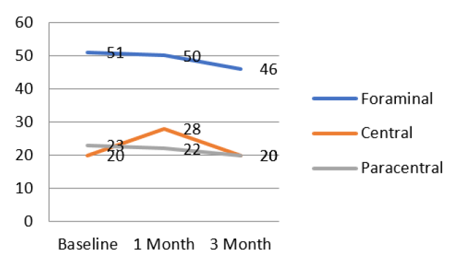
Comparison of VAS values in patients with lumbar disc herniation before and after PLDD surgery based on the type of lumbar disc herniation used ANOVA test on baseline data because it met the assumption of normality and Kruskall-Wallis test on ODI data 1 month after surgery and 3 months after surgery, because they did not meet the assumption of normality (Table 7).
Table 7: Kruskall-Wallis Test VAS Value in Lumbar Disc Herniated Patients Before and After PLDD Action Based on Herniation Type
Herniation Type
ODI Score (%)
Pre
1 Month
3 Month
Foraminal
51,00 (36-56)
20,00 (12-40)
23,00 (8-42)
Sentral
50,00 (32-60)
28,00 (8-40)
22,00 (10-42)
Parasentral
46,00 (38-68)
20,00 (10-55)
20,00 (8-60)
p-value
0,943a
0,628b
0,417b
Note: aANOVA- Test; bKruskal-Wallis-Test
The ODI score before (pre) action (p = 0.943) between patients with central, paracentral and foraminal herniation groups did not show a significant difference with p>0.05 (homogeneous) (Figure 4).
Figure 4: Graph of ODI Score (%) in Lumbar Disc Herniated Patients Before and After PLDD Actions Based on Disc Herniation Type
Then the decrease in the ODI score between patients in the central, paracentral and foraminal herniation group after PLDD action on 1 Month (p = 0.628) and 3 Months (p = 0.417) data did not show a significant difference with p>0.05. From the comparison of improvement in ODI scores after PLDD surgery according to the type of lumbar disc herniation mentioned above, it can be seen that the effectiveness of PLDD in ODI scores improvement was not affected by the type of herniation of an intact lumbar disc.
DISCUSSION
In this study, the patients were predominantly female. This is in accordance with a study by Sedighi et al. [4] who found that 68.4% of patients with lumbar disc herniation were female due to a lack of motivation for male patients to come to health facilities even though they had experienced symptoms and complaints of low back pain due to herniation. Lumbar disc [4]. However, the results are different from the research conducted by Alshehri et al. [5] which found that there was a male sex dominance in cases of lumbar disc herniation of 62.4% compared to women who were associated with physical activity and male work which tended to be heavier.
In this study, the lowest patient age was 21 years and the highest age was 65 years with an average age of 43.2 years. These results are in accordance with the research of Hashemi et al. [6] and Gibson et al. [7] where in this study the highest age prevalence was between 30-50 years with an average age of 49.8 years.
The type of lumbar disc herniation is dominated by the paracentral type, then the central type and the foraminal type. These results are in accordance with the research conducted by Hong and Ball [8] who found that the most common type of lumbar disc herniation was the paracentral type (88.7%) due to the anatomical structure of the posterior longitudinal ligament which is thick on the central side but is getting thinner towards the lateral so that become a weak point (locus minoris) during lumbar disc herniation.
The value of VAS in patients with lumbar disc herniation before and after PLDD in this study used the Friedman Test because the data were not normally distributed, if there was a significant difference, further Wilcoxon test was performed. Based on the Friedman test, it was found that there was a decrease in the value of VAS after PLDD action. The results of the statistical test obtained a value of p≤0.001 (p<0.05), which means that there is a significant difference in the VAS value between before and after the EEAT, 1 Month and 3 Months action. Then continued with the Wilcoxon test, the results of the VAS values did not get significantly different results, namely at post EEAT vs 1 Month (p = 0.808), post EEAT vs 3 Month (p = 0.211) and 1 Month vs 3 Month (p = 0.180). Based on this, it is known that there is a significant difference in the VAS value between before and after the Post EEAT, 1 Month and 3 Months action. These results are consistent with a study conducted by Choy et al. [2] who found a significant decrease in the value of VAS in lumbar disc herniation patients after PLDD which directly reduced intra disc pressure due to vaporization of laser energy, especially in the herniated area and tissue pressure from retraction of the intact annulus fibrosus, thereby reducing pressure on the nerve root of the spinal cord and resulting in a decreased VAS value.
The ODI score (%) in patients with lumbar disc herniation before and after PLDD action in this study used the Friedman Test because there were data that did not meet the assumption of normality, if there was a significant difference, further Wilcoxon test was performed. Based on the Friedman test, the results obtained p value 0.001 (p<0.05), which means that there is a significant difference in VAS values between before and after EEAT, 1 Month and 3 Months. Followed by the results of the Wilcoxon test, the results were significantly different, namely at Baseline vs. 1 Month (p≤0.001) and Baseline vs. 3 Month (p≤0.001). ODI Score (%) with no significant difference, namely at 1 Month vs. 3 Months (p = 0.399). There is a significant difference in the ODI score between before and after the ODI score after 1 Month and 3 Months action, while the ODI value between after 1 Month and 3 Months action has no significant difference. These results are consistent with research conducted by Erbas et al. which showed that PLDD measures can significantly reduce low back pain complaints so as to improve ODI scores in patients with lumbar disc herniation [9].
The level of satisfaction of patients with lumbar disc herniation who underwent PLDD surgery according to Macnab criteria, most of them were in the Good category, namely there were 20 patients (66.7%), while the Excellent category had 5 patients (16.7%), the Fair category had 4 patients (13.3%) and the least was in the Poor category. i.e. there is 1 patient (3.3%). This is in accordance with the results of a study conducted by Choy et al. [2] who received an assessment of the Macnab criteria with the results of good criteria of 70.2% and excellent of 18.3% in patients with lumbar disc herniation who had PLDD action. The research of Choy [3] and Erbas et al. also shows that there are patients with poor and fair satisfaction levels according to Macnab criteria of 3.2% and 8.3%, respectively [3,9]. This is thought to be the result of an advanced disc degeneration process and the water content in the nucleus pulposus has decreased considerably, so that with PLDD the pain reduction felt by the patient was not significant.
CONCLUSION
Based on the results of the study, 30 patients with lumbar disc herniation with intact disc underwent Percutaneous Laser Disc Decompression (PLDD) at Dr. Moewardi Surakarta. PLDD procedures in patients with intact lumbar disc herniation gave a significant change in the Visual Analogue Scale (VAS) and Oswestry Disability Index (ODI) scores compared to the VAS and ODI scores before PLDD, both in patients with herniation, central, paracentral and foraminal type discs, with most of the patients satisfied according to the Macnab criteria in the categories of Good (66.7%) and Excellent (16.7%). PLDD can be used as an option in the management of patients with low back pain due to an intact lumbar disc herniation.
There are some limitations in this study, namely the time to evaluate the results of clinical improvement is quite short, namely 1 month and 3 months. In this study, the sample distribution was uneven and unbalanced in each type of herniated lumbar disc that was intact. This type of research is an observational analytic retrospective cohort, so further research is needed with a prospective Randomized Controlled Trial (RCT) type in order to provide a reference so that PLDD action can become the standard of treatment for low back pain patients with intact lumbar disc herniation.
REFERENCES
Choy, D.S. et al. "Percutaneous laser disc nucleolysis of lumbar disc." N. Engl. J. Med, vol. 317, 1987, pp. 771–772.
Choy, D.S. et al. "About 23rd anniversary of Percutaneous Laser Disc Decompression (PLDD)." Photomed Laser Surg, vol. 27, 2009, pp. 535–538.
Choy, D.S. "Percutaneous laser disc decompression: an update." Photomed Laser Surg, vol. 5, 2004, pp. 393–406.
Sedighi, M. and A. Haghnegahdar. "Lumbar disk herniation surgery: Outcome and predictors." Global Spine Journal, vol. 4, no. 4, 2014, pp. 233–243. DOI: 10.1055/s-0034-1390010.
Alshehri, A.K. et al. "Awareness of disc herniation among general population in Aseer province, Saudi Arabia." Journal of Family Medicine and Primary Care, vol. 8, no. 3, 2019, pp. 1159–1163. https://doi.org/10.4103/jfmpc.jfmpc_462_18.
Hashemi, M. et al. "Effectiveness of intradiscal injection of radiopaque gelified ethanol (DiscoGel®) versus percutaneous laser disc decompression in patients with chronic radicular low back pain." The Korean Journal of Pain, vol. 33, no. 1, 2020, pp. 66–72. https://doi.org/10.3344/kjp.2020.33.1.66.
Gibson, J.N. "Most physical tests to identify lumbar-disc herniation show poor diagnostic performance when used in isolation, but findings may not apply to primary care." Evidence-Based Medicine, vol. 15, no. 3, 2010, pp. 82–83. https://doi.org/10.1136/ebm1071.
Hong, J. and P.A. Ball, "Resolution of lumbar disk herniation without surgery." New England Journal of Medicine, vol. 374, no. 16, 2016, p. 1564. DOI: 10.1056/nejmicm1511194.
Kerr, D. et al. "What are long-term predictors of outcomes for lumbar disc herniation? A randomized and observational study." Clinical Orthopaedics and Related Research, vol. 473, no. 6, 2015, pp. 1920–1930. https://doi.org/10.1007/s11999-014-3803-7.
Recommended Articles
Research Article
It Remains Unproven That the Variant M.8231C>A Causes Coronary Atherosclerosis
Finsterer J
Published: 15/07/2020
Download PDF
Research Article
Vision for a Brighter Kangra: Unmasking the Truth about Pink Eye – A Comprehensive Study on Types, Symptoms, and Proactive Prevention in Himachal Pradesh’s Kangra District
Deepti ,
...
Garima
Published: 11/11/2023
Download PDF
Research Article
Leigh Syndrome Should Not Be Diagnosed Exclusively Upon Cerebral MRI
Finsterer J
Published: 15/07/2020
Download PDF
Research Article
Release Kinetic Study of Matrix Type Transdermal Patch Using an Analgesic Drug
Mohit Saini,
...
Mukesh Kumar
Published: 10/08/2020
Download PDF
PO Box 101, Nakuru
Kenya.
Email: office@iarconsortium.org
Editorial Office:
J.L Bhavan, Near Radison Blu Hotel,
Jalukbari, Guwahati-India
Brilianto, B. I., Soetjahjo, B., Ermawan, R. & None, R. (2022). Evaluation of Visual Analogue Scale, Oswestry Disability Macnab Index and Criteria Post Action Percutaneous Laser Disc Decompression (Pldd) on Types of Lumbal Disc Herniation in Rsud Dr. Moewardi Surakarta. IAR Journal of Medical Sciences, 3(1), 1-5.
MLA
Brilianto, Bagus I., et al. "Evaluation of Visual Analogue Scale, Oswestry Disability Macnab Index and Criteria Post Action Percutaneous Laser Disc Decompression (Pldd) on Types of Lumbal Disc Herniation in Rsud Dr. Moewardi Surakarta." IAR Journal of Medical Sciences 3.1 (2022): 1-5.
Chicago
Brilianto, Bagus I., Bintang Soetjahjo, Rieva Ermawan and Romaniyanto . "Evaluation of Visual Analogue Scale, Oswestry Disability Macnab Index and Criteria Post Action Percutaneous Laser Disc Decompression (Pldd) on Types of Lumbal Disc Herniation in Rsud Dr. Moewardi Surakarta." IAR Journal of Medical Sciences 3, no. 1 (2022): 1-5.
Harvard
Brilianto, B. I., Soetjahjo, B., Ermawan, R. and None, R. (2022) 'Evaluation of Visual Analogue Scale, Oswestry Disability Macnab Index and Criteria Post Action Percutaneous Laser Disc Decompression (Pldd) on Types of Lumbal Disc Herniation in Rsud Dr. Moewardi Surakarta' IAR Journal of Medical Sciences 3(1), pp. 1-5.
Vancouver
Brilianto BI, Soetjahjo B, Ermawan R, Romaniyanto R. Evaluation of Visual Analogue Scale, Oswestry Disability Macnab Index and Criteria Post Action Percutaneous Laser Disc Decompression (Pldd) on Types of Lumbal Disc Herniation in Rsud Dr. Moewardi Surakarta. IAR Journal of Medical Sciences. 2022 Jan;3(1):1-5.



 Download PDF
 Download XML