Introduction: AlP is also known as celphos and is one of the most dreaded poisons one can ever encounter in toxicology. Due to non-availability of antidote and high mortality celphos poisoning has always been a big headache and menace for the intensivists throughout the world. Literature is full of different drugs and trials to counter its irreversible toxic effects, but hardly with any concrete success. In literature the usefulness of the gastric ventilation technique had been reported. Further clinical trials are required to assess the usefulness of this technique and this study is one of the attempts towards the same. Materials and Methods: This is a prospective descriptive study of patients of celphos (AlP) poisoning carried out in Oscar super-speciality hospital Sonipat Haryana from January to December 2019. Twenty patients were admitted in ICU of our hospital over the last 1-year period with an alleged history of intake of celphos. Gastric lavage along with gastric ventilation was done in all patients along with other supportive treatment. Results: Mean age of our patients was 22.2 years with male to female ratio of 3:2. Most of the patients presented with non-specific findings. During hospitalisation hypotension, arrythmias and respiratory failure were common. 60% of patients in this study need artificial ventilation while as ionotropic support was needed in 50% of patients. Mortality rate in this study was 65%. Mortality rate was statistically high in cases who had hypotension, multiorgan failure, delayed gastric lavage and where ingestion of poison was greater than 4 grams. Conclusion: AlP poisoning is one of the most dreaded poisons one can ever encounter in toxicology. In India it is commonly used by farmers as pesticide as it is cheap, effective, free from toxic residue and does not affect seed viability. As there is no specific antidote for AIP poisoning so most of the treatment is supportive. Gastric ventilation along with gastric lavage with the aim of removing phosphine gas from stomach before it is absorbed seems to be effective in decreasing mortality in these patients however before it becomes a routine procedure like gastric lavage in these patients further studies with controlled head-to-head trails and minimising confounding factors needs to be done.
Each year around 300,000 deaths occur worldwide due to pesticides. Organophosphate, organochlorines and aluminium phosphide (AlP) compounds are commonly used pesticides. AlP is also known as celphos and is one of the most dreaded poisons one can ever encounter in toxicology. The problem is more complicated in developing world where at some places pesticide poisoning causes more deaths than infections [1]. In India it is commonly used by farmers as pesticide as it is cheap, effective, free from toxic residue and does not affect seed viability. Between 1977 and 1987 barbiturates (33.3 %), organophosphates (23.8 %) and copper sulphate (14.3 %) were the most common causes of death by poisoning and between 1987 and1997 they were replaced by organophosphates (45%) and AlP (26.5 %) [2,3]. AIP also poses as a threat for chemical terrorism due to the immediate release of lethal phosphine gas.
The lethal dose of AlP is around 0.5 g. Acute poisoning with these compounds may be direct due to ingestion of the salts or indirect from accidental inhalation of phosphine generated during their approved use. It is not just limited to the agricultural society, but the incidence is increasing in the urban families also It is a highly toxic compound that releases phosphine gas on contact with moist surfaces and patients can present clinically with gastrointestinal (GI) haemorrhage, arrhythmias, shock, renal and hepatic failure, central nervous system disturbances and ultimately leading to death. The diagnosis of ALP usually depends on the clinical suspicion or history (self-report or by attendants). In case of doubt, diagnosis can be made easily by simple silver nitrate-impregnated paper test on gastric content or on breath. Chemical analysis for phosphine in blood or urine is not recommended as phosphine is rapidly oxidised to phosphite and hypophosphite.
Due to non-availability of antidote and high mortality celphos poisoning has always been a big headache and menace for the intensivists throughout the world. Literature is full of different drugs and trials to counter its irreversible toxic effects, but hardly with any concrete success. As there is no specific antidote for AIP poisoning so most of the treatment is supportive. The foremost supportive measures include Circulation, Airway and Breathing (CAB). The patient must be removed from the source of poisoning to avoid ongoing exposure. AlP can be absorbed directly through the skin, therefore the exposed must be washed with soap and water. Medical and first aid teams must take precautionary measures to avoid exposure to the poison. Gastric lavage with Potassium permanganate and mineral oil or coconut oil has been shown to reduce mortality [4]. In literature the usefulness of the gastric ventilation technique had been reported.5 A nasogastric tube is inserted to insufflate the air in the distal part of stomach, while an orogastric tube with wider lumen is inserted to serve as an outflow tube. An air-pump is used to blow air in the nasogastric tube while Phosphine-contaminated air comes out of the orogastric tube however there are no large case studies to further show its effectiveness. Further clinical trials are required to assess the usefulness of this technique and this study is one of the attempts towards the same.
This is a prospective descriptive study of patients of celphos (AlP) poisoning carried out in Oscar super-speciality hospital Sonipat Haryana from January to December 2019. Twenty patients were admitted in ICU of our hospital over the last 1-year period with an alleged history of intake of celphos. On admission to ICU, monitoring gadgets were attached for Heart Rate (HR), Non-Invasive Blood Pressure (NIBP), ECG and Pulse Oximetry (SpO2). Those patients who were unstable hemodynamically or had respiratory distress or who were in altered sensorium endotracheal intubation was done with appropriate size cuffed endotracheal tube and patients were put on mechanical ventilation if required. A narrower nasogastric tube, is inserted in the distal part of the stomach and is used as the air inflow tract and a wide-bore orogastric tube is inserted near the gastroesophageal junction as an air outflow tract. Ventilation is provided by blowing fresh air through nasogastric tube and PH3-contaminated air escapes via the orogastric tube due to the larger diameter and, therefore, lower resistance. To ensure adequate ventilation of the gastric space, the air pump provided air flow at 30L/min with a maximum pressure of 0.02Mpa. To prevent the potential for contamination of ICU personnel, the aspirated air was transferred to the external environment through a pipe and sucking fan. This procedure was repeated multiple times in initial 2 to 3 minute. Gastric lavage was initiated immediately after that with potassium permanganate and coconut oil and 50 ml of sodium bicarbonate solution and continued for the next half an hour, with simultaneous aspiration being done after every 2-3 minutes through nasogastric Ryle's tube while orogastric tube was retained in. Coconut oil was just heated to lukewarm temperature so as to make a miscible solution with sodium bicarbonate. When performing the procedure, patients are continually monitored including regular clinical observations and abdominal examination. Once gastric lavage was done for half an hour gastric ventilation was continued. The silver nitrate test was performed every hour to determine the presence of PH3 in the output tract. This test is performed by exposing filter paper moistened with fresh silver nitrate solution (0.1 M) to the gaseous outflow from the orogastric tube. On drying, the paper turns black in the presence of phosphine. Once this test shows negative results gastric ventilation was stopped and orogastric tube was removed. Apart from this all patients received steroids and magnesium sulphate. Further management was guided by clinical condition of patient and complications during hospitalisation of patients. Final outcome in this study was discharge from hospital in stable condition or death.
Out of 20 patients with alleged history of AIP ingestion females outnumbered males with male female ratio of 2:3. Most of the patients in this study were young below age of 25 years (75%) with youngest of 15 years and oldest of 52 years (Table 1).
Mean age of our patients was 22.2 years with standard deviation of +/- 4 .3 years. Majority of patients were from rural setting and unmarried. Suicide was the main intention of poisoning and majority of patients were unemployed. In this study mean dose of AIP was 5.8 grams and average time from ingestion to hospital was 5.2 hours. Traditional treatments were received by 8 patients (40%) with 5 patients were given salt water and remaining 3 patients were given herbal medicine to induce vomiting.
Clinical Features
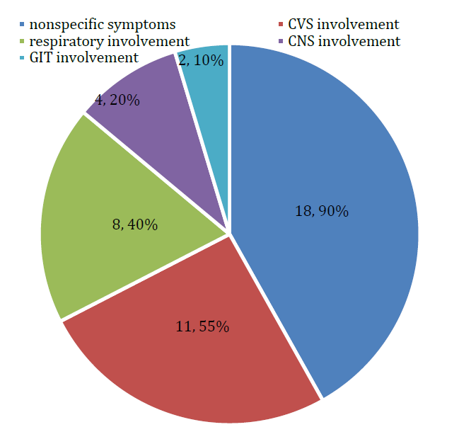
At presentation to hospital most patients (18; 90%) present with nonspecific finding in form of abdominal discomfort, nausea, vomiting, dizziness and anxiety features. Among organ specific findings CVS involvement in form of hypotension and arrythmias were seen in 11 (55%) patients followed by breathlessness and respiratory failure in 8 (40%) patients. CNS involvement in form of altered sensorium, coma or seizures was seen in 4 (20%) patients and 2(10%) patients had haematemesis (Figure 1).
During hospitalization arrythmias were seen in all patients mostly sinus tachycardia and 15 patients had hypotension at some point during hospitalisation. Respiratory failure developed in 16 patients while 12 patients had features of ARDS. Altered sensorium was seen in 18 patients while 6 of patients went into coma and 3 patients had seizures.10 patients had renal failure and 4 patient gastrointestinal bleed during hospitalization (Table 2).
Table 1: Demographic Data
Total number of patients | 20 |
Male -female ratio | 2:3 |
Mean age and standard deviation | 22.2 years, SD±4.3 years |
Rural vs urban | 4:1 |
intention of poisoning | Suicidal: 17(85%) Accidental: 3(15%) |
Mean dose of celphos | 5.8 grams |
Educational status | Illiterate: 4(20%) Up to high school: 10(50%) >High school: 6(30%) |
Employment status | Unemployed: 14(70%) Employed: 6 (30%) |
Average time from ingestion to hospital | 5.2 hours |
Traditional treatment given | 8 (40%) |

Figure 1: Clinical features on admission
Table 2: Complications
System involved | Frequency (%) |
Cardiovascular system | Hypotension:15(75%) Sinus tachycardia: 13(65%) Atrial fibrillation: 7(35%) |
Respiratory system | Respiratory failure: 16(80%) ARDS: 12(60%) |
Nervous system | Altered sensorium:14(70%) Coma:6(30%) Seizures:3(15%) |
Renal system | Renal failure:10(50%) |
Git system | Upper gastrointestinal bleed:4(20%) |
Table 3: Management in hospital
Treatment | Frequency (%) |
Gastric lavage and gastric ventilation | 20(100%) |
Inotropic support | 10(50%) |
Invasive ventilation | 12(60%) |
Non-invasive ventilation | 4(20%) |
Anti-arrhythmic treatment | 5(25%) |
Haemodialysis | 5(25%) |
PPI infusion | 4(20%) |
Anti-epileptic drugs | 4(20%) |
Average stay in hospital | 52 days |
Table 4: outcome
Variable | Mortality |
Overall mortality | 13(65%) |
Time of mortality | Within 72 hours of admission: 8 After 72 hours of admission: 5 |
Haemodynamic status | Patients with hypotension: 11 Patients with no hypotension: 2 |
organ dysfunction | Multiorgan dysfunction: 10 Single or no organ dysfunction: 3 |
Time between poison ingestion and gastric lavage | Greater than 4 hours: 8 Less than 4 hours: 5 |
Quantity of poison ingested | greater than 4 grams: 9 less than 4 grams: 4 |
Management
All patients were managed in ICU and received gastric ventilation and gastric lavage. out of 16 patients with respiratory failure 12 patients were managed with invasive ventilation and rest with non-invasive ventilation. Vasopressor support was needed in 10 patients while in other 4 patients’ hypotension was managed with intravenous fluids. All 5 patients with atrial fibrillation were given intravenous digoxin. Out of 10 patients of renal impairment 5 needed haemodialysis and rest were managed conservatively. Proton pump inhibitors were used in all patients while four patients with gastrointestinal bleed were managed with PPI infusion for 3 days (Table 3).
Outcome
Total 12 patients in this study expired in hospital while 8 patients were discharged in stable condition. Majority of patients expired within 72 hours after poison ingestion. Among 14 patients who had hypotension 11 expired and patients in coma mortality rate was 100%. Mortality was high in patients who developed ARDS and need artificial ventilation. Patients with multiorgan dysfunction had 95% mortality as compared to 40% mortality in patients with no or one organ dysfunction. Mortality was almost 100% when time from ingestion of poisoning and gastric lavage was greater than 4 hours and when dose of poison was more than 4 gms (approximately 2 tablets of wheat pill) (Table 4).
In India AIP poisoning is a public health concern because of its extensive use and ready availability. In this study young male population were common victims and most of the patients were unmarried, less educated and unemployed. Majority of our patients were from rural areas because of easy availability of aluminium phosphide in rural areas. Although most patients seek medical attention but traditional treatments are still practiced in rural areas and is responsible in delay in seeking medical attention. Suicidal intention is the main cause of poisoning than accidental ingestion and there was no homicidal case in this study. Rural Young males were reported as common victims by others also like Gupta et al. [6] and Karamjit et al. [7]. The manner of death was reported to be suicidal in 87% of the cases by Dalbir et al. [8], 76% in a study by Chugh et al. [9] and 100% in Jain et al. [10].
Following oral ingestion, AlP reacts with water and stomach acid to produce phosphine gas, which may account in large part for its observed toxicity. The exact mechanism of action of phosphine is still not clear. Some authors have claimed it to be an inhibitor of cytochrome oxidase [11], while others showed reduction in catalase activity leading to free-radical toxicity [12,13]. AIP poisoning affects virtually every organ of body. In this study non-specific symptoms in form abdominal discomfort, nausea, vomiting, dizziness and anxiety features were seen in most patients. Among organ systems cardiovascular system was most common organ system to be involved followed by central nervous and gastrointestinal systems. Arrythmias, cardiogenic shock, respiratory failure and encephalopathy were common complications during hospitalisation. Similar findings were reported by others with early symptoms include nausea, vomiting, retrosternal and epigastric pain, dyspnoea, anxiety, agitation and garlic breath and early signs of fatal toxicity are shock and peripheral circulatory failure [14].
Unfortunately, due to no known specific antidote, management remains primarily supportive care. As each poison has a definite elimination time, so also is the case with AlP. Early arrival, resuscitation, diagnosis, intensive monitoring and supportive therapy may result in good outcome. Gastric lavage with Potassium permanganate, coconut oil and bicarbonate solution is widely practised and supported by most clinicians however Nasri Nasrabadi and Marashi published their finding that phosphine is a hard nucleophile and the free oxygen radicals from the resolution of KMnO4 do not interact with each other [15-17]. Therefore, there is no well proven basis to conclude that KMnO4 is efficient against ALP poisoning. Slurry of activated charcoal is also used to adsorb phosphine from the gastrointestinal (GI) tract in most of the literature. Marashi et al. [18] concluded that activated charcoal has a wide internal surface area consisting of pores (10 Å to 20 Å). It efficiently adsorbs toxins of moderate molecular weight (100 Da to 800 Da). The molecular weight of ALP is about 58 Da, therefore, role of activated charcoal in ALP poisoning is again doubtful.
Gastric ventilation with the aim of removing phosphine gas from stomach before it is absorbed is reported in literature however how effective is it in decreasing mortality in these patients is not known as there are no large head-to-head trails or case series on this reported anywhere in literature [5]. In this study all patients were subjected to gastric ventilation along with gastric lavage, immediately after admission to hospital. The mortality rate in present study was 65%. The mortality rate in clinical reports is stated to vary between 37-100% by different authors [19-27]. In present study mortality was comparatively higher in first 72 hours of admission mainly due to arrhythmia. Death rate was more in patients with hypotension (p=0.17) and in patients with multiorgan failure (p=0.03). Similarly, mortality was more in patients where gastric lavage and gastric ventilation were delayed more than 4 hours (p=0.04) and in patients when quantity of poison ingested was more than 4 grams (p=0.01). in literature poor prognostic factors mentioned are shock, altered mental status, high APACHE II score, acute kidney injury, low prothrombin rate, hyper-leucocytosis, requirement of mechanical ventilation, lack of vomiting after ingestion, hyper-glycemia, time lapsed after exposure arterial pH, serum bicarbonate level and ECG abnormalities [28-36].
AlP poisoning is one of the most dreaded poisons one can ever encounter in toxicology. In India it is commonly used by farmers as pesticide as it is cheap, effective, free from toxic residue and does not affect seed viability. As there is no specific antidote for AIP poisoning so most of the treatment is supportive. Gastric ventilation along with gastric lavage with the aim of removing phosphine gas from stomach before it is absorbed seems to be effective in decreasing mortality in these patients however before it becomes a routine procedure like gastric lavage in these patients, further studies with controlled head-to-head trails and minimising confounding factors need to be done.
Acknowledgment
Authors would like to thank Mr. Manvender Singh, Mr. Pawan Kumar Yadav and other nursing staff of Oscar Super Speciality Hospital Sonipat for all the support during this study.
Eddleston, Michael et al. “Pesticide poisoning in the developing world-a minimum pesticides list.” The Lancet, vol. 360, 2002, pp. 1163-1167.
Singh, Dalbir et al. “Spectrum of unnatural fatalities in the Chandigarh zone of north-west India: a 25 year autopsy study from a tertiary care hospital.” Journal of Clinical Forensic Medicine, vol. 10, 2003, pp. 145-152.
Gupta, S. and S.K. Ahlawat. “Aluminum phosphide poisoning: a review.” Journal of Toxicology: Clinical Toxicology, vol. 33, 1995, pp. 19-24.
Shadnia, Sh. et al. “Successful treatment of acute aluminium phosphide poisoning: possible benefit of coconut oil.” Human and Experimental Toxicology, vol. 24, no. 4, 2005, pp. 215-218.
Hassanian-Moghaddam, H. and A. Shahbazi. “Gastric ventilation: a new approach to metal phosphide fumigant ingestion.” Clinical Toxicology, vol. 50, no. 5, 2012, pp. 435-437.
Gupta, B.D. et al. “Current trend of poisoning in Jamnagar.” Journal of the Indian Academy of Forensic Medicine, vol. 28, no. 3, 2006, pp. 90-92.
Karamjit, Singh et al. “Poisoning trends in the Malwa region of Punjab.” Journal of Punjab Academy of Forensic Medicine and Toxicology, vol. 3, 2003, pp. 26-29.
Dalbir, Singh et al. “Aluminium phosphide poisoning - autopsy and histopathological findings.” Journal of Forensic Medicine and Toxicology, vol. 12, no. 1-2, 1985, pp. 16-20.
Chugh, S.N. “Aluminium phosphide poisoning - present status and management.” Journal of the Association of Physicians of India, vol. 40, 1992, pp. 401-405.
Jain, A.K. et al. “Aluminium phosphide poisoning - autopsy findings.” Journal of the Indian Academy of Forensic Medicine, vol. 27, no. 1, 2005, pp. 35-39.
Chefurka, W. et al. “The effect of phosphine on electron transport in mitochondria.” Pesticide Biochemistry and Physiology, vol. 6, 1976, pp. 350-362.
Bolter, C.J. and W. Chefurka. “Extramitochondrial release of hydrogen peroxide from insect and mouse liver mitochondria using the respiratory inhibitors phosphine, myxothiazol and antimycin and spectral analysis of inhibited cytochromes.” Archives of Biochemistry and Biophysics, vol. 278, no. 1, April 1990, pp. 65-72.
Chugh, S.N. et al. “Free radical scavengers and lipid peroxidation in acute aluminium phosphide poisoning.” Indian Journal of Medical Research, vol. 104, August 1996, pp. 190-193.
Singh, R.B. et al. “Hypermagnesemia following aluminum phosphide poisoning.” International Journal of Clinical Pharmacology, Therapy and Toxicology, vol. 29, 1991, pp. 82-85.
Bumbrah, G.S. et al. “Phosphide poisoning: a review of literature.” Forensic Science International, vol. 214, 2012, pp. 1-6.
Mehrpour, O. et al. “Severe hypoglycemia following acute aluminum phosphide (rice tablet) poisoning; a case report and review of the literature.” Acta Medica Iranica, vol. 50, 2012, pp. 568-571.
Nasri-Nasrabadi, Z. and S.M. Marashi. “Comments on ‘A systematic review of aluminium phosphide poisoning.’” Archives of Industrial Hygiene and Toxicology, vol. 63, 2012, p. 551.
Marashi, S.M. et al. “A common misconception in the management of aluminium phosphide poisoning.” Archives of Industrial Hygiene and Toxicology, vol. 64, 2013, pp. 475-476.
Chopra, J.S. et al. “Aluminium phosphide poisoning: a prospective study of 16 cases in one year.” Postgraduate Medical Journal, vol. 62, no. 734, December 1986, pp. 1113-1115.
Kabra, S.G. and R. Narayanan. “Aluminium phosphide: worse than Bhopal.” The Lancet, vol. 1, no. 8598, June 1988, pp. 1333.
Sepaha, G.C. et al. “Acute aluminium phosphide poisoning.” Journal of the Indian Medical Association, vol. 83, no. 11, November 1985, pp. 378-379.
Raman, R. et al. “Prevalence of fatalities due to aluminium phosphide poisoning in Southern Rajasthan (an epidemiological study).” Journal of Forensic Medicine and Toxicology, year not stated.
“The electrocardiographic changes in quick phos poisoning.” Heart Journal, vol. 37, no. 3, May-June 1985, pp. 193-195.
Khosla, S.N. et al. “Systemic involvement in aluminium phosphide poisoning (a report of 10 cases).” Journal of the Association of Physicians of India, vol. 34, no. 3, March 1986, pp. 227-230.
Mishra, U.K. et al. “The spectrum of phosphine toxicity in India.” Journal of the Association of Physicians of India, vol. 37, 1989, pp. 68.
Chugh, S.N. et al. “Adult respiratory distress syndrome (ARDS) following aluminium phosphide poisoning.” Journal of the Association of Physicians of India, vol. 37, 1991, pp. 27.
Chugh, S.N. “Aluminium phosphide poisoning with special reference on its diagnosis and management.” Journal of the Indian Association of Clinical Medicine, vol. 1, 1995, pp. 20-22.
Singh, R.B. et al. “Cardiovascular manifestations of aluminium phosphide intoxication.” Journal of the Association of Physicians of India, vol. 37, 1989, pp. 590-592.
Chugh, S.N. et al. “Electrocardiographic abnormalities in aluminium phosphide poisoning with special reference to its incidence, pathogenesis, mortality and histopathology.” Journal of the Indian Medical Association, vol. 89, 1991, pp. 32-35.
Chugh, S.N. et al. “Magnesium levels in acute cardiotoxicity due to aluminium phosphide poisoning.” Indian Journal of Medical Research, vol. 94, 1991, pp. 437-439.
Siwach, S.B. et al. “A study of serum and tissue magnesium content in patients of aluminium phosphide poisoning and critical evaluation of high dose MgSO₄ therapy in reducing mortality.” Journal of the Association of Physicians of India, vol. 42, 1994, pp. 107-110.
Mehrpour, O. et al. “Hyperglycemia in acute aluminum phosphide poisoning as a potential prognostic factor.” Human and Experimental Toxicology, vol. 27, 2008, pp. 591-595.
Singh, S. et al. “Aluminium phosphide poisoning: a clinicopathologic study.” Journal of Toxicology: Clinical Toxicology, vol. 34, 1996, pp. 703-706.
Shadnia, S. et al. “A retrospective 7-years study of aluminum phosphide poisoning in Tehran: opportunities for prevention.” Human and Experimental Toxicology, vol. 28, 2009, pp. 209-213.
Louriz, M. et al. “Prognostic factors of acute aluminium phosphide poisoning.” Indian Journal of Medical Sciences, vol. 63, 2009, pp. 227-234.