Pseudo exfoliative glaucoma (PEXG) is a sight-threatening disease that develops resulting from Pseudo-exfoliation syndrome (PEX). The clinical impact of the diagnosis of early glaucomatous change can be understood. About 30-50% of retinal ganglion cells may be lost before any visual field changes are detected due to the deposition of PEX material. Some studies have reported the presence of difference in RNFL thickness measurement between the eyes with the PEX. The purpose of this study was to evaluate the RNFL thickness using SD-OCT system among the patients with PEX in comparison with patients affected with PEXG attending OPD clinic in the department of ophthalmology in IGMC Shimla. Patients having pseudo-exfoliation with glaucoma showed significant visual field defects as compared to their counterparts (p<0.001). The overall mean RNFL Thickness(µm) in the two groups was 89.47±13.52 µm and 72.73±13.54 µm and the difference between the means of RNFL thickness between the two groups was found to be statistically significant (p<0.001). Measurement of RNFL thickness in early stages of Pseudo-exfoliation with glaucoma can help to modify the course of management.
Keywords
PEXG
PEX
Visual Field Defects
Retinal Nerve Fibre Layer Thickness
INTRODUCTION
Pseudo exfoliative glaucoma (PEXG) is a sight-threatening disease that develops resulting from Pseudo-exfoliation syndrome (PEX). The latter is also found to be in association with various systemic disorders, such cardiovascular disease, cerebrovascular disease, sensorineural hearing loss and Alzheimer’s dementia [1]. In particular, ocular involvement in Pseudo-exfoliation (PEX) primarily manifests with anterior segment findings. PEX material accumulation has also been found on the lens capsule, pupillary border, the iris, non-pigmented ciliary epithelium, lens zonules, trabecular meshwork and corneal endothelial cells in the posterior ciliary artery, vortex vein and central retinal artery walls. The material has also been demonstrated along the vascular endothelium, corneal epithelial basement membrane and stroma. Moreover, various clinical studies have reported that PEX material affects ocular blood flow and vascular resistance [2].
On assessing the macula, the clinical impact of the diagnosis of early glaucomatous change can be understood [3]. Nearly half of retinal ganglion cells are located in the macula [4].In this context, evaluation of retinal nerve fibre layer (RNFL) thickness is important because glaucomatous retinal atrophy mostly affects the RNFL. About 30-50% of retinal ganglion cells may be lost before any visual field changes are detected due to the deposition of PEX material [5,6]. Some studies have reported the presence of difference in RNFL thickness measurement between the eyes with the PEX [7,8].
However, few studies have compared the RNFL thicknesses in pseudo-exfoliative patients without glaucomatous damage and pseudo-exfoliative patients with glaucoma damage and no studies in a North Indian population are reported to date. The purpose of this study was to evaluate the RNFL thickness using SD-OCT system among the patients with PEX in comparison with patients affected with PEXG.
MATERIALS AND METHODS
This cross-sectional analytical study was conducted in the Department of Ophthalmology, Indira Gandhi Medical College and Shimla. The subjects were enrolled and investigated between July 2018 and June 2019 i.e. one year. Assuming a difference of average RNFL thickness of 11.69mm 85.52±19.7mm in pseudo exfoliation without glaucoma (PEXG) patients and 97.21±20.21mm in normal healthy controls), Confidence level 95%, Absolute error as 5% and power of study 80%, a sample size of 46 was calculated. All consecutive patients presenting to ophthalmology OPD and diagnosed with pseudo exfoliation without glaucoma and pseudo exfoliation with glaucoma were included in the study till the completion of sample size.
Patient who did not consent or having a history or evidence of ocular disease such as age-related macular degeneration, diabetic retinopathy, central serous chorioretinopathy, epiretinal membrane, macular dystrophy etc., media opacity interfering with visualization and OCT images capturing such as corneal opacity or dense cataract were excluded from the study. Subjects with systemic diseases or conditions, such as diabetes mellitus, cardiovascular disease, dyslipidaemia, renal failure, malignancy, autoimmune diseases, haematological diseases, chronic obstructive pulmonary disease, uncontrolled arterial hypertension, history of transient ischemic attack or stroke and a history of smoking, as they might affect RNFL thickness, were also excluded. The data was entered and cleaned using Microsoft Excel Spreadsheet 2007. The data was analysed using SPSS v22. The data was checked for normalcy. The quantitative variables were expressed as mean and standard deviation whereas the qualitative variables were expressed as frequencies and proportions. After testing for the normalcy of the data, independent t-test was to compare RNFL thickness between the two groups. A p-value <0.05 was considered to be statistically significant. Prior permission was taken from ethical committee of Indira Gandhi Medical College, Shimla to go ahead with the study.
RESULTS
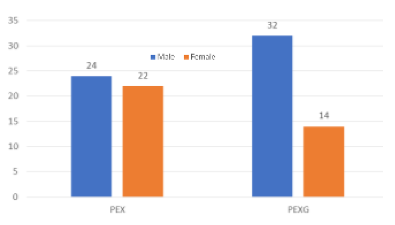
There were 46 study participants in each group of which there were 24 males and 22 females in the PEX group and, 32 males and 14 females in PEXG group (Figure 1).
Figure 1: Gender distribution of the study participants
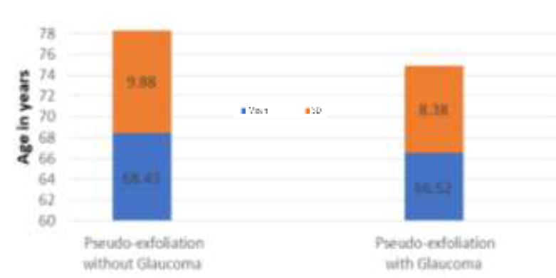
The mean age of patients with pseudo-exfoliation without Glaucoma was 68.43±9.88 years ranging from 38 years to 88 years whereas the mean age of the patients having Pseudo-exfoliation with Glaucoma was 66.52±8.38 years ranging from 50 years to 81 years. The difference in the mean age of the two groups was not found to be statistically significant (p=0.810), (Figure 2).
Figure 2: Age distribution of the study participants
The visual activity was in the range from 6/6 to FC 2/60 in the left eye as well as in the right eye. An intra ocular pressure (IOP) of less than 10 mm Hg was found in more than three-fourth of the participants while the rest had an IOP ranging from 10-20 mm and none had an IOP of more than 21 mm Hg in the left eye, whereas an IOP of <10 mm Hg was found to be in nearly 68% of the patients, while one-third of the patients had an IOP ranging between 10-20 mg Hg whereas the rest had an IOP of >21 mm Hg in the right eye. Vertical cup disc between 0.3 -0.4 was found in 39 patients while a cup disc between 0.5 and 0.6 was found to be among the rest of the study participants (Table 1).
Table 1: Visual activity of the study participants in two groups (N=92)
Side
Visual Acuity
Pseudo-exfoliation
p-value
without Glaucoma
with Glaucoma
Left Eye
6/6- 6/12
22(47.83%)
23 (50%)
<0.001
6/18-6/36
10 (21.74%)
9 (19.57%)
6/60 or worse
14 (30.43%)
14 (30.43%)
Right Eye
6/6- 6/12
14 (30.43%)
0
<0.001
6/18-6/36
14 (30.43%)
0
6/60 or worse
18 (39.13%)
46 (100%)
In patients of pseudo-exfoliation without glaucoma, the mean RNFL thickness(µm) in Left Nasal, Left Temporal, Left Superior, Left Inferior, Right Nasal, Right Temporal, Right Superior, Right Inferior quadrant was 74.70±20.506, 67.61±20.477, 118.59±23.053, 113.07± 23.449, 70.35±20.068, 96.46±31.758, 106.78±26.168, 106.78±26.168 respectively and the overall mean RNFL thickness was found to be 89.47±13.52 µm. In patients od pseudo-exfoliation with glaucoma, the mean RNFL Thickness(µm) in Left Nasal, Left Temporal, Left Superior, Left Inferior, Right Nasal, Right Temporal, Right Superior, Right Inferior quadrant was 67.15±20.196, 50.98±14.751, 84.93±28.497, 83.33 ±28.157, 69.00±16.780, 82.07 ±29.826, 87.70±30.577 and 87.70±30.577 respectively.
RNFL thickness was seen to be decreased in all PEX patients with glaucoma in all quadrants; however, statistically significant differences were detected in all quadrant except left nasal (p=0.079) and right nasal quadrants (p=0.728). The overall mean RNFL Thickness (µm)
in the two groups was 89.47±13.52 µm and 72.73±13.54 µm and the difference between the means of RNFL thickness between the two groups was found to be statistically significant (p<0.001), (Table 2).
Table 2: RNFL thickness of the study participants of two groups (N=96)
The Part
Pseudo-exfoliation without Glaucoma
Pseudo-exfoliation with Glaucoma
p-value
Mean
SD
Mean
SD
Left Nasal
74.70
20.506
67.15
20.196
0.079
Left Temporal
67.61
20.477
50.98
14.751
<0.001
Left Superior
118.59
23.053
84.93
28.497
<0.001
Left Inferior
113.07
23.449
83.33
28.157
<0.001
Right Nasal
70.35
20.068
69.00
16.780
0.728
Right Temporal
96.46
31.758
82.07
29.826
0.005
Right Superior
106.78
26.168
87.70
30.577
0.002
Right Inferior
106.78
26.168
87.70
30.577
0.028
Over all Mean
89.47
13.52
72.73
13.54
<0.001
DISCUSSION
The measurement of Retinal Nerve Fibre Layer thickness can provide vitally important information regarding glaucomatous changes in the initial stages [9]. Even after having such knowledge, there is a meagre scientific literature available that compares the change in the thickness between patients of pseudo-exfoliation without glaucoma and those with glaucoma. In our study, patients of pseudo-exfoliation without glaucoma showed relatively greater overall mean RNFL thickness as compared to those in patients of pseudo-exfoliation with glaucoma using SD-OCT There was however weak evidence to determine the difference of thickness change in the left nasal and right nasal quadrants between the two groups.
In our study, patients having pseudo-exfoliation with glaucoma showed significant visual field defects as compared to their counterparts. Our results demonstrated a significant reduction in the thickness in the all except nasal quadrant. Aydin et al. [10] conducted a study in which they found that the RNFL thickness in the superior quadrant was thinner in pseudo-exfoliative patients with glaucoma.
Eltutar et al. also reported that there was a significant reduction in the thickness in the superior quadrants of the patients of pseudo-exfoliation with glaucoma. However, one metanalysis [11] does not seem to be in line with our study, in which the RNFL thickness was significantly lower in all the quadrants in the patients of pseudo-exfoliation with glaucoma. This could be possibly due to a separate population characteristics based on the sample size, ethnicity and the presence of systemic diseases among them.
The thinning of the RNFL could be attributed to the presence of systemic vascular diseases as obtained from the literature. We, however, had excluded all the patients who were suffering from any cardiovascular diseases so this possibility can be ruled out from our study. However, there could be local factors, for example, tissue weakness leading to decreased stiffness of lamina cribrosa and adjoining scleral tissues especially in the elderly. A study conducted by Wang et al. [12] reported high prevalence of cardiovascular disease in patients of pseudo-exfoliative syndrome. Structural changes in lamina cribrosa may also influence hemodynamic changes, such as increased venous outflow resistance or turbulence that may have led to thin RNFL [13].
CONCLUSION
The visual acuity was significantly poor in all the patients of pseudo-exfoliation with glaucoma. RNFL thickness was significantly lower in the patients of pseudo-exfoliation with glaucoma. Measurement of RNFL thickness in early stages of Pseudo-exfoliation with glaucoma can help to modify the course of management. Further, analytical studies to understand the causes of thickness changes of RNFL as well as how early does it need to be detected in order to prevent a permanent damage to the vision.
REFERENCE
Ritch, R. et al. “Why is glaucoma associated with exfoliation syndrome?” Progress in Retinal and Eye Research, vol. 22, no. 3, 2003, pp. 253-275.
Tekin, K. et al. “Monitoring and management of the patient with pseudo-exfoliation syndrome: current perspectives.” Clinical Ophthalmology, vol. 13, 2019, pp. 453-464.
Lalezary, M. et al. “Baseline optical coherence tomography predicts the development of glaucomatous change in glaucoma suspects.” American Journal of Ophthalmology, vol. 142, no. 4, 2006, pp. 576-582.e571. https://doi.org/10.1016/j.ajo.2006.05.004.
Curcio, C.A. and K.A. Allen. “Topography of ganglion cells in human retina.” Journal of Comparative Neurology, vol. 300, no. 1, 1990, pp. 5-25. https://doi.org/10.1002/cne.903000103.
Desai, M.A. and R.K. Lee. “The medical and surgical management of pseudo-exfoliation glaucoma.” International Ophthalmology Clinics, vol. 48, no. 4, 2008, pp. 95-113.
Nagrale, P. et al. “A study of eyes with pseudo-exfoliation, its association with cataract and its implications in cataract surgery.” International Journal of Contemporary Medical Research, vol. 5, no. 9, 2018, pp. 1-15.
Kozobolis, V.P. et al. “Retinal nerve fibre layer thickness in patients with exfoliation, exfoliative glaucoma and primary open angle glaucoma.” European Journal of Ophthalmology, vol. 20, no. 1, 2010, pp. 142-148.
Lim, S.H. et al. “Comparison of the retinal nerve fibre layer and ganglion cell complex thickness in Korean patients with unilateral exfoliation syndrome and healthy subjects.” Eye, vol. 34, no. 8, 2020, pp. 1419-1425.
Plateroti, P. et al. “Pseudo-exfoliation syndrome and pseudo-exfoliation glaucoma: a review of the literature with updates on surgical management.” Journal of Ophthalmology, vol. 37, no. 3, 2015, pp. 1-9.
Aydin, D. et al. “Evaluation of retinal nerve fibre layer and ganglion cell complex thickness in unilateral exfoliation syndrome using optical coherence tomography.” Journal of Glaucoma, vol. 25, no. 6, 2016, pp. 523-527.
Yu, J.G. et al. “Retinal nerve fibre layer thickness changes in the pseudo-exfoliation syndrome: a meta-analysis of case-control studies.” Ophthalmic Research, vol. 59, no. 1, 2018, pp. 14-23.
Wang, W. et al. “Ocular pseudo-exfoliation syndrome and vascular disease: a systematic review and meta-analysis.” PLoS One, vol. 9, no. 3, 2014, e92767.
Jonas, J.B. “Central retinal artery and vein collapse pressure in eyes with chronic open angle glaucoma.” British Journal of Ophthalmology, vol. 87, no. 8, 2003, pp. 949-951.
Recommended Articles
Research Article
Leigh Syndrome Should Not Be Diagnosed Exclusively Upon Cerebral MRI
Finsterer J
Published: 15/07/2020
Download PDF
Research Article
Vision for a Brighter Kangra: Unmasking the Truth about Pink Eye – A Comprehensive Study on Types, Symptoms, and Proactive Prevention in Himachal Pradesh’s Kangra District
Deepti ,
...
Garima
Published: 11/11/2023
Download PDF
Research Article
It Remains Unproven That the Variant M.8231C>A Causes Coronary Atherosclerosis
Finsterer J
Published: 15/07/2020
Download PDF
Research Article
Bioactive ingredients screening and antibacterial activity of Citrus aurantifolia leaf extract against some enteric isolates
Abdallah M.S,
Kabiru Sama’ila
Published: 10/08/2020
Download PDF
PO Box 101, Nakuru
Kenya.
Email: office@iarconsortium.org
Editorial Office:
J.L Bhavan, Near Radison Blu Hotel,
Jalukbari, Guwahati-India
S., S., R. L., S. & V., S. (2021). Comparison of retinal nerve fibre layer thickness in patients residing in the western Himalayas: A cross-sectional analytical study.. IAR Journal of Medical Sciences, 2(1), 1-3.
MLA
S., Sharma, Sharma R. L. and Sharma V.. "Comparison of retinal nerve fibre layer thickness in patients residing in the western Himalayas: A cross-sectional analytical study.." IAR Journal of Medical Sciences 2.1 (2021): 1-3.
Chicago
S., Sharma, Sharma R. L. and Sharma V.. "Comparison of retinal nerve fibre layer thickness in patients residing in the western Himalayas: A cross-sectional analytical study.." IAR Journal of Medical Sciences 2, no. 1 (2021): 1-3.
Harvard
S., S., R. L., S. and V., S. (2021) 'Comparison of retinal nerve fibre layer thickness in patients residing in the western Himalayas: A cross-sectional analytical study.' IAR Journal of Medical Sciences 2(1), pp. 1-3.
Vancouver
S. S, R. L. S, V. S. Comparison of retinal nerve fibre layer thickness in patients residing in the western Himalayas: A cross-sectional analytical study.. IAR Journal of Medical Sciences. 2021 Jan;2(1):1-3.



 Download PDF
 Download XML