Introduction: The calcaneus is the most frequently injured tarsal bone. Many calcaneal fractures are work-related as they result from a fall from height, especially in males’ age 35–45 years. These fractures frequently result in long-term disability with potentially severe economic impact on the patient. Operative treatment of displaced calcaneal fractures has been a controversial due to tissue condition and wound infection. In this study, our objective is to investigate the outcome of percutaneous fixation of displaced calcaneal fractures with cannulated cancellous screws. Materials and Methods: A retrospective review of 15 patients with calcaneal fractures who had percutaneous reduction and screw fixation for displaced fracture of calcaneus was done. The patients were assessed with plain radiography. In developing countries like India, cost of medical treatment is often a concern, so computed tomography scan was not done as most of the patients were poor and did not afford the treatment. The fractures were classified according to Essex‑Lopresti classification system. Functional assessment involved the use of calcaneal fracture scoring system. Results: The age of the patients ranged from 25 to 45 years (mean: 35 years). Time to union averaged 14.5 weeks (range 12–17). The treatment outcome as measured by calcaneal fracture scoring system ranged from 66 to 90 (mean 78). Conclusion: Percutaneous fixation of fracture calcaneum, offers the prospect of fewer complications and better outcome.
Calcaneus fractures are the most common of all tarsal fractures (60%) and account for almost 2% of all adult fractures. Approximately 75% of these injuries are intra-articular, and almost all occur due to an axial load such as a fall from a height or a motor vehicle accident. Approximately 10% of patients will have a spine fracture as well due to the axial load. Approximately 10% of injuries are bilateral and fewer than 5% are open. Many calcaneal fractures are work-related, as they result from a fall from height, especially in males’ age 35–45 years [1-2]. These fractures frequently result in long-term disability with potentially severe economic impact on the patient as many patients fail to return to their original occupation after the trauma. Since the original description of these fractures, they have been recognized as being problematic [3-4]. The disabling nature of severe calcaneus fracture and its huge economic burden has long been known and it remains so today. In 1916, Cotton and Henderson stated that “ordinarily speaking, the man who breaks his heel bone is done so far as his industrial future is concerned [5]”. Calcaneal fractures constitute one of the most contradictory chapters of traumatology of the musculoskeletal apparatus. No other type of fracture is associatedwith such a wide range of different views regarding its management. The irregular bony anatomy and the complicated joint mechanics between the tarsal bones and the delicate soft tissue envelope in which they sit have made these fractures a challenge. Many classification schemes, operative techniques, and postoperative regimens have been proposed, but a true consensus has not been reached. Lack of consensus is documented also by the fact that so far about 140 therapeutic methods have been suggested for its treatment. Historically, treatment of displaced calcaneal fractures has varied from nonoperative management with or without closed reduction, to open reduction with internal fixation by various surgical approaches, to primary arthrodesis [6-11]. Historically, most fractures were treated non operatively because open reduction and internal fixation were associated with high complication rates and did not result in significantly improved outcomes. Despite very strict indication criteria for open reduction and internal fixation, the number of reported infection rate ranges between 5% and 20%, and amputations of limbs are not an exception due to chronic osteomyelitis and permanent disability [12]. However, non operative treatment also has many complications; it includes inability to maintain reduction. Consequently, there will be broadening of heel, muscle imbalance, loss of motion, peroneal impingement, impaired gait, intractable pain, and early development of subtalar arthritis, and permanent disability [13-14]. Following this line of reasoning, percutaneous reduction and fixation could be the ideal option for displaced fracture of calcaneus addressing both the above issues. Thus, we present a study to evaluate the results of percutaneous reduction and fixation of displaced fracture calcaneus.
A retrospective review of 15 patients with calcaneal fractures who had percutaneous reduction and screw fixation between jan 2019 and jan 2020 was done. Out of these 10 were male and 5 were female. The patients were assessed with plain radiography. Due to financial constraint CT scan was not possible in all patients. Standard views for calcaneus were performed along with anteroposterior view of the foot and the ankle to exclude associated fractures. Only displaced fracture of the calcaneus was included in the study. The fractures were classified according to Essex‑Lopresti classification system Essex‑Lopresti (X‑ray‑based classification ‑ lateral view foot) [15]. Extra‑articular Intra‑articular o Tongue type o Articular depression type. After preoperative assessment, patients underwent operative procedures within 6 days; in case of excessive swelling, surgery was delayed for 12-14 days to allow soft tissue swelling to resolve. In such cases, the foot was placed in a posterior plaster splint and the limb was elevated. Surgery was undertaken after the edema had settled completely and the wrinkle sign appeared.
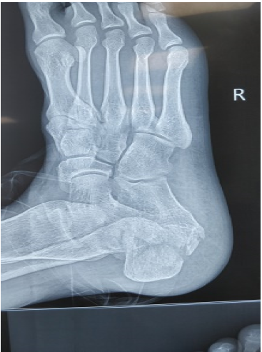
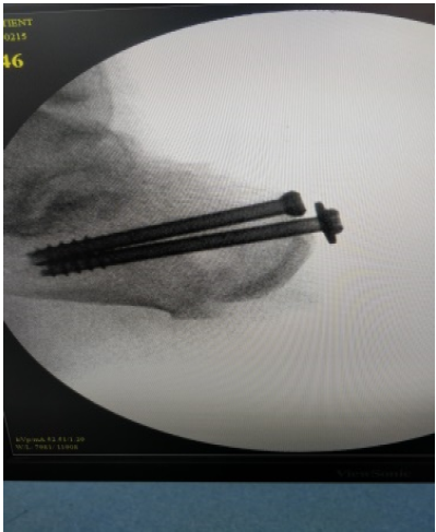
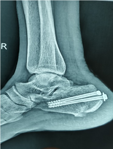
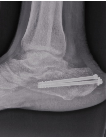
Surgery was performed without tourniquet under spinal anesthesia; all patients were given injection ceftriaxone with sulbactam for antibiotic prophylaxis 20 min before starting surgery. The patient was placed in lateral decubitus position with the affected leg upward. Using the image intensifier, the fracture was visualized both in lateral and axial view. Percutaneous reduction of fracture fragments was done using Steinmann pin and k‑wires, and fracture was fixed with 6.5 mm cancellous cannulated screws (Figures1,2,3 and 4). Postoperatively, a below-knee plaster cast is applied with the foot at a right angle. All patients were kept no weight-bearing for 6 weeks. Ankle mobilization was started after the plaster slab was discarded in 6 weeks. The patients were followed up at 6 weeks, 3 and 6 months in the OPD. Partial weight-bearing was allowed in 6 weeks’ time, and full weight- bearing on the affected limb was allowed after 3 months post‑operative. Functional assessment involved the use of calcaneal fracture scoring system of Kerr et al. [16] and Maryland foot scoring system by asking patients about discomfort/pain during activity and rest, ability to stand and walk, ability to return to work, duration of absence from work, and any change in the size of the shoe. An overall rating of excellent (90–100), good (80–89), fair (65–79), and poor (below 64) was assigned to each fracture. Patients were also enquired about their satisfaction with their treatment outcome.

Figure 1: Pre-Operative Xray Image Showing Displaced Intraatricular Fracture of Calcaneus

Figure 2: Intra Operative C Arm Image

Figure 3: 1 Month Follow Up Xray Image Showing Well Reduced and Uniting Fracture

Figure 4: 6 Month Follow Up Xray Image Showing Well United Fracture
The age of the patients ranged from 25 to 45 years (mean: 35 years). Ten patients were male and 5 were female. The right foot was affected in 8 patients, the left in 7 patients. All the patients had sustained this injury as result of fall from height. All fractures were subdivided according to Essex‑Lopresti classification system based on X‑ray findings. Ten fractures were tongue type and remaining five were joint depression type. Average time delay from injury to surgery was 7 days (range, 4–14 days). Mean follow-up was 6 months. All fractures healed without any major complication. Two of the patients developed superficial wound infection which healed with dressings and antibiotics. Time to union, judged both clinically and radiologically, averaged 14.5 weeks (range 12–17). The treatment outcome as measured by calcaneal fracture scoring system ranged from 66 to 90 (mean 78). Twelve of 15 patients (80%) were satisfied with the treatment. None of the patients required subtalar fusion till the last follow-up.
Calcaneus fractures comprise 2% of all fractures, and 60%–75% of these fractures are intra-articular [17]. Calcaneus fractures are generally due to high-energy traumas such as a fall from height and traffic accidents. In our study patients were between 25 and 45 years of age, and in all the cases, the cause of fracture was fall from height. Essex‑Lopresti classified displaced intra‑articular calcaneal fractures as either tongue type or joint depression type, based on the configuration of a secondary fracture line through the posterior facet as seen on plain radiograph. He suggested that non displaced fractures be treated non surgically. Displaced tongue fractures are best treated by percutaneous pin fixation, using a large posterior reduction tool for closed reduction. Joint depression fractures require surgical reduction because of fracture impaction [15]. Essex‑Lopresti classification is the most commonly used X‑ray‑based classification system for calcaneal fractures, and we have assessed the fractures only on X‑rays due to economic constraints, so we have used the same in our study. Options for treating displaced calcaneal fractures are conservative or operative. Kitaoka et al. [18] reviewed the results of the walking analyses of 16 of 27 patients, who were not subjected to reduction and were treated with plaster. Many patients in their study showed differences in walking, especially on uneven grounds, which showed that conservative treatment of displaced calcaneus fractures results in permanent functional disorders, at least to some extent. O’Farrell et al. [19] treated 12 patients with surgical methods and 12 patients with conservative methods. They concluded that the surgical treatment was superior. Similarly, Leung et al. [20] compared 44 patients treated by surgical methods with 19 patients treated conservatively according to the results of 3-year follow-up on average. Comparing pain, ease of movement, return to job, and swallowed back of the foot, they found that the results of the group treated surgically were significantly better. Thus, from the above studies, it is clear that surgical treatment is the only viable alternative for displaced fractures of calcaneum. For patients who are to undergo surgical treatment, timing of the surgery is one of the most important determinants for the outcome of treatment and determined by subsidence of edema and appearance of wrinkle sign, persistence of which may lead to soft tissue healing problems and high infection rate, while surgery after 3 weeks of injury causes difficulty in reduction, due to early consolidation of fracture. Hence, it is better to delay surgery till soft tissue heals, and during this pre surgical period, patients should be managed by splinting with proper padding, limb elevation, and soft tissue care. Thus, most authors suggest that the surgical intervention should take place after the edema in the foot has regressed and the soft tissue envelope is conducive. Surgical results are dependent upon the timing of surgery; according to Sanders, the correct timing is between 7 and 10 days from trauma because if treated earlier, there is a risk of compartmental syndrome or soft tissue damage and if treated later, the results could be otherwise unsatisfactory [21-23]. Average time delay from injury to surgery was 7 days in our study which is probably the optimum time for surgical intervention. We applied below-knee plaster splint with elevation of limb till the swelling and edema subsided. The balance between the percentage of anatomical reduction and the amount of soft tissue damage should be carefully weighted in the management of fracture calcaneus, especially the severely comminuted type. The choice lies between trail of full reduction with extensive soft tissue damage or reasonable reduction with minimal soft tissue insult. The treatment outcome in our study as measured by calcaneal fracture scoring system ranged from 66 to 90 (mean 78) with only two patients having minor complication like superficial skin infection. Similar results were also seen by authors like Essex‑Lopresti [15] and Tim Schepers et al. who concluded that percutaneous reduction by skeletal distraction and screw fixation can be considered a useful treatment modality, especially in severely comminuted fractures, expected wound complications in open fractures, and patients with diabetes or peripheral vascular occlusive disease.
Thus, we conclude that percutaneous reduction and fixation with cancellous screws is a suitable mode of treatment for displaced intra-articular calcaneal fractures.
Financial Support and Sponsorship
Nil.
Conflicts of Interest
There are no conflicts of interest.
Barei, D.P. et al. “Fractures of the calcaneus.” Orthopedic Clinics, vol. 33, no. 1, 2002, pp. 263–285.
Nicklebur, S., Dixon, T.B. and Probe, R. “Calcaneus fractures.” EMedicine, 2004.
Sanders, R. “Displaced intra-articular fractures of the calcaneus.” Journal of Bone and Joint Surgery, vol. 82, 2000, pp. 225–250.
Rammelt, S. and Zwipp, H. “Calcaneus fractures: Facts, controversies and recent developments.” Injury, vol. 35, no. 5, 2004, pp. 443–461.
Cotton, F.J. and Henderson, F.F. “Results of fractures of the os calcis.” American Journal of Orthopedic Surgery, vol. 14, 1916, pp. 290–298.
Stehlík, J. et al. “Calcaneal fractures.” Galén, 1st ed., 2010.
Abidi, N.A. et al. “Wound-healing risk factors after open reduction and internal fixation of calcaneal fractures.” Foot and Ankle International, vol. 19, no. 12, 1998, pp. 856–861.
Bridgman, S.A. et al. “Interventions for treating calcaneal fractures.” The Foot, vol. 12, no. 1, 2002, pp. 47–61.
Buckley, R.E. and Meek, R.N. “Comparison of open versus closed reduction of intra-articular calcaneal fractures.” Major Fractures of the Pilon, the Talus, and the Calcaneus, 1993, pp. 195–205.
Buckley, R. et al. “Operative compared with nonoperative treatment of displaced intra-articular calcaneal fractures.” Journal of Bone and Joint Surgery, vol. 84, no. 10, 2002, pp. 1733–1744.
Buckley, R.E. and Tough, S. “Displaced intra-articular calcaneal fractures.” Journal of the American Academy of Orthopaedic Surgeons, vol. 12, 2004, pp. 172–178.
Al-Mudhaffar, M., Prasad, C.V.R. and Mofidi, A. “Wound complications following operative fixation of calcaneal fractures.” Injury, vol. 31, no. 6, 2000, pp. 461–464.
Zwipp, H. et al. “Osteosynthesis of displaced intraarticular fractures of the calcaneus.” Clinical Orthopaedics and Related Research, no. 290, 1993, pp. 76–86.
Randle, J.A. et al. “Should calcaneal fractures be treated surgically?” Clinical Orthopaedics and Related Research, vol. 377, 2000, pp. 217–227.
Essex-Lopresti, P. “The mechanism, reduction technique, and results in fractures of the os calcis.” British Journal of Surgery, vol. 39, no. 157, 1952, pp. 395–419.
Kerr, P.S., Prothero, D.L. and Atkins, R.M. “Assessing outcome following calcaneal fracture.” Injury, vol. 27, no. 1, 1996, pp. 35–38.
de Souza, L.J. and Rutledge, E. “Grouping of intraarticular calcaneal fractures relative to treatment options.” Clinical Orthopaedics and Related Research, vol. 420, 2004, pp. 261–267.
Kitaoka, H.B. et al. “Displaced intra-articular fractures of the calcaneus treated non-operatively.” Journal of Bone and Joint Surgery, vol. 76, no. 10, 1994, pp. 1531–1540.
O’Farrell, D.A. et al. “Fractures of the os calcis: Improved results with internal fixation.” Injury, vol. 24, no. 4, 1993, pp. 263–265.
Leung, K.S., Yuen, K.M. and Chan, W.S. “Operative treatment of displaced intra-articular fractures of the calcaneum.” Journal of Bone and Joint Surgery, vol. 75, no. 2, 1993, pp. 196–201.
Zwipp, H. et al. “Calcaneus fractures.” Surgical Techniques in Orthopaedics and Traumatology, Elsevier SAS, 2000.
Sanders, R. and Gregory, P. “Operative treatment of intra-articular fractures of the calcaneus.” Orthopedic Clinics of North America, vol. 26, no. 2, 1995, pp. 203–214.
Levin, L.S. and Nunley, J.A. “The management of soft-tissue problems associated with calcaneal fractures.” Clinical Orthopaedics and Related Research, no. 290, 1993, pp. 151–156.