Contents
Download PDF
pdf Download XML
110 Views
32 Downloads
Share this article
Research Article | Volume 1 Issue 1 (Jan-June, 2020) | Pages 1 - 7
A Retrospective and a Multicenter Analysis of Acute Appendicitis in Children: Complications and Surgical Treatment
 ,
 ,
 ,
 ,
 ,
1
Department of Pediatrics, Lebanese University-Faculty of Medical Sciences, Lebanon
2
Department of Internal Medicine, American University of Beirut Medical Center, Lebanon
3
Department of Pediatric Surgery, Lebanese University-Faculty of Medical Sciences, Lebanon
Under a Creative Commons license
Open Access
Received
May 3, 2020
Revised
June 6, 2020
Accepted
July 19, 2020
Published
Aug. 10, 2020
Abstract

Background: Acute appendicitis (AA) is the inflammation of the vermicular appendage. It is one of the most common causes of abdominal pain and surgical abdominal emergencies in children. Nowadays and according to literature, the method of treatment remains a dialectical subject. The long delay before admission to hospital is associated with increased rates of perforation. In this study, we evaluate how physicians in Lebanon act in front of acute appendicitis. Materials and Methods: Retrospective, observational, multicenter study on all pediatric patients who underwent appendectomy in three Lebanese hospitals: Nabih Berri Governmental University Hospital, Bahman Hospital and Rafic Hariri University Hospital during year 2017. Results: The study included 102 patients, mean age 10.7 years. Up to 48 Hours of delay, no correlation was found between delay in surgery and the risk of developing a complicated form of the disease such as perforation and peritonitis and postoperative complications up to 30 days after discharge for group 1 (surgery <24 hours) and group 2 (surgery between 24 to 48 hours). In group 1 the complication rate was 13.7% identical to that of group 2 which was 13.68 %. Patients with AA showed a male/female ratio of 0.7 (p <0 .0001). Treatment with antibiotherapy before surgery achieved better outcomes. The treatment option was independent from the clinical presentation. The polled difference in Length of Stay (LOS) showed a trend for shorter LOS in the Immediate Surgery (IS) group. Conclusion: While the complication rate was the same in children who underwent Immediate appendectomy (within the first 24 hours) and patients who were observed and treated with intravenous antibiotics and good hydration followed by delayed appendectomy throughout 24 to 48 hours, Immediate surgery was associated with a shorter hospitalization.

Keywords
INTRODUCTION

Acute appendicitis, defined as the inflammation of the appendix vermicular. Acute appendicitis is one of the most common causes of acute abdominal pain and surgical emergencies in children as in adults. In the United States, appendicitis in children has an incidence of 70,000 cases per year. About 7% of people in the United States undergo appendectomy during their lifetime. It is more common in the second decade of life, between 10 and 19 years of age. Then, its incidence continues to decrease, even if acute appendicitis may occur in adults and elderly [1]. It occurs through an occlusion of the appendix lumen. Lymphoid hyperplasia caused by inflammatory bowel diseases, infections, parasites, stool, or more rarely a foreign body and neoplasm, is the most common cause of occlusion [2]. The stages of appendicitis are: catarrhal appendicitis, ulcerative and suppurative appendicitis, abscessed appendicitis and phlegmnous appendicitis [3]. In general, the prognosis is excellent. The percentage of perforation at the time of diagnosis is 20-35%. Upon Appendix rupture, the patient risks having intra-abdominal abscesses and small bowel obstruction and would require an extended hospital stay. In children, the mortality rate is 0.1 to 1% [4]. As soon as the diagnosis of acute appendicitis (perforated or non-perforated) is made, an appendectomy is indicated. There are currently 2 approaches in surgical treatment, the first being an emergency appendectomy <24 hours (Immediate surgery) and the second, consists of antibiotic therapy followed by a delayed appendectomy (delayed surgery within 24 to 48 hours) [5]. Appendicular disease is a common cause of emergency hospitalization and appendectomy is one of the emergency procedures of contemporary medicine [2,6,7]. It should be noted that 6.7% of girls and 8.6% of boys are at risk of developing appendicitis during their lifetime [3,5,2] Today, the treatment method remains a dialectical subject. Indeed, some studies find that appendicitis can lead to several complications if not performed within the first 24 hours of onset. Perforation is a good example, where increasing hospitalization stay and costs increases the burden for patient, health care, payers and society [8]. The long observation period in hospital before appendectomy is linked to an increase in the perforation rate of children and adults [9]. Others suggest that a delayed surgery is safe and is preferred, although clinical results are no better than immediate appendectomy, In fact surgeons choose the appropriate time for surgery, taking into account many factors. However, in Lebanon, it is not known how doctors respond to acute appendicitis and whether they consider it as a surgical emergency or they start with antibiotic therapy followed by appendectomy within the second 24 hours and no previous assessment was performed to check if this delayed approach influences the rate of complications, thus it is important to describe the current practice in Lebanon in face of appendicitis in pediatric population and to compare the two approaches of immediate ( within the first 24 hours) versus delayed intervention ( observe for 24 hours with IV antibiotics & good hydration then intervene surgically the next day: 24-48 hours) as well as analyze the difference in outcomes to shape an evidence based approach for an informed therapeutic decision making.


Literature Review

Historical Context and Generalities: Clinicians supported the following proposition: inflammation of the worm-like appendix caused common intra-abdominal disease just in the late 1880s, when Reginald Fitz reviewed 257 autopsy cases at Harvard and coined the term "appendicitis", universally recognized today. Complex appendicitis has been signaled out as being considerable in regard to the consequences of simple appendicitis which is treated by appendectomy in the advanced stage of the disease before the production of a perforation. New York surgeon Charles Mc. Burney noted that the site of maximum sensitivity was "in the average adult, almost exactly two inches from the anterior iliac spine," on a line drawn from this process through the navel," it's now designated as eponymous point" [10].


Current Practice

Some features of the management of appendicitis have become common. As an example, we replaced an open appendectomy with an incision in the lower right quadrant by a routine laparoscopic approach for simple and complex appendicitis. The laparoscopic approach has been equated with a reduction of the risk of wound infections and bowel obstruction, excellent postoperative pain control, shorter hospital stays and faster return to daily life [11]. For the moment, most appendectomies are performed by laparoscopy in the United States but by laparotomy in Lebanon. The general consensus is that antibiotics should be given to a child with simple acute appendicitis before the incision and discontinued after the operation [12]. The need to continue antibiotics postoperatively during the treatment of perforated appendicitis is universally accepted. However, the choice of antibiotic, the duration of administration and the possible role for oral medications vary and change depending on the surgeon. Justifications and evidence support the other stages of diagnosis and management of appendicitis [13]. An equivoqual observation is undoubtedly the role of imagery and the choice of the type of imagery, marked by a controversy and a series of propositions and counter-propositions. The decision to operate has long been based on clinical data, without being influenced by imagery.


Diagnostic Approach

The pathophysiological process, the stretching of the obstructed hollow organ, leads to stimulation of the visceral nerves. The child experiences mild abdominal pain. Since the appendix belongs to the middle intestine. its sensation depends on the visceral nerves which send the painful sensation back to the T10 dermatome [14]. As the inflammation spreads through the wall of the appendix, it stimulates the somatic nerves, causing peritoneal signs. The evolution of localized peritoneal signs (peritonitis of the right lower quadrant) points the diagnosis. If the inflammation lasts, bacterial contamination of the peritoneum is caused by necrosis of the perforated appendix wall. The bacterial content that surrounds the intestine or omentum forms a phlegmon or abscess. Unconfined contamination leads to diffusion and then to peritonitis and septicemia associated with increased morbidity and mortality. Clinicians treating children, especially the very young, are hurdled by the lack of a clear history and the difficulty of establishing peritoneal signs during a physical exam. The diagnostic measures are supplemented by a Complete Blood Count with Differential (CBCD), a Urine Analysis (U/A) and an abdominal x-ray. Until the last decade, abdominal radiography was the only accessible imaging tool that aids and facilitates diagnosis. The absence of a radiopaque fecal impaction reduced its usefulness. A very high number of white blood cells (WBC) confirms the diagnosis of appendicitis but it was neither specific nor sensitive [14]. A significantly high number of white blood cells was compatible with complex appendicitis, but was no longer a sophisticated tool.


GUE has been widely indicated when a patient had possible appendicitis to rule out urinary tract pathology. However, its role has been blurred by the fact that red blood cells are often considered to be part of acute appendicitis, as the inflamed appendix can touch the ureter or bladder. With an extensive history, physical examination, CBCD, U/A, an occasional abdominal X-Ray, clinicians distinguish whether the child has appendicitis or not. The process was–and remains- active, accompanied by an active observation period with serial examinations [15]. The goal is to get a quick diagnosis of appendicitis early enough in the disease to prevent progression of perforation. Some experts have recommended that surgeons remove the unaffected appendages, assuming that if the surgeon removes too few unaffected appendages, some patients may encounter perforation. Indeed, it is not necessary to have a high rate of negative appendectomies in order to avoid cases of perforation. The lack of diagnosis of appendicitis is the most common cause of prosecution of emergency care physicians treating children, prompting them to use (CT scans) to confirm the diagnosis before operating.


Imaging

The reliability of the imagery explains its impact on the management of appendicitis. This has been demonstrated by ultrasound [13], or computed tomography [14], studies. In particular, ultrasound has been shown to reduce the time between arrival and surgical management, but there was no improvement in the morbidity of appendectomy or reduction in the length of hospital stay. The study published by Rao [14], had some repercussions even in the mainstream press and showed that performing an emergency CT scan considerably improved the therapeutic management and made it possible to achieve substantial savings, two thirds of the savings; being due to the prevention of unnecessary appendectomies and a third to the reduction in days of hospitalization for surveillance. These potential gains remain to be quantified in other countries depending on the cost of hospitalizations, apart from a precise diagnosis of appendicitis; the interest of imagery is also to allow an alternative diagnosis in front of pain in the right iliac fossa. In patients referred for suspected appendicitis, appendicitis is present only in 25 to 50% of cases. In almost a third of cases, imagery allows differential evidence, most often digestive (75% of cases), more rarely genital (15% of cases) or urinary (10% of cases). The semiology in imaging of these different alternative diagnoses to be evoked in front of a right iliac fossa pain must be known [15,16]. But according to the new literature, we find that there was a 41% decrease in the use of CT (from 90% of children undergoing appendectomy to 48%) and an increase in the use of ultrasound. and the percentage of children undergoing appendectomy without imaging. The rate of negative appendectomy has not increased [16]. These results confirm that reducing dependence on CT scans is safe and effective when children are in well-trained hands and suggests that a clinical practice guide may be helpful. The selective use of computed tomography should target patients for whom the diagnosis remains unclear despite less invasive approaches and patients with particularly advanced appendicitis, since the presence of a significant abscess can modify the therapeutic approach.


Time of Appendectomy

Reginald Fitz has established for over 125 years the goal for clinicians who manage appendicitis: have the appendectomy done at the right time, before the perforation. Acute appendicitis was the most common abdominal emergency operation in children. Perforated appendicitis carries significantly more threat than simple appendicitis. The circumstances of a delayed diagnosis could provide possible prosecution. As a result, appendectomies were performed at all hours of the day and night as an emergency. When a child had complex appendicitis, a period of time to provide intravenous hydration and antibiotics was widely accepted [17]. However, in the case of simple appendicitis, the operation was considered as an emergency that should be done immediately due to the presumed risk of allowing simple appendicitis to become complex with worsen morbidity, yet some studies report that a delayed appendectomy during the day can provide a better working environment [18]. However, others suggested that it increases the risk of pathological forms [19,20]. This study aims to assess the impact of the appendectomy time interval window on the development of complex appendicitis and postoperative complications. In the era of antibiotics and long after Fitz's work, this approach to emerging surgery for simple appendicitis was changed. To avoid pressure on resources, when appendicitis is diagnosed at night, a common practice is to administer antibiotics and intravenous hydration and perform the operation the next morning. This approach assumes that antibiotics stop the progression of the disease, allowing a safe operation led by a full staff and a rested team.


Finally

A pediatric population study has confirmed the safety of delaying appendectomy until overnight when patients are admitted late in the evening. In addition, many studies have shown that excessive loading can increase fatigue among hospital staff and have pointed out that lack of sleep has negative effects on residents' cognitive abilities and clinical performance, which could increase morbidities and mortalities. There was no association between surgical site infection rates and delayed surgery. Our study says that appendectomy can be safely delayed up to 48 hours in children. According to Chen and Al [21], the clinical results of delayed appendectomy are no better than those of immediate appendectomy. Chen's findings demonstrated that delaying an appendectomy more than 8 hours after admission did not increase the risk of perforation and postoperative complications rate [22]. Horney and Al found that a delay in surgery of less than 48 hours did not influence the pathological outcome of acute appendicitis [20]. The surgical site infection rate did not increase if surgery was delayed for less than 48 hours. Several surgical centers for children have confirmed the safety of this approach [23]. Once the appendix is perforated, the appendectomy is no longer urgent: it is better to prepare the patient and administer intravenous fluids as well as antibiotics to reduce the operating risk. This course of action is even currently applied for non-perforated appendicitis. Therefore, at present, all appendicitis must be prepared and operated under optimal conditions and not in an immediate emergency setting [18].


Treatment of Appendicitis with Antibiotics

There is a suspicion of complex appendicitis when you have a history of pain that has been dragging for more than three days, intense abdominal tenderness, high fever and leukocytosis. Ultrasound may reveal an abscess. In these cases, you should request a scan for possible drainage of a collection. With drainage, the patient should be given antibiotics with hydration and a cold appendectomy should be performed after 2 months in general [19]. This management is not the gold standard currently in the management of all appendicitis, but especially in perforated appendicitis.


Surgical Treatment

In general, appendectomy is done under general anesthesia in the supine position. The incision in uncomplicated cases is made at the Mc. Burney point. Then we divide the muscles obliquely like the Mc. Burney technique or transversely like the Rocky-Davis technique [20]. In the case of perforated appendicitis, a lower midline laparotomy should be performed. The caecum must be located by tracing the tænia libera (anterior tænia), the most visible of the three tænias coli, if one at the base of the appendix, the appendix is difficult to identify. This leads to a medial extension of the incision (Fowler-Weir) or a higher extension of the lateral incision, this is appropriate if further evaluation of the abdominal cavity or right colon is warranted. A laparoscopic technique through an incision in the lower quadrant has been described. In case of pathology of the upper abdomen, a midline incision is necessary [24]. Laparoscopic appendectomy is also performed under general anesthesia. The patient should be placed in the supine position with his left arm folded and securely attached to the operating table [22]. The surgeon and the assistant must stand to the left of the patient facing the appendix. The ascending colon must be positioned to the patient's right or at the foot of the operating table. Standard laparoscopic appendectomy typically uses three ports. Typically, a 10 or 12 mm trocar is placed at the umbilicus, while two 5 mm ports are placed suprapubic and in the left iliac fossa [23].


Objectives

The primary objective of this study is to find the best course of action possible when managing acute appendicitis in a pediatric patient. The secondary objectives of this study are to compare the consequences of early and delayed appendectomy, to describe the way in which Lebanese doctors act in the face of acute appendicitis and to know the advantages and disadvantages of each therapeutic approach.

MATERIALS AND METHODS

Subjects

Children underwent appendectomy in the following three Lebanese hospitals: NBGUH (Governmental University Hospital of Nabih Berri), Bahman Hospital, RHUH (University Hospital of Rafic El Hariri) in 2017. We included in this study all cases of children aged 1 to 18 years, treated by laparotomy. Cases treated just with antibiotic therapy and those with congenital diseases were excluded from this study.


Methods

Type of the Study: This is a retrospective, multicenter and observational study performed on all patients who underwent appendectomy in the three Lebanese hospitals already mentioned.


Data Collection

The following data were collected on the basis of patient records: demography, the time between the onset of symptoms and arrival at the hospital, the time from arrival until the diagnosis of appendicitis, the time between diagnosis and operation, initial vital signs, laboratory results on admission, postoperative laboratory results, radiological results, postoperative complications, length of stay in hospital, hospital costs and readmissions within 30 days of surgery. After collecting these data, we compared between the results of immediate surgery and delayed surgery. To follow the patients, we contacted the parents, after taking their oral consent, using the telephone number mentioned in the files and to find out if the patient was readmitted to the hospital within 30 days of surgery.


Statistical Analysis

Statistical analyzes were performed using SPSS version 20 Software. For quantitative variables, descriptive statistics present the mean, standard deviation, minimum and maximum of each variable. For qualitative variables, descriptive statistics calculate the size and percentage of each variable. The chi-2 test is used for the bivariate analysis of qualitative variables and the Student test, to compare the mean values of the quantitative variables. The significance level considered is 5% (p-value <0.05).


Ethical Considerations

Note that to access the patient files, we took an authorization signed by the dean of our faculty and we presented it to the medical director of each hospital who granted us access to the archives section to collect data essential for our study from patient records.

RESULTS

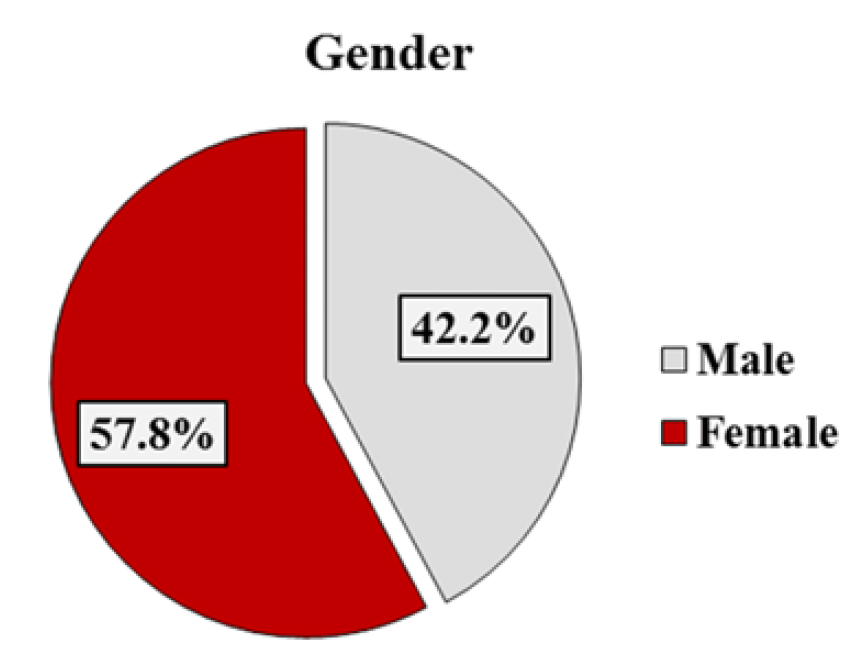
The study population consisted of 102 patients. The prevalence of appendicitis in children by sex was in the three hospitals 42.2% in boys and 57.8% in girls (Figure 1).


Figure 1: Prevalence of Abdominal Pain at Time of Presentation


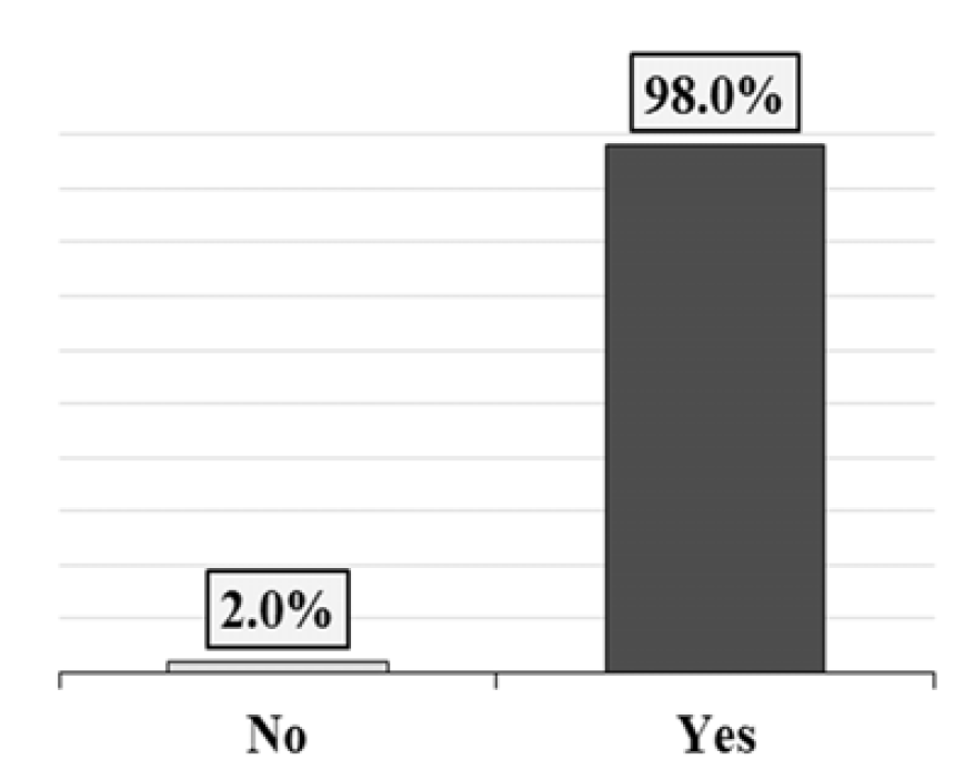
98% of patients have abdominal pain at the time of presentation to the emergency room, while only 2% do not have abdominal pain (Figure 2).


Figure 2: Prevalence of Abdominal Pain at the Time of Diagnosis


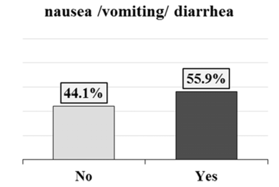
Digestive signs (nausea, vomiting and / or diarrhea) were present in more than half of the studied population (55.9%) (Figure 3).


Figure 3: Prevalence of Digestive Signs at the Time of Diagnosis


Diagnostic Methods

Clinical Examination: A total of 89 patients, or 87.3%, presented with tenderness to palpation at the Mc. Burney point (Table 1).


Table 1: Percentage of Children with Positive Mc. Burney

Percentage

Frequency

Physical exam

12.7

13

No

87.3

89

Yes

100

102

Total


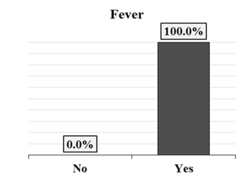
The fever is the most common sign in the diagnosis of appendicitis. In our series, 100% of patients had a fever at the time of diagnosis (Figure 4).


Figure 4: Percentage of Patients with Fever at the Time of diagnosis


Balance Sheet

Regarding the workup, the most sensitive marker of inflammation is CRP, which increases before white blood cells. In this series of 102 patients, CRP was normal in 31.4% of cases, high in 44.1% of cases and very high in 24.5% of cases (Table 2).


Table 2: Raising of CRP in the Case of Acute Appendicitis

PercentageFrequencyCRP
31.432Normal
44.145High
24.525Very High
100102Total


Imaging Test

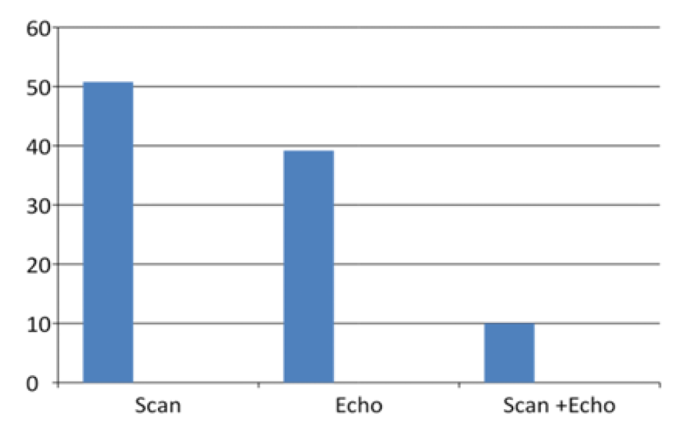
Currently, the diagnosis of appendicitis is no longer purely clinical. We rely on imaging methods to confirm or refute the diagnosis. The two most commonly used tools are abdominal and pelvic ultrasound and CT. In our study, CT was used more frequently than ultrasound. 50.8% of patients underwent a scan to confirm the diagnosis of acute appendicitis. 39.2% were diagnosed by ultrasound and 10% were diagnosed by ultrasound and CT scan.


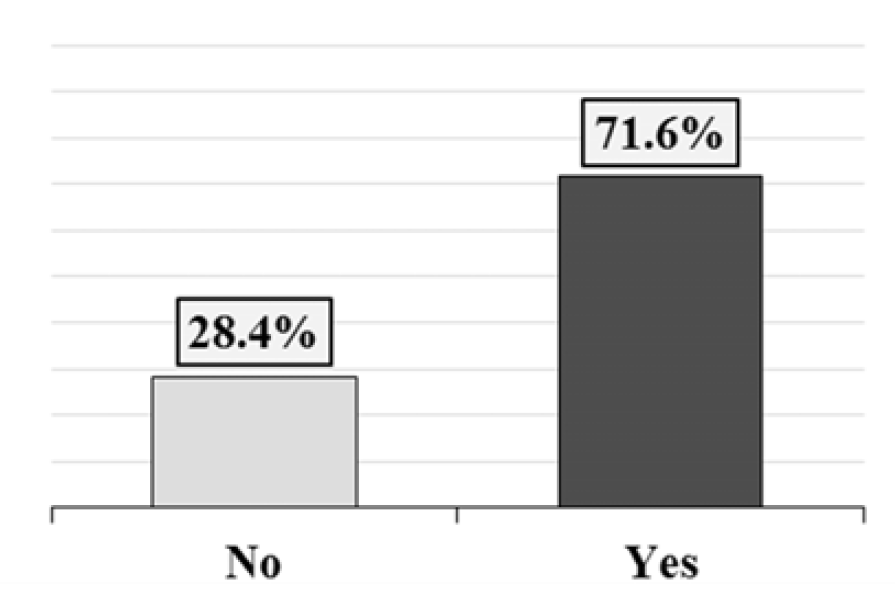
71.6% of patients underwent surgery in the first 24 hours after admission, while in 28.4% of cases; surgery was postponed and performed after 24 hours after diagnosis and admission (Figure 5).


Figure 5: Prevalence of Imaging


Complications

Ulceration of appendicitis was common. It was present in 35.3% of the cases but only 29.4% of these 35.3% presented necrosis during the operation. Patient follow-up for one month showed a readmission rate of 2.9% for different reasons. The overall complication rate was 13.7% for all complications combined. Among these patients, 9.8% underwent the operation within 24 hours. Of the patients who underwent the operation within 24 hours, 86.3% did not experience complications. 16% of patients operated after 24 hours had complications during their follow-up (Table 3,4).


Table 3: Sensitivity and Specificity of the Imaging

EchoScanParameters
77%90%Sensitivity
82%89%Specificity


Table 4: Complications Depending on the Time to Operation

p-valueTotalSurgery 24-48 hrs.Surgery <24 hrs.Complications


0.990>0.05

14410FrequencyYes
13.7%3.9%9.8%Percentage


0.99

882563FrequencyNo
86.3%24.5%61.8%Percentage
 1022973FrequencyTotal
100.0%28.4%71.6%Percentage


Figure 6: Percentage of Patients Who Underwent Immediate Versus Postponed Appendectomy

DISCUSSION

Appendicitis is considered one of the most common diseases that require emergency surgery after the onset of symptoms. For long it has been considered that to decrease the rate of morbidity and mortality, it is necessary to make rapid appendectomy (within 24 hrs) and not to postpone it [25]. Our study proved that a short appendectomy delay of less than 48 hours is possible since it does not increase the rate of perforation and postoperative complications. The postoperative complications taken into account are: the development of an infection of operating site, an abdominal abscess or sepsis up to 30 days after surgery. Traditionally, it was thought that each of appendicitis would develop into a perforated appendicitis. The fear of the development of a perforation if the appendectomy was postponed led to considering it as a surgical emergency with minimal delay (within 24 hrs of onset). However, this fear dates back to over 100 years, when perforated appendicitis had a very high mortality rate. Immediate and therefore sometimes nocturnal surgery are associated with several potential drawbacks, since in current practice many hospitals do not have an operating room available 24 hours a day, nor optimal imaging methods exist at night. In addition, delaying the appendectomy creates additional time to confirm the clinical diagnosis with suitable tests [26]. Several studies have shown that delayed appendectomies are associated with poor results. For example, a delay of more than 12 hours in hospital time, the time between the hospital visit and the operation, is an independent risk factor for perforation according to "Busch et al.,". “Girado et al.,” reported that the rate of complications increases with delayed surgery, 24 hours later after the onset of symptoms [26]. In contrast, other studies have reported that delayed appendectomy does not increase the rate of complications [27]. Recent studies suggest that appendicitis can be managed with a semi-elective strategy after antibiotic therapy. Some studies have reported that performing the operation during the day can provide a better working environment. Despite the support of these studies for delay in the surgical operation, the ideal delay remains ambiguous (30). However, other studies have suggested other risk factors influencing the rate of complications. For example, Chen and Al [21], reported that complications increase in the elderly (>55 years of age), having a higher number of neutrophils, a higher temperature, a high perforation rate and a longer residence time, while the time from ER registration to appendectomy is not significantly associated with the development of complications [21]. Our study confirms these results, like Teixeira and Al and Abou-Nukta and Al, who reported that the delay in appendectomy (below 48 hrs?) does not increase the risk of perforation [28,29].


Study Limitations

We encountered limitations in our study. We quote the size of the sample which is relatively small. As well as the symptoms of appendicitis are vague, non-specific and subjective. This is why the time of onset of symptoms reported by the patient was considered to be the time of onset of appendicitis.


Study Perspectives

For more than a century, appendectomy has been considered as the standard treatment for acute appendicitis, allowing healing and avoiding progression to more serious complications such as perforation and peritoneal and generalized diffusion of the infection. However, even if appendectomy is considered a radical treatment, it is not without several complications such as infection of the surgical site, intra-abdominal abscess, bridle occlusion, sepsis, etc… In recent years, several studies have been published with a purpose was to assess the effectiveness of antibiotic therapy alone, as a conservative treatment and thus avoid surgical treatment, the question that arises is: Can the antibiotic treatment replace surgical treatment in few years and thus eliminate its complications without influencing the morbidity and mortality rate?

CONCLUSION

The treatment of acute appendicitis is appendectomy after the fluid resuscitation, analgesia and intravenous antibiotics. Intravenous resuscitation is usually done with normal saline solution and must take into account the water deficit (based on clinical signs of dehydration and tissue perfusion) and maintenance needs. In patients with generalized peritonitis, plasma electrolytes should be controlled and fluids and electrolytes adjusted accordingly. Antibiotics are given to prevent the formation of intra-abdominal abscesses, sepsis and wound infection. Common antibiotic regimens cover gram-negative and anaerobic bacteria and include Cefuroxime plus Metronidazole or Amoxicillin /Clavulanic acid. Antibiotics are started quickly once the diagnosis of acute appendicitis is made and always before the start of surgery. Children with uncomplicated acute appendicitis usually do not need intravenous antibiotics for more than 24 hours after the operation. Those with perforated appendicitis need longer treatment, but there is no consensus on the choice, way of administration or duration of antibiotic use after perforated appendicitis. In such cases, intravenous antibiotics are continued until the fever subsides, the patient tolerates diet and the clinical signs have disappeared. This study highlights the optimal treatment for AAC which is not yet specified. It is obvious that each method of treatment has advantages and disadvantages. The main conclusion of our study is that a short delay in appendectomy of less than 48 hours does not increase the rate of pre or postoperative complications, nor the death rate, even among children living in developing countries.

REFERENCES
  1. Ferris, M. et al. “The global incidence of appendicitis: A systematic review of population-based studies.” Annals of Surgery, vol. 266, no. 2, 2017, pp. 237–241.

  2. Addiss, D.G. et al. “The epidemiology of appendicitis and appendectomy in the united states.” American Journal of Epidemiology, vol. 132, no. 5, 1990, pp. 910–925.

  3. Ceresoli, M. et al. “Acute appendicitis: epidemiology, treatment and outcomes analysis of 16,544 consecutive cases.” World Journal of Gastrointestinal Surgery, vol. 8, no. 10, 2016, pp. 693.

  4. Humes, D.J. and J. Simpson. “Acute appendicitis.” BMJ, vol. 333, no. 7567, 2006, pp. 530–534.

  5. Epstein, M.D. “Complications of acute appendicitis.” Ohio Medical Journal, vol. 86, no. 8, 1990, pp. 602–607.

  6. Luckmann, R. and P. Davis. “The epidemiology of acute appendicitis in California: Racial, gender and seasonal variation.” Epidemiology, 1991, pp. 323–330.

  7. Lee, J.H. et al. “The epidemiology of appendicitis and appendectomy in south Korea: National registry data.” Journal of Epidemiology, vol. 20, no. 2, 2010, pp. 97–105.

  8. Singh, M. et al. “Complicated appendicitis: analysis of risk factors in children.” African Journal of Paediatric Surgery, vol. 11, no. 2, 2014, pp. 109–113.

  9. Pieper, R. and L. Kager. “The INCIDENCE OF ACUTE APPENDICITIS AND APPENDECTOMY: An epidemiological study of 971 cases.” Acta Chirurgica Scandinavica, vol. 148, no. 1, 1982, pp. 45.

  10. Hori, T. et al. “Laparoscopic appendectomy for acute appendicitis: how to discourage surgeons using inadequate therapy.” World Journal of Gastroenterology, vol. 23, no. 32, 2017, pp. 5849.

  11. Salminen, P. et al. “Antibiotic therapy vs appendectomy for treatment of uncomplicated acute appendicitis: The Appac randomized clinical trial.” JAMA, vol. 313, no. 23, 2015, pp. 2340–2348.

  12. Mostbeck, G. et al. “How to diagnose acute appendicitis: Ultrasound first.” Insights into Imaging, vol. 7, no. 2, 2016, pp. 255–263.

  13. Nshuti, R. et al. “Clinical presentation of acute appendicitis in adults at the Chris Hani Baragwanath academic hospital.” International Journal of Emergency Medicine, vol. 7, no. 1, 2014, pp. 12.

  14. Yang, H.R. et al. “Laboratory tests in patients with acute appendicitis.” ANZ Journal of Surgery, vol. 76, nos. 1–2, 2006, pp. 71–74.

  15. Elangovan, S. “Clinical and laboratory findings in acute appendicitis in the elderly.” Journal of the American Board of Family Practice, vol. 9, no. 2, 1996, pp. 75–78.

  16. Rao, P.M. et al. “Helical CT technique for the diagnosis of appendicitis: prospective evaluation of a focused appendix CT examination.” Radiology, vol. 202, no. 1, 1997, pp. 139–144.

  17. Bongard, F. et al. “Differential diagnosis of appendicitis and pelvic inflammatory disease: A prospective analysis.” American Journal of Surgery, vol. 150, no. 1, 1985, pp. 90–96.

  18. Hansen, L.W. and S.E. Dolgin. “Trends in the diagnosis and management of pediatric appendicitis.” Pediatrics in Review, vol. 37, no. 2, 2016, pp. 52–57.

  19. Podda, M. et al. “Antibiotics-first strategy for uncomplicated acute appendicitis in adults is associated with increased rates of peritonitis at surgery.” The Surgeon, vol. 15, no. 5, 2017, pp. 303–314.

  20. Nyström, P.-O. Appendicitis. Zuckschwerdt, 2001.

  21. Chen, C.C. et al. “Appendectomy timing: Will delayed surgery increase the complications?” Journal of the Chinese Medical Association, vol. 78, no. 7, 2015, pp. 395–399.

  22. Costa-Navarro, D. et al. “Laparoscopic appendectomy: Quality care and cost-effectiveness for today’s economy.” World Journal of Emergency Surgery, vol. 8, no. 1, 2015, pp. 45.

  23. Nowzaradan, Y. et al. “Laparoscopic appendectomy: Treatment of choice for suspected appendicitis.” Surgical Laparoscopy & Endoscopy, vol. 3, no. 5, 1993, pp. 411–416.

  24. Sandell, E. et al. “Surgical Decision-Making in Acute Appendicitis.” BMC Surgery, vol. 15, no. 1, 2015, pp. 69.

  25. Kim, J.Y. et al. “Early versus late surgical management for complicated appendicitis in adults: A multicenter propensity score matching study.” Annals of Surgical Treatment and Research, vol. 97, no. 2, 2019, pp. 103–111.

  26. Bickell, N.A. et al. “How time affects the risk of rupture in appendicitis.” Journal of the American College of Surgeons, vol. 202, no. 3, 2006, pp. 401–406.

  27. Giraudo, G. et al. “Prompt or delayed appendectomy? Influence of timing of surgery for acute appendicitis.” Surgery Today, vol. 43, no. 4, 2013, pp. 392–396.

  28. Teixeira, P.G. et al. “Appendectomy timing: Waiting until the next morning increases the risk of surgical site infections.” Annals of Surgery, vol. 256, no. 3, 2012, pp. 538–543.

  29. Abou-Nukta, F. et al. “Effects of delaying appendectomy for acute appendicitis for 12 to 24 hours.” Archives of Surgery, vol. 141, no. 5, 2006, pp. 504–507.

Recommended Articles
Research Article
It Remains Unproven That the Variant M.8231C>A Causes Coronary Atherosclerosis
Published: 15/07/2020
Download PDF
Research Article
Vision for a Brighter Kangra: Unmasking the Truth about Pink Eye – A Comprehensive Study on Types, Symptoms, and Proactive Prevention in Himachal Pradesh’s Kangra District
...
Published: 11/11/2023
Download PDF
Research Article
Leigh Syndrome Should Not Be Diagnosed Exclusively Upon Cerebral MRI
Published: 15/07/2020
Download PDF
Research Article
Release Kinetic Study of Matrix Type Transdermal Patch Using an Analgesic Drug
...
Published: 10/08/2020
Download PDF
Chat on WhatsApp
Flowbite Logo
PO Box 101, Nakuru
Kenya.
Email: office@iarconsortium.org

Editorial Office:
J.L Bhavan, Near Radison Blu Hotel,
Jalukbari, Guwahati-India
Useful Links
Order Hard Copy
Privacy policy
Terms and Conditions
Refund Policy
Shipping Policy
Others
About Us
Team Members
Contact Us
Online Payments
Join as Editor
Join as Reviewer
Subscribe to our Newsletter
+91 60029-93949
Follow us
MOST SEARCHED KEYWORDS
Copyright © iARCON International LLP . All Rights Reserved.