Objective: This research aimed to assess the correlation between the occurrence of middle ear effusion in pediatric patients with adenoid hypertrophy and identify the variables that contribute to the development of middle ear effusion in children residing in Baqubah city. Method: Its A controlled; prospective research was conducted on newly diagnosed cases of adenoid hypertrophy at the ENT clinic of Baqubah Teaching Hospital from March to October 2022. eighty cases observed during the study period were between the ages of 3 and 12 years. For diagnosis of adenoid hypertrophy, each case underwent nasal endoscopy and skull lateral soft tissue X-ray examinations. The parents and patients were questioned to gather information about accompanying signs and symptoms, such as nasal obstruction, rhinorrhea, mouth breathing, snoring, obstructive sleep apnea and hearing problem were documented in these patients. Tympanometry was used to diagnose the occurrence of middle ear effusion, Tympanogram result being classified as Type A, Type B, or Type C. Both types B and C indicate the occurrence of middle-period effusion, while type A consider normal. Results: The research findings indicate that the average age of the 80 cases was six years, with a higher proportion of females (female to male ratio of 1.6:1). Out of the 80 patients (160 ears), 70 ears (43.75%) exhibited type B tympanograms, 50 ears (31.25%) exhibited type A tympanogram and 40 ears (25%) exhibited type C tympanograms. Out of the entire population of patients with adenoid hypertrophy, the occurrence of otitis media with effusion was 56.25%. Among these cases, 30 (66.67%) had bilateral middle ear effusion, while 15 (33.33%) had unilateral middle ear effusion, including both type B and C tympanogram, with type B and C being the most prevalent. The results also showed that patients with Grade 3 and 4 adenoid hypertrophy were prevalent. Furthermore, there is a substantial correlation between Grade 3 and 4 adenoid hypertrophy and the occurrence of middle ear effusion. Conclusion: The research identified a significant association between the presence of middle ear effusion in children and adenoid hypertrophy, specifically in boys. The association was particularly strong in pediatric patients who had grade III and IV adenoid hypertrophy. The majority of patients had bilateral middle ear effusion, with hearing impairment being the most prevalent symptom. Given these data, we advise that all children with adenoid enlargement undergo screening for middle ear effusion and get suitable treatment to attain the most favorable result.
Keywords
Otitis Media With Effusion
Adenoid Hypertrophy
Baqubah Teaching Hospital
INTRODUCTION
Adenoid hypertrophy and otitis media with effusion are prevalent conditions in pediatric otolaryngology. Pathological Adenoid hypertrophy is caused by repeated stimulation from bacteria, viruses and allergies, leading to clinical manifestations, such as nasal congestion, excessive nasal discharge, breathing through the mouth, obstructive sleep apnea, snoring and a characteristic facial appearance known as "adenoid face", Chronic airway obstruction is a significant contributing factor to the development or worsening of middle ear effusion, [1,2].
Adenoid hypertrophy is common in children and its symptoms manifest more quickly in younger individuals owing to the higher occurrence of upper respiratory tract infections and the smaller size of the nasopharynx. The adenoids undergo physiological hypertrophy from birth until the age of 6, after which they progressively decrease in size until they fully vanish at the age of 16. However, in certain cases, they may linger into adulthood [3].
Friedman's criteria, (Tonsil grading system), were used to categorize Adenoid hypertrophy, the grading system for tonsils is as follows: grade 0 (patients who have undergone tonsillectomy), grade 1 (tonsils located within the tonsillar fossa), grade 2 (tonsils protruding beyond the tonsillar pillars), grade 3 (tonsils extending beyond the tonsillar pillars but not reaching the midline) and grade 4 (tonsils extending up to the midline) [4,5]. Tonsil hypertrophy was characterized as tonsil grade II or above [6].
Otitis media with effusion refers to the persistent buildup of mucus, serous, or glue-like fluid in the middle ear and sometimes in the mastoid air cell system. The duration required for the presence of the fluid to classify the illness as chronic is often considered to be 12 weeks. [7], Otitis media with effusion is often seen in youngsters due to the accompanying hearing loss and occasionally follows a previous sickness and ear pain resulting from an episode of acute otitis media [8]. While the precise origin of otitis media with effusion remains unknown, it is commonly recognized that Eustachian tube occlusion is a significant contributing factor [9,10].
Inflammation in the nasal passages caused by a viral infection in the upper respiratory tract, allergic rhinitis, enlarged adenoids, or a mass at the back of the nasal cavity may disrupt the normal functioning of the Eustachian tube and lead to the accumulation of fluid in the middle ear. [11].
Multiple studies have been conducted to uncover the cause of otitis media with effusion. The main causes of otitis media with effusion include respiratory tract infections, adenoid hypertrophy, craniofacial abnormalities, mechanical blockage of the nasopharynx and allergy and immunological factors. Possible variables contributing to middle-period effusion include gender, climatic circumstances, environment, humidity, socioeconomic level, length of nursing, living in a crowded household, attending nursery or kindergarten, exposure to passive smoking and gastric reflux [12,13].
The presence of adenoid tissue contributes to the development of otitis media with effusion through several mechanisms, including its abnormally large size, interference with nasopharyngeal ventilation, blockage of the Eustachian tube due to mass effect, buildup of secretions, potential for infection, inflammation-induced swelling and release of allergic inflammatory mediators by mast cells in the adenoid tissue [14,15].
Several research has been conducted to evaluate the association between adenoid hypertrophy and middle ear effusion. One of these investigations demonstrates, the occurrence of middle ear effusion was highest in the younger individuals with adenoid hypertrophy grade II and grade III. and the predominant symptoms seen were auditory impairment, snoring and nasal blockage, with Greater grade of adenoid hypertrophy are crucial for the continued presence of middle ear effusion and may lead to the failure of conservative treatment [16], while another study revealed to, Adenoid Hypertrophy can be relevant in the pathogenesis of otitis media with effusion due to its anatomic position [15].
Several previous studies have been undertaken on a subject comparable to ours. However, these studies still need to examine the prevalence of middle ear effusion in pediatric patients in the city of Baqubah. Hence, our research aims to investigate the variables that lead to the occurrence of middle ear effusion in pediatric patients with adenoid hypertrophy in Baqubah City.
MATERIALS AND METHODS
Study Populations
The research included eighteen pediatric patients with recently identified adenoid hypertrophy at the ENT clinic of Baqubah Teaching Hospital. The study took place between 22nd March and 1st October 2022, Patients were selected within the age range of 3-12 years; from the total number of patients, the female number was 50 while the male number was 30, at a ratio equal to 1.6:1, The research included pediatric patients who reported clinical symptoms and signs of adenoid hypertrophy, The question was posed to parents and patients in order to gather information on the signs and symptoms, which included nasal obstruction, rhinorrhea, mouth breathing, snoring, obstructive sleep apnea and hearing problems.
Grouping Criteria
The patients included in the study were separated into two groups: one with adenoid hypertrophy and another with adenoid hypertrophy accompanied by middle ear effusion. This division was based on the presence of middle ear effusion. Patients who were diagnosed with middle ear effusion were included in the group of individuals with adenoid hypertrophy and middle ear effusion. Only pediatric patients who did not have middle ear effusion were included in the adenoid hypertrophy group. Pediatric patients who were diagnosed with middle ear effusion were further split into two groups: those with unilateral middle ear effusion and those with bilateral middle ear effusion.
Investigation
The diagnostic procedure for adenoid hypertrophy included the use of nasal endoscopy and lateral soft tissue X-ray of the skull, tonsil grading was employed to measure the extent of adenoid hypertrophy. The grading was determined based on the degree of choanal opening occlusion caused by the adenoid, as follows:
Grade 0: Refers to people who have had a tonsillectomy
Grade 1: The tonsils are located inside the tonsillar fossa and cause obstruction of less than 25% of the choanal orifice
Grade 2: The tonsils protrude beyond the tonsillar pillars: The adenoid gland occupies around 25% to 50% of the choanal orifice
Grade 3: The tonsils protrude beyond the tonsillar pillars but do not reach the center line
The adenoid fills 50%-75% of the choanal aperture. while the patient is in grade 4, with tonsils that extend up to the midline. The adenoid is blocking 75% to 100% of the choanal orifice., [17], Patients with grade 3 and 4 adenoid enlargement were sent for a hearing evaluation test using tympanometry.
The tympanogram result is classified into three categories: Type A, Type B, or Type C. Results classified as Type A are deemed within the usual range. Both types B and C indicate the occurrence of middle-period effusion [18].
Ethical Approval
The research on ethics received clearance from the Medical Faculty at the University of Diyala, Baqubah and the patient's medical history was gathered with their consent.
Statistical Analysis
The quantitative data were reported using measures such as standard deviation, standard error of the mean and percentage. The χ2 test was used to compare the various variables in the research. The statistical significance was assessed at a significance level of p<0.05 using SPSS, version 21 (SPSS Inc, Chicago, Illinois) [19].
RESULTS AND DISCUSSION
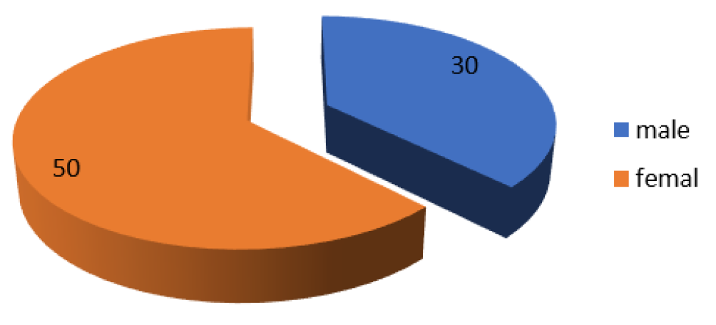
The research comprised 80 patients, consisting of 30 boys and 50 girls, there was a considerable difference between the gender groups in the research. Table 1, Figure 1, the average age of the patients participating was six years, the investigation revealed a significant difference between infected individuals with adenoid hypertrophy and middle ear effusion and patients with adenoid hypertrophy but without middle ear effusion (p<0.05). The findings indicate that out of the 80 pediatric patients with adenoid hypertrophy, 55 were diagnosed with middle ear effusion. In contrast, the remaining 25 pediatric patients only had adenoid hypertrophy without middle ear effusion.
Table 1: Distribution of Genders Among a Total of 80 Patients
Total
Male
Female
p-value
SD
SE
80
30
50
0.157*
14.142
10.000
%
37.5
62.5
-
-
-
*Significant at level (p<0.05), SD = Stander Deviation, SE = Stander Error of Mean
Figure 1: Distribution of Genders Among a Total of 80 Patients
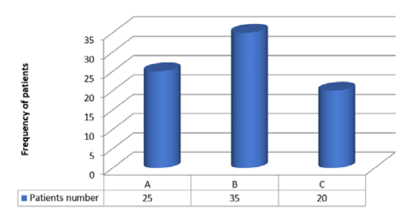
The results of tympanogram categorization showed that out of 80 pediatric patients with adenoid hypertrophy, 25 patients (31.25%) were classed as A and 35 patients (43.75%) were categorized as B. while the last group, consisting of 20 patients, was classified as category C. The difference between the two groups was statistically significant, with a p-value (p<0.05) (Table 2, Figure 2).
Table 2: Grades of Adenoid Hypertrophy Seen in Individuals Diagnosed with Adenoid Hypertrophy. n = 80
Grade
Frequency
%
p-value
SD
SE
0
4
5
0.220*
12.981
5.805
I
6
7.5
-
-
-
II
10
12.5
-
-
-
III
29
36.25
-
-
-
IV
31
38.75
-
-
-
*Significant at Level (p<0.05), SD = Stander Deviation, SE = Stander Error of Mean
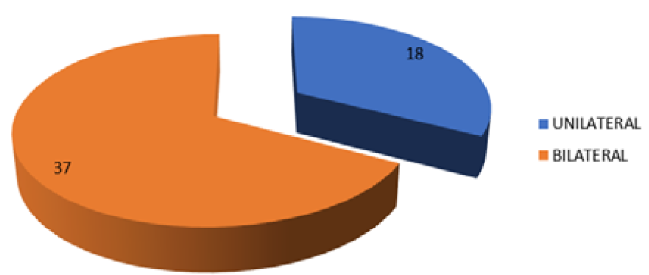
Figure 2: Patients' Unliteral and Bilateral Middle Ear Effusion Distribution n = 55
The results also showed a significant difference between individuals with unilateral middle ear effusion compared to those with bilateral middle ear effusion (p<0.05), The findings indicate that out of the total number of patients with middle ear effusion, 18 patients exhibited unilateral effusion, accounting for 33% of the cases. The other patients, totaling 37, had bilateral middle ear effusion, representing 67% of the total number of patients with middle ear effusion (Table 3, Figure 3).
Table 3: Patient Distribution with Middle Ear Effusion in Adenoid Hypertrophy n = 80
Grade
ADENOID HYPERTROPHY
Adenoid hypertrophy with middle ear effusion
p-value
0
3
1
0.287*
I
3
3
-
II
3
7
-
III
8
21
-
IV
8
23
-
Total number
25
55
-
SD
2.739
10.296
-
SE
1.225
4.604
-
*Significant at Level (p<0.05), SD = Stander Deviation, SE = Stander Error of Mean
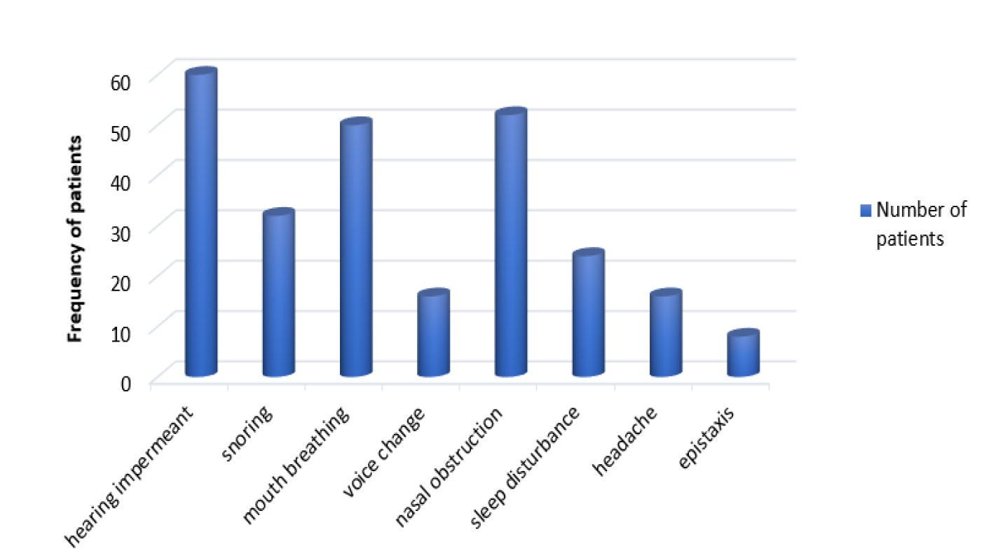
Figure 3: Symptoms in 80 Adenoid Hypertrophy Patients
On the other hand, the findings of the symptoms analysis showed a statistically significant change in the symptoms of adenoid hypertrophy (p<0.05). A study involving 80 patients with adenoid hypertrophy revealed that all 80 patients reported hearing problems. Additionally, 44 patients experienced mouth breathing, 50 patients had nasal obstruction, 52 patients snored, 32 patients complained of sleep disturbance, 24 patients exhibited voice changes, 16 patients reported headaches and eight patients had epistaxis (Table 4, Figure 4).
Table 4: Tympanometric Distribution of Adenoid Hypertrophy Patients, n = 80
Category of tympanogram
Patients number
Percentage of patients
p-value
A
25
31.25
0.199
B
35
43.75
-
C
20
25
-
SD
7.638
-
-
SE
4.410
-
-
*Significant at Level (p<0.05), SD = Stander Deviation, SE = Stander Error of Mean
Figure 4: Tympanometric Distribution of Adenoid Hypertrophy Patients, n = 80
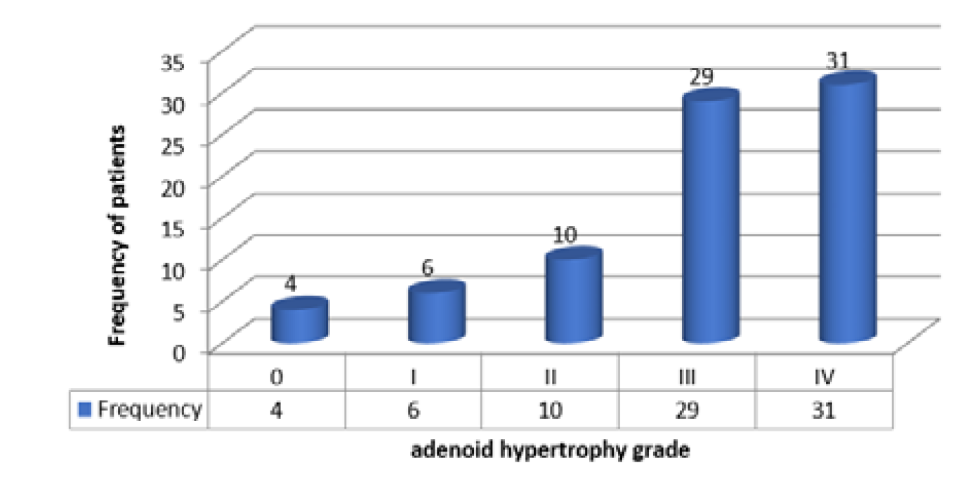
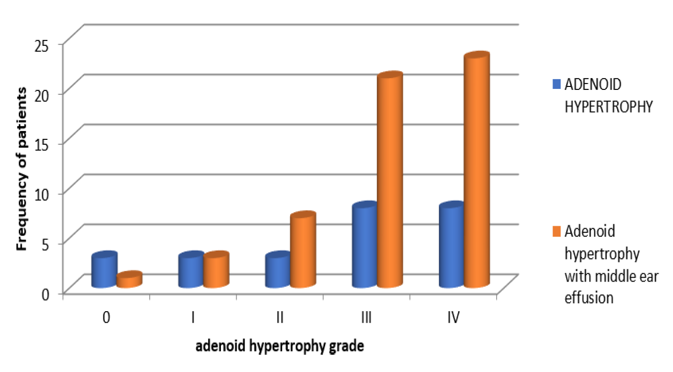
The results demonstrated the categorization of patients based on tonsil grading, patients with grad zero was 4 , three with adenoid hypertrophy and one with adenoid hypertrophy with middle ear effusion , while number of patients with grad one was 6, 3 patients with adenoid hypertrophy and 3 patients with adenoid hypertrophy with middle ear effusion , grad two patients involved 10 patients, 3 patients with adenoid hypertrophy and 7 patients with adenoid hypertrophy with middle ear effusion, while number of patients in grad three was 29 divided to 8 patients with adenoid hypertrophy and 21 patients with adenoid hypertrophy with middle ear effusion , grad 4 patients included 31 patients ,8 patients with adenoid hypertrophy and 23 patients with adenoid hypertrophy with middle ear effusion. Statistically there was a significant difference between groups (p<0.05) (Table 5,6, Figure 5,6).
Table 5: Patients' Unliteral and Bilateral Middle Ear Effusion Distribution. N = 55
Number of Patents with middle ear effusion
unliteral
bilateral
p-value
SD
SE
55
18
37
0.157*
13.435
9.500
Percentage
33%
67%
-
-
-
*Significant at Level (p<0.05), SD = Stander Deviation, SE = Stander Error of Mean
Figure 5: Grades of Adenoid Hypertrophy Seen in Individuals Diagnosed with Adenoid Hypertrophyn = 80
The findings of our investigation indicate a substantial correlation between adenoid hypertrophy and the gender of the patients, the higher prevalence of adenoid hypertrophy in males may be attributed to the greater frequency of upper respiratory infection episodes experienced by boys compared to girls [20]. Additional study findings corroborate our results, indicating that males are more often affected by adenoid hypertrophy compared to girls [21].
Our study result conduct to the most common grade of adenoid hypertrophy with middle ear effusion in study patients' sample was in grade III and grad IV, this result is compatible with other study that shows, the incidence of middle ear effusion in the adenoid grade IV group was 26.86%, which was significantly higher than that in the grade III group (14.5%), The incidence of middle ear effusion in the adenoid grade IV group was 1.662 times than that in the grade III group. A high adenoid grade is a risk factor for adenoid hypertrophy complicated by middle ear effusion [1].
Table 6: Symptoms in 80 Adenoid Hypertrophy Patients
symptom
Number of patients
percent age
p-value
hearing impermeant
60
75
0.243*
snoring
32
40
-
mouth breathing
50
62.5
-
voice change
16
20
-
nasal obstruction
52
65
-
sleep disturbance
24
30
-
headache
16
20
-
epistaxis
8
10
-
SD
19.492
-
-
SE
6.891
-
-
* Significant at level (p<0.05), SD=stander deviation; SE=stander error of mean
Figure 6: Patient Distribution with Middle Ear Effusion in Adenoid Hypertrophy
On other side, tympanogram result shows 43.75% for patients with adenoid hypertrophy shows type B and 31.25% shows type C, while 25 % show type A, grade A proportion represented patients with grade 0,I and II of adenoid hypertrophy, this result indicated incidence of middle ear effusion is most commonly in patients with adenoid hypertrophy grade III and IV , this conclusion similar to the conclusion of other studies showing , grade 4 adenoid hypertrophy showed a highly significant correlation with type B tympanometry either as unilateral or as bilateral finding [22]. (While others reveled to grade 3 adenoid hypertrophy showed higher number of cases of middle ear effusion [23].
Study result shows incidence of middle ear effusion is more commonly as bilateral comparing with unilateral, several studies revealed to resemble outcomes, as a study shows, 14% of cases presented with unilateral middle ear effusion, while 86% of cases showed a bilateral middle ear effusion [24].
Patients involved in the study shows a different symptom including mouth breathing, nasal obstruction, snoring, sleep disturbance, voice change, headache and epistaxis, because Middle ear effusion is a major cause of hearing loss in children [25]. The majority of patient involved in our study complain from hearing impermeant.
The research provided, however, had several limitations. The sample sizes were constrained due to the small number of patients available for the research from Baqubah city, which has a limited number of people; in addition, the research was confined to a certain number of participants due to the restricted time of investigation, which was between March and October. These factors contributed to the restriction in the number of patient samples studied.
CONCLUSION
study result conduct to a significant correlation between incidence of middle ear effusion in pediatric patient with adenoid hypertrophy mostly in male and in pediatric patients with grade III and IV of adenoid hypertrophy, with the majority cases was bilateral middle ear effusion and the most common symptom involved was hearing impermeant, according to this relation adenoid hypertrophy and middle ear effusion that exhibit by our study, we advise to, any children with adenoid enlargement should be screened for middle ear effusion and managed accordingly for best outcome to be achieved in the children.
Authors’ Contributions
Conception and Design: Ali Lafta Salman, Qays Jaafar Khalaf
Data Collection and Compilation: Ali Lafta Salman
Data Analysis and Interpretation: Qays Jaafar Khalaf
Drafting of the Article: Qays Jaafar Khalaf
Reviewing the Paper Critically for Key Ideas: Ali Lafta Salman, Qays Jaafar Khalaf
Proficiency in Statistical Analysis: Ali Lafta Salman, Qays Jaafar Khalaf
Article's Ultimate Endorsement and Guarantor: Ali Lafta Salman
Acknowledgements:
The authors express gratitude for the assistance provided by the management and staff of Baqubah Teaching Hospital/ENT Clinic.
REFERENCES
Chen, W. et al. “Analysis of factors that influence the occurrence of otitis media with effusion in pediatric patients with adenoid hypertrophy.” Frontiers in Pediatrics, vol. 11, 2023, pp. 1098067.
Dogru, M. et al. “Does adenoid hypertrophy affect disease severity in children with allergic rhinitis.” European Archives of Oto-Rhino-Laryngology, vol. 274, 2017, pp. 209–213. https://doi.org/10.1007/s00405-016-4196-x
Althobaiti, T. et al. “Adenoid hypertrophy in adults: Causes and precipitating factors.” IJMDC, vol. 4, no. 11, 2020, pp. 1792–1796.
Gao, W. et al. “Analysis of the cause of retrolingual obstruction in patients with moderate–severe obstructive sleep apnea.” European Archives of Oto-Rhino-Laryngology, 2023, pp. 1–9.
Lee, J. et al. “Assessment of friedman classification by measuring actual tonsil size during surgery.” Journal of Rhinology, vol. 27, no. 2, 2020, pp. 122–126.
Evcimik, M.F. et al. “Adenoid hypertrophy in children with allergic disease and influential factors.” International Journal of Pediatric Otorhinolaryngology, vol. 79, 2015, pp. 694–697. https://doi.org/10.1016/j.ijporl.2015.02.017
Gurunathan, R.K. and M. Perry. “The ear and associated structures: Part II.” Diseases and Injuries to the Head, Face and Neck: A Guide to Diagnosis and Management, 2021, pp. 1511–1587.
Browning, G. “Otitis media with effusion.” Scott-Brown’s Otolaryngology, 7th ed., Hodder Arnold, 2008, pp. 877–880.
Wayes, A.M. Investigation of Biofilms Associated with Chronic Otitis Media with Effusion and Adenoids Hypertrophy. Newcastle University, 2020. Doctoral dissertation.
Minovi, A. and S. Dazert. “Diseases of the middle ear in childhood.” GMS Current Topics in Otorhinolaryngology, Head and Neck Surgery, vol. 13, 2014.
Eliçora, S.Ş. et al. “Risk factors for otitis media with effusion in children who have adenoid hypertrophia.” International Journal of Pediatric Otorhinolaryngology, vol. 79, 2015, pp. 374–377. https://doi.org/10.1016/j.ijporl.2014.12.030
Juma, A.S. and M.H. Ubeid. “A microbiological survey of ear infections in inhabitants of erbil city.” Cihan University-Erbil Scientific Journal, vol. 6, no. 2, 2022, pp. 62–67.
Davcheva-Chakar, M. et al. “Adenoid vegetations—reservoir of bacteria for chronic otitis media with effusion and chronic rhinosinusitis.” Pril (Makedon Akad Nauk Umet Odd Med Nauki), vol. 36, 2015, pp. 71–76. https://doi.org/10.1515/prilozi-2015-0080
Songu, M. et al. “Risk factors for otitis media with effusion in children with adenoid hypertrophy.” Acta Otorhinolaryngologica Italica, vol. 40, no. 2, 2020, p. 133.
Galić, M.Z. and M. Klančnik. “Adenoid size in children with otitis media with effusion.” Acta Clinica Croatica, vol. 60, no. 3, 2021, pp. 532–538.
Patel, A. et al. “Comparison of adenoid assessment by flexible endoscopy and mirror exam.” International Journal of Pediatric Otorhinolaryngology, vol. 134, 2020, pp. 110073.
Rosenfeld, R.M. et al. “Clinical practice guideline: otitis media with effusion (update).” Otolaryngology–Head and Neck Surgery, vol. 154, suppl. 1, 2016, pp. S1–S41. https://doi.org/10.1177/0194599815623467
Mann-Whitney, U. “SPSS for windows (Release 9.0).” The Neuroradiology Journal, vol. 20, 2007, pp. 175–178.
Kalcioglu, M.T. et al. “Prevalence of and factors affecting otitis media with effusion in children in the region from balkans to caspian basin: A multicentric cross-sectional study.” International Journal of Pediatric Otorhinolaryngology, vol. 143, 2021, pp. 110647.
Sharad, K.A. et al. “Association of adenoid hypertrophy with nasal and middle ear pathologies in adults.” Indian Journal of Otolaryngology and Head & Neck Surgery, 2023, pp. 1–4.
AbdelTawab, H.M. and S.M.S. Tabook. “Correlation between adenoid hypertrophy, tympanometry findings and viscosity of middle ear fluid in chronic otitis media with effusion, southern Oman.” Ear, Nose & Throat Journal, vol. 100, no. 3, 2021, pp. NP141–NP146.
Timna, C.J. and D. Chandrika. “Role of adenoid hypertrophy in causation of chronic middle ear effusion.” International Journal of Otorhinolaryngology and Head and Neck Surgery, vol. 4, no. 1, 2018, pp. 203–209.
Nwosu, C. et al. “Tympanometric findings among children with adenoid hypertrophy in port harcourt, Nigeria.” International Journal of Otolaryngology, vol. 2016, 2016, Article ID 1276543.
Samuels, T.L. et al. “Association of gel-forming mucins and aquaporin gene expression with hearing loss, effusion viscosity and inflammation in otitis media with effusion.” JAMA Otolaryngology–Head & Neck Surgery, vol. 143, 2017, pp. 810–817
Recommended Articles
Research Article
A study on the feasibility of online classes for medical education
Seema Bose,
Atindra Krishna Ghosh
Published: 15/02/2021
Download PDF
Research Article
Amyand’s Hernia: A Rare form of Common Entity
Download PDF
Research Article
Prevalence of Typhoid Fever among Patients Attending General Sani Abatcha Specialist Hospital Damaturu, Nigeria
Ahmed Idriss Jajere,
...
Usman Umar Faruk
Published: 10/10/2020
Download PDF
Research Article
A case report of management of a Case of Hyperalgesia by Ultrasound guided Fluoroscopically Verified Transforaminal Nerve Root Block
Sunil Dutt,
...
Anil Kumar
Published: 29/06/2024
Download PDF
PO Box 101, Nakuru
Kenya.
Email: office@iarconsortium.org
Editorial Office:
J.L Bhavan, Near Radison Blu Hotel,
Jalukbari, Guwahati-India
Salman, A. L. & Khalaf, Q. J. (2024). The incidence of Middle ear effusion as a complication of adenoid hypertrophy in children attending Baqubah Teaching Hospital. IAR Journal of Medical Case Reports, 5(1), 1-6.
MLA
Salman, Ali L. and Qays J. Khalaf. "The incidence of Middle ear effusion as a complication of adenoid hypertrophy in children attending Baqubah Teaching Hospital." IAR Journal of Medical Case Reports 5.1 (2024): 1-6.
Chicago
Salman, Ali L. and Qays J. Khalaf. "The incidence of Middle ear effusion as a complication of adenoid hypertrophy in children attending Baqubah Teaching Hospital." IAR Journal of Medical Case Reports 5, no. 1 (2024): 1-6.
Harvard
Salman, A. L. and Khalaf, Q. J. (2024) 'The incidence of Middle ear effusion as a complication of adenoid hypertrophy in children attending Baqubah Teaching Hospital' IAR Journal of Medical Case Reports 5(1), pp. 1-6.
Vancouver
Salman AL, Khalaf QJ. The incidence of Middle ear effusion as a complication of adenoid hypertrophy in children attending Baqubah Teaching Hospital. IAR Journal of Medical Case Reports. 2024 Jan;5(1):1-6.



 Download PDF
 Download XML