Background: Acute pancreatitis is a common clinical entity, common causes being chronic alcohol consumption and gallstones. Acute episode of pancreatitis is uncommonly reported with a case of primary hyperparathyroidism. So, here we present a 33-year-old male who presented with acute abdomen caused by acute pancreatitis with no history of gallstones and alcohol consumption. He was further investigated for other uncommon causes of pancreatitis and found to have hyperparathyroidism caused by parathyroid adenoma. Hence, we suspect hypercalcemia induced by parathyroid adenoma can be a causative factor for the acute pancreatitis patients. Case Summary: We are reporting a case of 33-year-old male who presented with chief complaint of severe pain in epigastric region associated with vomiting and mild abdominal distension. With clinical, biochemical and image modalities a confirmed diagnosis of acute pancreatitis was made. On detailed investigations he was found to have hypercalcemia and increased parathyroid hormone levels. Patient was managed conservatively initially but due to repeated episodes of pain abdomen and persistent severe hypercalcemia with significant ECG changes showing bradycardia and prolonged QT interval, parathyroid adenoma was excised and renal stones were removed. After parathyroid excision patient improved drastically and parathyroid hormone levels came to a normal range post-operatively but hypocalcemia was seen post operatively which was medically managed successfully. Conclusion: Acute pancreatitis is a uncommon first clinical manifestation of hyperparathyroidism induced hypercalcemia.
Keywords
Pancreatitis
Parathyroid
Hypercalcemia and hormone
INTRODUCTION
Primary hyperparathyroidism is a generalized disorder of calcium, phosphate and bone metabolism caused by increased secretion of parathyroid hormone levels. Patient with hyperparathyroidism is usually asymptomatic for years but may present with recurrent kidney stones, peptic ulcer, mental changes and less frequently due to bone resorption [1-3]. Prevalence of hypercalcemia presenting as acute pancreatitis as first clinical manifestation is between 1.5% and 7% [4,5]Normal serum calcium levels range from 8.2 to 10.2 gm/dl. Hypercalcemia caused by undiagnosed PHPT may be the only causative factor in acute pancreatitis.
Case Presentation
A 33-year-old male presented to emergency department with severe, non radiating epigastric pain associated with nausea, vomiting and abdominal distension for 2 days. There was no history of fever, chills or cough or breathlessness.
There is no history of hypertension, diabetes mellitus or any cardiovascular disease. Patient is non-alcoholic and non-smoker.
There is no history of any previous gastrointestinal disease of biliary system problems.
On examination temperature was normal, blood pressure was 120/86 mmHg, pulse rate 112/minute and saturation was 98% at room air. There was mild upper abdominal tenderness on examination.
Lab investigations showed hemoglobin of 9.4 gm/dl, white blood cells count of 17.6 X 10⁶/µL (4.5-5.5 X 10⁶/µ/L), platelet count 3.03 lakh. Hsc-Reactive protein-368.4 mg/dl, amylase-631 U/L, lipase- 631 U/L, urea-85 mg/dl, creatinine-2.84mg/dl (0.6-1.17mg/dl). Liver function tests, lipid profile were within normal limits.
Imaging Findings
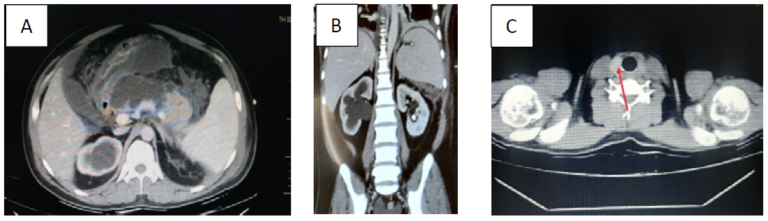
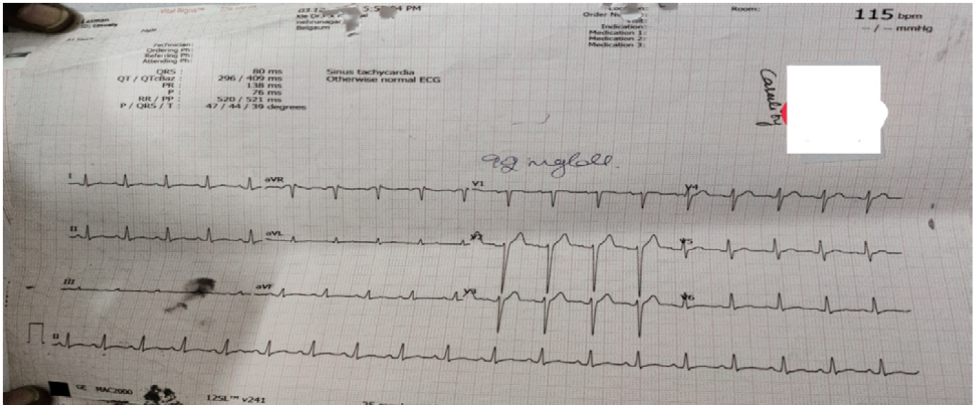
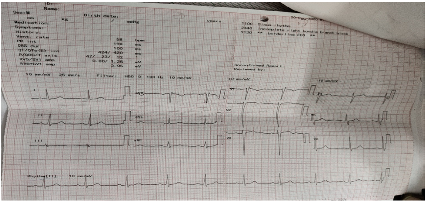
Ultrasonography revealed bulky pancreas, normal liver, gallbladder and no intrabilliary abnormalities. MRCP was done to rule out pancreatic divisum which found to be normal. CT abdomen plain showed bulky pancreas with significant ill-defined peripancreatic collection (Figure 1a). Right kidney showed moderate hydronephrosis and upper hydroureter due to mid ureteric calculus, left Electrocardiography on admission showed sinus tachycardia and QT interval of 296 milliseconds (Figure 2). Serial ECG showed QT prolongation and incomplete RBBB due to persistent hypercalcemia (Figure 3).
Further Diagnostic Work-Up
To find out other uncommon causes of pancreatitis thyroid profile and calcium levels were sent, calcium levels were found to be increased (11.5 mg/dl), thyroid function tests were with in normal range. Parathyroid hormone levels were sent to find out reason for hypercalcemia and found to be 94.4 pg/ml (15-65 pg/ml). Ultrasound neck was performed which showed normal thyroid. There was well defined hypoechoic area with central echogenicity noted posterior to left lobe of thyroid gland in its superior aspect measuring 1.0 X 0.5 cm suggestive of parathyroid adenoma. CT neck also showed left parathyroid adenoma (Figure 1c).
Final Diagnosis
On the basis of clinical features, all biochemical parameters and imaging modalities diagnosis of acute pancreatitis with peripancreatic collection, hypercalcemia, hyperparathyroidism and bilateral renal calculi with right hydronephrosis was made.
Treatment
Patient was treated conservatively with intravenous fluids for initial 2 days. Patient’s pain abdomen improved after 5 days of treatment. Serial calcium levels were on higher side, hydration, loop diuretics and small doses of glucocorticoids was started to decrease calcium levels. During the period of hospital stay patient developed recurrent episodes of pain abdomen in left hypochondrium. Due to persistent pain abdomen and persistent severe hypercalcemia for 3 days even after hydration with daily six liters of fluid intake, loop diuretics, zoledronic acid injection and glucocorticoids, patient was counselled to undergo parathyroid excision. Antihypertensive agent was started in view of newly diagnosed hypertension. Patient had successful, uneventful parathyroidectomy of superior parathyroid gland and bilateral double J stent was put to remove ureteral obstruction. Parathyroid levels came to a normal level after parathyroidectomy. Patient developed symptomatic hypocalcemia with bilateral hands and feet numbness after parathyroid removal. Hypocalcemia was treated with intravenous calcium gluconate and calcitriol supplement. Numbness improved after 2 days of treatment.
Outcome And Follow Up
Patient was discharged after four days of surgery with calcium and pancreatic enzymes supplement. Calcium levels came to a normal range after 10 days of supplements.
Figure 2: ECG: On admission ECG showed sinus tachycardia
Figure 3: ECG: Bradycardia and increased QT interval due to hypercalcemia
DISCUSSION
Primary hyperparathyroidism is a common endocrine disorder caused by raised PTH levels secreted by overactive parathyroid gland [6]. Postmenopausal women over the age of 50 years have a greater probability of developing PHPT compared with young individuals [7]. Parathyroid gland adenomas (80%-85%) are most common pathological cause of PHPT and rare causes include parathyroid hyperplasia, carcinoma, multiple endocrine neoplasia type 1 and 2A, and parathyroid cysts [8]. Most of the patients are asymptomatic and usually have mild symptoms. These patients can have variety of clinical manifestations but hypercalcemia being the most common condition [9]. Hypercalcemia is a common and potentially fatal metabolic disorder that is most often attributable to PHPT or malignancy-associated disease [10]. An elevation in PTH is one of the principal factors in the initiation of hypercalcemia. Excessive accumulation of serum calcium and decompensation of the renal system promote the development of hypercalcemia. Early recognition and preoperative management of hypercalcemic crisis is crucial as it can cause mental disturbance, metabolic encephalopathy, renal insufficiency, gastrointestinal symptoms, and cardiac dysrhythmia which can be very fatal [11].
Acute pancreatitis is an inflammatory process, which has sudden onset due to the premature activation of proteolytic zymogens within the exocrine pancreas [12]. Gallstones and chronic alcohol consumption are among most common causes of acute pancreatitis.
Association between PHPT and acute pancreatitis is a topic of debate for decades and it’s an uncommon clinical manifestation in patients with PHPT. Prevalence of acute pancreatitis in PHPT is just 1%-8% [13].
Three mechanisms are involved in the development of PHPT-induced acute pancreatitis. One is PHPT-induced high serum calcium level, which can lead to acceleration of the conversion of trypsinogen to trypsin in the pancreas resulting in pancreatic autodigestion and subsequent acute pancreatitis [14]. Secondly, the accumulation of calcium can promote the formation of ductal obstruction, pancreatic calculi, and subsequent attacks of acute pancreatitis [15]. Thirdly, genetic variants in serine protease inhibitor Kazal type 1 and cystic fibrosis transmembrane conductance regulator genes in combination with hypercalcemia markedly increase the risk of developing acute pancreatitis in patients with PHPT [16].
Calcium levels are of extreme importance in finding cause, disease severity of the illness. Early recognition of hypercalcemia not only leads to etiology but also prevents fatal complications if treated early. The treatment of hypercalcemia includes hydration with saline, forced diuresis, bisphosphonates, calcitonin, oral phosphates, glucocorticoids, and dialysis. But surgical resection is still the most effective treatment. Parathyroidectomy may not only relieve the abdominal symptoms of acute pancreatitis but also prevent the recurrence of hypercalcemia.
In our case patient had raised creatinine and urea levels in the beginning which improved with hydration, but calcium levels were increased over a period of 5 days of presentation and patient developed recurrent pain abdomen with persistent hypercalcemia. In view of persistent severe hypercalcemia, prolonged QT interval, development of incomplete Right bundle branch block and gastrointestinal symptoms, superior parathyroid gland was excised. There was drastic improvement of pain abdomen and gradually QT interval also became normal and normal sinus rhythm was obtained.
CONCLUSION
It becomes a diagnostic challenge to diagnose a PHPT-induced acute pancreatitis since it has a very non-specific presentation. Therefore, we should always suspect possibility of hyperparathyroidism in patient presenting with no history of alcohol consumption and gallstones. As surgical excision of parathyroid gland is a definite therapeutic intervention for such patients and it decreases recurrent acute pancreatitis episodes also, early recognition is of paramount importance.
REFERENCE
Silverberg, S.J. et al. “Asymptomatic Primary Hyperparathyroidism.” Journal of Clinical Densitometry, vol. 16, 2013, pp. 14–21. https://doi.org/10.1016/j.jocd.2012.11.005
Rejnmark, L. et al. “Nephrolithiasis and Renal Calcifications in Primary Hyperparathyroidism.” Journal of Clinical Endocrinology & Metabolism, vol. 96, 2011, pp. 2377–2385. https://doi.org/10.1210/jc.2011-0569
Starup-Linde, J. et al. “Renal Stones and Calcifications in Patients with Primary Hyperparathyroidism: Associations with Biochemical Variables.” European Journal of Endocrinology, vol. 166, 2012, pp. 1093–1100. https://doi.org/10.1530/EJE-12-0032
Egea Valenzuela, J. et al. “Acute Pancreatitis Associated with Hypercalcemia: A Report of Two Cases.” Revista Española de Enfermedades Digestivas, vol. 101, 2009, pp. 65–69. https://doi.org/10.4321/S1130-01082009000100009
Kanno, K. et al. “A Case of Esophageal Small Cell Carcinoma Associated with Hypercalcemia Causing Severe Acute Pancreatitis.” Fukushima Journal of Medical Science, vol. 53, 2007, pp. 51–60. https://doi.org/10.5387/fms.53.51
Bandeira, F. et al. “Diagnosis and Management of Primary Hyperparathyroidism—A Scientific Statement from the Department of Bone Metabolism, the Brazilian Society for Endocrinology and Metabolism.” Arquivos Brasileiros de Endocrinologia & Metabologia, vol. 57, 2013, pp. 406–424. https://doi.org/10.1590/S0004-27302013000600002
DeLellis, R.A. “Parathyroid Tumors and Related Disorders.” Modern Pathology, vol. 24, suppl. 2, 2011, pp. S78–S93. https://doi.org/10.1038/modpathol.2010.132
Bai, H.X. et al. “The Association of Primary Hyperparathyroidism with Pancreatitis.” Journal of Clinical Gastroenterology, vol. 46, 2012, pp. 656–661. https://doi.org/10.1097/MCG.0b013e31825c446c
Machenahalli, P., and K. Shotliff. “Problem Based Review: The Patient with Hypercalcaemia.” Acute Medicine, vol. 14, 2015, pp. 138–141.
Ziegler, R. “Hypercalcemic Crisis.” Journal of the American Society of Nephrology, vol. 12, suppl. 17, 2001, pp. S3–S9.
Edelson, G.W., and M. Kleerekoper. “Hypercalcemic Crisis.” Medical Clinics of North America, vol. 79, 1995, pp. 79–92. https://doi.org/10.1016/S0025-7125(16)30085-2
Tsang, S.W. et al. “Inhibition of Pancreatic Oxidative Damage by Stilbene Derivative Dihydro-Resveratrol: Implication for Treatment of Acute Pancreatitis.” Scientific Reports, vol. 6, 2016, p. 22859. https://doi.org/10.1038/srep22859
Foroulis, C.N. et al. “Ectopic Paraesophageal Mediastinal Parathyroid Adenoma, a Rare Cause of Acute Pancreatitis.” World Journal of Surgical Oncology, vol. 2, 2004, p. 41. https://doi.org/10.1186/1477-7819-2-41
Haverback, B.J. et al. “Trypsin, Trypsinogen and Trypsin Inhibitor in Human Pancreatic Juice.” The American Journal of Medicine, vol. 29, 1960, pp. 421–433. https://doi.org/10.1016/0002-9343(60)90038-3
Cope, O. et al. “Pancreatitis, a Diagnostic Clue to Hyperparathyroidism.” Annals of Surgery, vol. 145, 1957, pp. 857–863. https://doi.org/10.1097/00000658-195706000-00007
Felderbauer, P. et al. “Pancreatitis Risk in Primary Hyperparathyroidism: Relation to Mutations in the SPINK1 Trypsin Inhibitor (N34S) and the Cystic Fibrosis Gene.” The American Journal of Gastroenterology, vol. 103, 2008, pp. 368–374. https://doi.org/10.1111/j.1572-0241.2007.01695.x
Recommended Articles
Research Article
A study on the feasibility of online classes for medical education
Seema Bose,
Atindra Krishna Ghosh
Published: 15/02/2021
Download PDF
Research Article
Amyand’s Hernia: A Rare form of Common Entity
Download PDF
Research Article
Prevalence of Typhoid Fever among Patients Attending General Sani Abatcha Specialist Hospital Damaturu, Nigeria
Ahmed Idriss Jajere,
...
Usman Umar Faruk
Published: 10/10/2020
Download PDF
Research Article
A case report of management of a Case of Hyperalgesia by Ultrasound guided Fluoroscopically Verified Transforaminal Nerve Root Block
Sunil Dutt,
...
Anil Kumar
Published: 29/06/2024
Download PDF
PO Box 101, Nakuru
Kenya.
Email: office@iarconsortium.org
Editorial Office:
J.L Bhavan, Near Radison Blu Hotel,
Jalukbari, Guwahati-India
Hazare, S. D. & Yadav, I. (2022). Acute Pancreatitis in a Patient with Parathyroid Adenoma- An Uncommon Case Report. IAR Journal of Medical Case Reports, 3(1), 1-4.
MLA
Hazare, Santosh D. and Indu Yadav. "Acute Pancreatitis in a Patient with Parathyroid Adenoma- An Uncommon Case Report." IAR Journal of Medical Case Reports 3.1 (2022): 1-4.
Chicago
Hazare, Santosh D. and Indu Yadav. "Acute Pancreatitis in a Patient with Parathyroid Adenoma- An Uncommon Case Report." IAR Journal of Medical Case Reports 3, no. 1 (2022): 1-4.
Harvard
Hazare, S. D. and Yadav, I. (2022) 'Acute Pancreatitis in a Patient with Parathyroid Adenoma- An Uncommon Case Report' IAR Journal of Medical Case Reports 3(1), pp. 1-4.
Vancouver
Hazare SD, Yadav I. Acute Pancreatitis in a Patient with Parathyroid Adenoma- An Uncommon Case Report. IAR Journal of Medical Case Reports. 2022 Jan;3(1):1-4.



 Download PDF
 Download XML