Background: The present study was done to evaluate the histo-pathological finding and tumor burden among prostate cancer patients. Material and Methods: Forty-four patients with suspected PCa on clinical assessment and/or raised serum PSA levels (>4ng/ml) were prospectively recruited. Patient who underwent prostatic biopsy prior to 68Ga-PSMA PET/CT and mpMRI, deranged renal function tests or coagulogram and refused consent were excluded. Forty-four patients underwent 68Ga-PSMA PET/CT and 33 patients underwent mpMRI and biopsy. Results: Mean age of patients was 66.61±9.3 years and median PSA level was 13.4 ng/ml (IQR 13.9). Malignancy was detected in 16/33 (48.5%) patients who underwent biopsy. Tumors were low grade PCa in 9 cases and high grade PCa in 7 cases. Prostatitis was detected in 4 cases. [Granulomatous prostatitis (n=1), Acute/chronic prostatitis (n=3)]. In all 16 patients diagnosed of having malignancy on biopsy, adenocarcinoma was detected in all on histopathology. Prostatic intra-epithelial neoplasia (PIN) was not detected in any case. The range of tumour burden in patients was from <5 to 70%. One patient with tumour burden of less than 5% was found to be negative on 68Ga PSMA PET/CT. Conclusions: The present study concluded that all the cases of Prostate Cancer were having adenocarcinoma and had high tumor burden. Histopathological examination of prostatic specimens has important role in the diagnosis and management of various prostatic lesions.
Keywords
Evaluation
Histo-Pathological Finding
Tumor Burden
Prostate Cancer
INTRODUCTION
Prostate cancer is the second most common cancer and fifth foremost cause of death in men. According to estimates, worldwide burden of carcinoma prostate will grow by 1.7 million new PCa cases and 499000 new deaths by 2030. Most common type of prostate cancer is prostatic adenocarcinoma (PCa) [1-3].PCa is detected during screening or evaluation of patients with lower urinary tract symptoms. Abnormal Digital Rectal Examination (DRE) and/or higher serum Prostate Specific Antigen (PSA) levels are indicative of high suspicion of PCa. Screening using serum PSA levels helps in early detection of PCa but it can be false positive in prostatitis and non-cancer related benign prostatic hypertrophy. Reported sensitivity and specificity of serum PSA is 86% and 33%, respectively. Twelve-core trans-rectal biopsy is routinely done for confirmation of diagnosis but it can miss 38% multifocal PCa cases [4–6].
Aims and Objectives
To evaluate the histo-pathological finding and tumor burden among prostate cancer patients
MATERIALS AND METHODS
Study Design
Prospective pilot study
Place of Study
This study has been conducted at the Department of Nuclear Medicine, PGIMER Chandigarh in collaboration with the Department Urology, Radiodiagnosis and Histopathology, PGIMER, Chandigarh
Period of Study
The study was done during the period between January, 2018 and June, 2019.
Inclusion Criteria
Patients having suspicion of carcinoma prostate on clinical assessment and/or raised serum PSA levels (>4ng/ml)
Patient not suffering from bleeding diathesis or renalfailure
Patient consent to participate in thestudy
Exclusion Criteria
Patient with prior diagnosed PCa or who underwent prostatic biopsy priorto68Ga-PSMA PET/CT and mpMRI
Patients with deranged renal function tests orcoagulogram
Patient refused to participate in thestudy
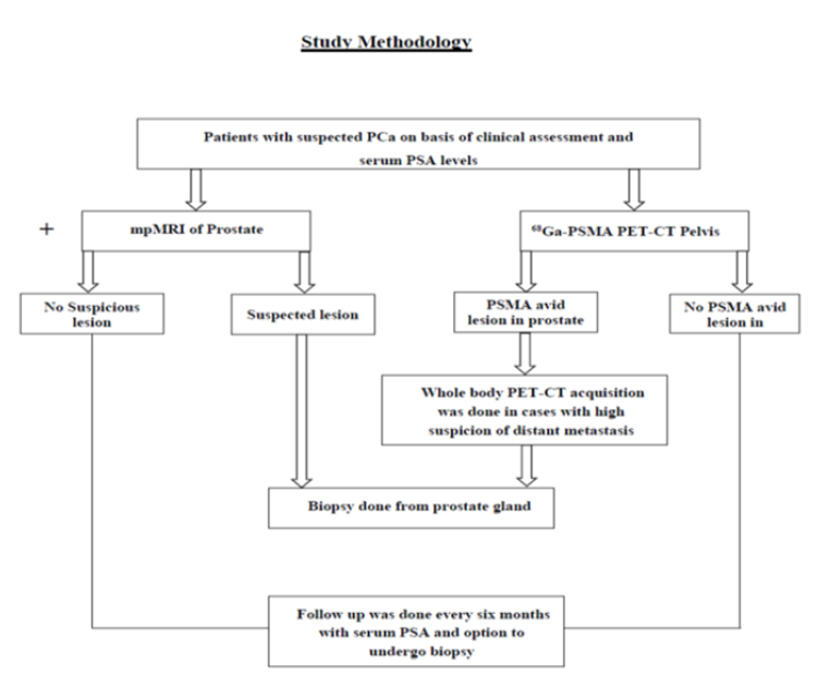
Study Population: A total of 44 patients suspected for PCa on the basis of clinical assessment and/or raised PSA were recruited for the study (Figure 1).
Patients suspected for prostate adenocarcinoma and/or serum PSA levels >4ng/ml and willing to participate in the study were recruited. Study was approved by Institutional Ethics Committee, PGIMER Chandigarh. Written informed consent was obtained. After clinical assessment, mpMRI and68Ga-PSMA PET/CT were done. Biopsy was done in cases with high clinical suspicion of PCa.
Statistical Analysis
Qualitative variables are described using number and percentages. Normally distributed continuous quantitative data is described using mean and standard deviation. Skewed quantitative data is described using median and interquartile range. The normality of data will be checked by measures of Kolmogorov-Smirnov tests of normality. The statistical analysis was conducted using the IBM SPSS STATISTICS (version 23.0)
RESULTS
In this prospective study, a total of 44 patients suspected to have PCa were recruited on the basis of clinical assessment and serum PSA levels. All patients were subjected to 68Ga-PSMA PET/CT acquisition. Out of total 44, 33 patients also underwent mpMRI (Figure 1).
Figure 1: Study Methodology
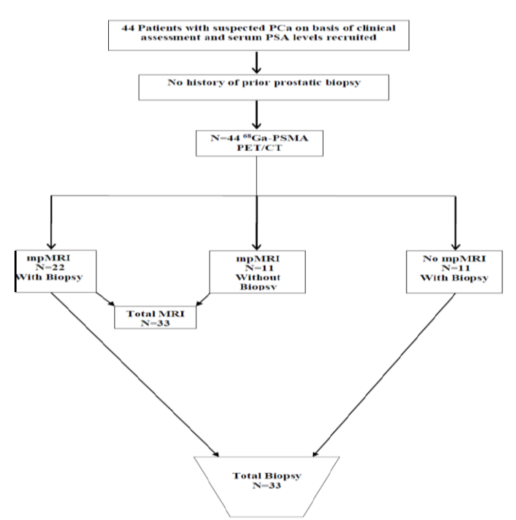
Out of the total 44 patients, 33 underwent biopsy. The remaining 11 patients did not undergo biopsy after revised clinical assessment based on negative 68Ga-PSMA PET/CT and mpMRI and were kept on follow up. Therefore, 33 patients underwent 68Ga-PSMA PET/CT and biopsy. Only 22 out of these 33 patients underwent mpMRI.
Figure 2 shows the flow chart depicting the study population.
Figure 2: Patient Flow Chart
Total 33 patients who underwent biopsy with complaints of LUTS or raised serum PSA levels were recruited.
Mean age was 66.7±9.3 years. The median pre-biopsy serum PSA level value was 13.4 ng/ml (IQR - 13.93) (Table 1).
Table 1: Patient Characteristics
Number of patients
N = 33
Age (mean±SD)
66.61±9.3 years
Weight (mean±SD)
72.12±12.3 Kg
PSA median (IQR)
13.4 ng/ml (13.9)
Family history of PCa
n=1
LUTS
n=24
Incidentally detected raised PSA
n=7
Other symptoms
n=2
Nodule on DRE
Present (n = 5)
Absent (n = 28)
The 33 patients underwent 12 core biopsy or cognitive targeted biopsy from the prostate. In six patients with negative initial biopsy but high clinical/imaging suspicion, repeat biopsies were attempted. Out of these, two came out to be positive for malignancy on repeat biopsy. In total, malignancy was detected in 16/33 (48.5%) patients on biopsy. In 17/33 (51.5%), no evidence of malignancy was noted (Table 2).
Table 2: Histopathological Findings
Parameters
n
No evidence of malignancy/prostatitis
13/4 = 17
Adenocarcinoma
16
Gleason’s score 6 (Grade group 1)
6
Gleason’s score 7 3+4 (Grade group II)
4+3 (Grade group III)
3
1
Gleason’s score 8 (Grade group IV)
5
Gleason’s score 9 (Grade group V)
1
In 17 patients, no evidence of malignancy was detected on biopsy. Out of these, Prostatitis was detected in 4 patients (Granulomatous prostatitis (n = 1), Acute/chronic prostatitis (n = 3)).
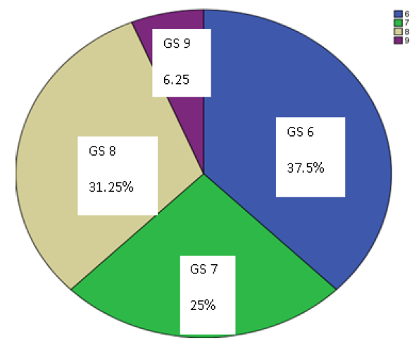
In 16 patients, malignancy was detected on biopsy. The detailed histopathological findings are summarized in Table 2. No tumor other than adenocarcinoma was detected. Prostatic intra-epithelial neoplasia (PIN) was not detected in any case. Figure 3 shows distribution of patients according to Gleason’s Score.
Figure 3: Pie Chart Showing Distribution of Percentage of Patients according to Gleason’s Score (GS 6-9).
In cases that underwent 12core biopsy with finding of PCa on histopathology, tumour burden was also assessed. The range of tumour burden in patients was from <5 to 70%. One patient with tumour burden of less than 5% was found to be negative on 68Ga PSMA PET/CT.
DISCUSSION
Diagnosis of PCa is suspected in patients having LUTS, raised serum PSA levels (>4ng/ml) or abnormal DRE findings. Transrectal 12 core biopsy is regularly done to confirm PCa.
In the present study, all 16 patients diagnosed of having malignancy on biopsy, adenocarcinoma was detected in all on histopathology. Prostatic intra-epithelial neoplasia (PIN) was not detected in any case. Tumors were low grade PCa in 9 cases and high grade PCa in 7 cases. Prostatitis was detected in 4 cases. The range of tumour burden in patients was from <5 to 70%.
In consistant to our findings, in the study done [7], the mean PSA level was 746 ng/ml with a range of 13 - 9224ng /ml. Advanced T3 and T4 tumors accounted for 25.4% and 73.2% respectively. Adenocarcinoma was the only histological form. Gleason score was less than 7 in 41(49.4 %) cases. Another study [8], also found that all the cases of prostate carcinoma were adenocarcinoma and the most frequent Gleason score was 9.
CONCLUSION
The present study concluded that all the cases of Prostate Cancer were having adenocarcinoma and had high tumor burden. Histopathological examination of prostatic specimens has important role in the diagnosis and management of various prostatic lesions.
REFERENCE
Center, M.M. et al. “International variation in prostate cancer incidence and mortality rates.” European Urology, vol. 61, no. 6, 2012, pp. 1079–1092.
Ferlay, J. et al. “Cancer incidence and mortality worldwide: Sources, methods and major patterns in GLOBOCAN 2012.” International Journal of Cancer, vol. 136, no. 5, 2015, pp. E359–E386.
Ferlay, J. et al. “Estimates of worldwide burden of cancer in 2008: GLOBOCAN 2008.” International Journal of Cancer, vol. 127, no. 12, 2010, pp. 2893–2917.
Bax, C. et al. “Innovative diagnostic methods for early prostate cancer detection through urine analysis: A review.” Cancers, vol. 10, no. 4, 2018, pp. 123.
Hoffman, R.M. et al. “Prostate-specific antigen testing accuracy in community practice.” BMC Family Practice, vol. 3, no. 1, 2002, pp. 19.
Oh, S.W. and G. J. Cheon. “Prostate-specific membrane antigen pet imaging in prostate cancer: Opportunities and challenges.” Korean Journal of Radiology, vol. 19, no. 5, 2018, pp. 819–831.
Kirakoya, B. et al. “Clinico-pathological features of prostate cancer at the university hospital yalgado ouedraogo, ouagadougou, Burkina faso.” Journal of the West African College of Surgeons, vol. 4, no. 4, 2014, pp. 70–81.
Bhatta, S. and S. Hirachan. “Prostatic lesions: Histopathological study in a tertiary care hospital.” Journal of Manmohan Memorial Institute of Health Sciences, vol. 4, no. 1, 2018, pp. 12–19.
Recommended Articles
Research Article
Imaging Evaluation of Ultrasonography Versus Magnetic Resonance,
Imaging in Diagnosis of Carpal Tunnel Syndrome among Female Patients with Positive Nerve Conduction Study
Download PDF
Research Article
The Efficacy of 5- Fluorouracil Cream and 25% Podophyllin Solution in Treatment of Plantar Warts in Alternative Day Regimen ; An Open Therapeutic Trial.
Download PDF
Research Article
An evaluation of Profile of Medico-legal autopsies conducted at Dr. Radhakrishnan Govt. Medical College, Hamirpur, Himachal Pradesh
Download PDF
Research Article
Silent Threats: Evaluating Knowledge of Pelvic Inflammatory Disease Among Reproductive-Age Women in District Kangra
Pankaj sharma,
Kritika Katoch
Published: 27/11/2024
Download PDF
PO Box 101, Nakuru
Kenya.
Email: office@iarconsortium.org
Editorial Office:
J.L Bhavan, Near Radison Blu Hotel,
Jalukbari, Guwahati-India
Sharma, V., Kumar, S. & Chadha, R. (2022). Evaluation of Histo-Pathological Finding and Tumor Burden among Prostate Cancer Patients at Tertiary Care Hospital. International Academic Research Journal of Internal Medicine & Public Health, 3(2), 1-3.
MLA
Sharma, Viper, Sunil Kumar and Rishabh Chadha. "Evaluation of Histo-Pathological Finding and Tumor Burden among Prostate Cancer Patients at Tertiary Care Hospital." International Academic Research Journal of Internal Medicine & Public Health 3.2 (2022): 1-3.
Chicago
Sharma, Viper, Sunil Kumar and Rishabh Chadha. "Evaluation of Histo-Pathological Finding and Tumor Burden among Prostate Cancer Patients at Tertiary Care Hospital." International Academic Research Journal of Internal Medicine & Public Health 3, no. 2 (2022): 1-3.
Harvard
Sharma, V., Kumar, S. and Chadha, R. (2022) 'Evaluation of Histo-Pathological Finding and Tumor Burden among Prostate Cancer Patients at Tertiary Care Hospital' International Academic Research Journal of Internal Medicine & Public Health 3(2), pp. 1-3.
Vancouver
Sharma V, Kumar S, Chadha R. Evaluation of Histo-Pathological Finding and Tumor Burden among Prostate Cancer Patients at Tertiary Care Hospital. International Academic Research Journal of Internal Medicine & Public Health. 2022 Jul;3(2):1-3.


 Download PDF
 Download XML