Since the wide range of industrial, medicinal, and therapeutic uses, gold nanoparticles (AuNPs) have sparked a lot of interest in the rapid development of nanotechnology. In this study, AuNPs were synthesized using the green approach with Pista (SH-1), Badam (SH-2), Walnut (SH-3), Groundnut (SH-4), and Tamarind (SH-5) nutshell extract in this work. Preliminary confirmation of the AuNPs production was attained by a visual color change from light brown to violet color. The UV Vis spectroscopic study of the AuNPs stability indicates the highest stability in Walnut SH-3 and Tamarind SH-5 up to 37th day at pH 7. UV- Visible Spectroscopy, Energy-dispersive X-ray spectroscopy (EDX) and Transmission electron microscope (TEM) were used to characterize the synthesized AuNPs. Physiochemical characterization of AuNPs exhibited absorbance at 550 nm for synthesized AuNPs nearly for all the extracts used, TEM image revealed that the obtained AuNPs are spherical and EDX analysis showed that obtained AuNPs were in pure form. We performed growth assessment on zebra fishes for up to 5 d by monitoring weight at various concentrations of AuNPs of walnut SH-3. It showed no histological abnormalities at organ levels in adult zebrafish up to 16 days. Our findings provided there is no notable cytotoxic evidence found in the synthesized AuNPs.
Keywords
Gold Nanoparticle
Nutshell Extract
Zebrafish
Cytotoxicity
INTRODUCTION
Gold is often used as a significant raw material in a variety of biomedical applications, including dental prosthesis, drug delivery microchips, endovascular stents, reconstructive surgery, cancer therapy, and as a food additive, owing to its non-reactive nature [1]. Bulk gold is harmless, and it may be found in ionic or molecular forms (as gold salts), allowing for the production of gold nanomaterials [2]. Gold in nanoscale form has numerous characteristics that distinguish it from gold in bulk or molecular form, rendering it a very useful tool in nanomedical application [3]. Simple synthesis, better particle reactivity, a larger surface area to volume ratio, the capacity to undergo surface changes, and strong optical qualities are some of these features [4]. AuNPs will have to be essential nanoscale elements in different technologies due to their unique characteristics, chemical stability, and ability to exhibit a variety of forms, particle sizes, and surface chemistry [5]. The applications are based on the durability and distinctive qualities of the molecules, as well as their capacity to readily displace into target cells, resulting in improved drug release and therapeutic efficacy [6]. Nevertheless, there is cause for worry since AuNPs of various sizes, shapes, and surface charges are produced throughout production and development for a variety of applications and may pose a health risk. However, due to the vagueness of short- and long-term therapeutic impacts, there is currently a scarcity of fresh discoveries to use [7]. It is essential to understand the entire spectrum of AuNP biocompatibility, as it is for any medication or other medical innovation, and to guarantee that the risk possibility is limited [8].
AuNPs have unique optical and electrical properties that make them appealing as drug delivery vehicles, biomarkers for cellular imaging, and targeted therapies [9]. Researchers have recently been interested in nanomedicine-based target-specific drug delivery [10].
AuNPs have potential applications in nanomedicine, including plasmon-based labeling, imaging, diagnostics, angiogenesis, antimicrobial agents, photothermal and radiofrequency-mediated thermal therapies, and delivery vehicles for genetic materials, imaging agents, and medications treatment of cancers, hepatitis, and tuberculosis [11]. The Zebrafish genome is quite similar to the human genome [12]. The effects of AuNPs and other nanoparticles on zebrafish were studied, indicating that Zebrafish (Danio rerio) serves as a useful animal model for evaluating nanomaterials consequences from multiple routes of exposure, at different life stages and concentration pressures [13]. The majority of research on the effects of AuNP on zebrafish utilized waterborne exposure, which did not reflect true ambient concentrations. Contamination levels in the water column reached up to 250 mg/L, and the majority of AuNPs applied were functionalized with organic compounds [14]. Another research looked at the bioavailability and toxicity of AuNPs (14 nm) from spiked marine sand (16 mg/g and 55 mg/g dry weight) on zebrafish after 20 days of exposure [15]. The effects on earthworms of sediment spiked with AuNPs and that utilized 5, 20, and 50 mg AuNPs/kg dry weight were examined in this maritime sedimentation research, where contamination amounts did not represent natural values [16]. The novelty of this study stems from the fact that NP-spiked sediment was employed as a contamination source for a vertebrate model residing in the upper water column for the first time [17].
Only a few researchers have utilized aquatic sediment as a source of NP contamination, and they have all used invertebrate animal models like annelids [18-19]. Dietary exposure to AuNPs in zebrafish showed a harmful effect at considerably lower doses. The sediment serves as a significant storage reservoir, has a high complexing capacity, and pollutants were mostly deposited as particles in suspension in the water column [19]. In this paper, we evaluated the effect of green synthesis gold nanoparticles using Pista (Pistacia vera), Badam (Prunus dulcis), Walnut (Juglans regia), Groundnut (Arachis hypogaea), and Tamarind (Tamarindus indica) shell extract were on adult zebrafish, physical characterization by UV spectrometry, TEM, and EDX. stability checking was evaluated on different days.
MATERIALS AND METHODS
Chemicals: Gold (I) chloride (AuCl) was acquired from Sigma-Aldrich, USA. All reagents used were of analytical grade. Without modification, different chemicals and media components were acquired from Hi-Media (Mumbai, India).
Extract Preparation
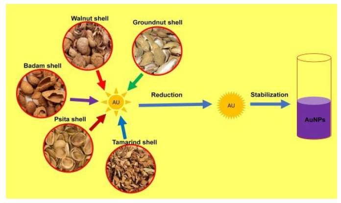
Pista (Pistacia vera), Badam (Prunus dulcis), Walnut (Juglans regia), Groundnut (Arachis hypogaea), and Tamarind (Tamarindus indica) whole nuts were acquired from the local market, Karaikudi, Tamilnadu, India. The whole nuts were washed with deionized water and the shells were removed carefully. The removed shells were pulverized using a kitchen blender to obtain a rough powder. 10 g of each shell powder was taken into a clean beaker containing 90 mL of deionized water. The beaker was kept on a heating mantle by covering the lid of the beaker using aluminum foil to avoid evaporation. After 30 min of heating at 75 °C, allow the aqueous extract to cool to room temperature and filter using Whatman Filter Paper No.1. The filtered aqueous extract was bottled and stored in a refrigerator for subsequent experiments. (Figure 1) represents the process of AuNPs preparation.
Figure 1: Preparation Chart for Plant Reduced Aunp Extracts from Pista Shell, Badam Shell, Walnut Shell, Groundnut Shell and Tamarind Shell.
Synthesis of AuNPs
10mM gold precursor AuCl was added to each 20mL of the shell extract containing Erlenmeyer flask and allowed it overnight at ambient temperature under static conditions to undergo reaction. AuNPs production was confirmed by the color change from light yellow to purple color after a few hours. The obtained emulsion was refrigerated for a subsequent experiment.
Optimization Studies
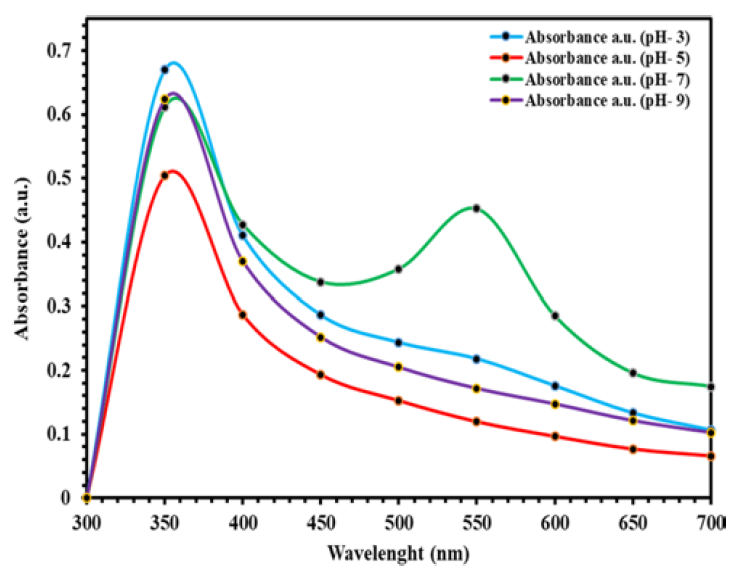
Optimization studies under varying pH levels were determined for the extracted sample. The extracted sample (1mL) was initially mixed with distilled water (4mL) in a ratio of 1:4. Furthermore, the green synthesis of gold nanoparticles was performed with the addition of 10 µl of AuCl in the solution. Further, the solution was incubated in the dark for 24 h. The pH was varied using different buffers (pH values of 3, 5, 7 and 9) while measuring the readings under varying pH ranges.
Characterization of Aunps
TEM and EDX Analysis: AuNP preparations of five different sources were transferred into the Petri plate and dried for about 2 h. The EDX and TEM analysis was done by placing the sample on the carbon-coated grid, placed into the chamber. The chamber was closed and the purge timer and cooled down after 5 minutes. The coverslip with the label facing up on the sticky tape on labeled stubs. The samples were dried for 30 min. The TEM images and the EDX results were obtained [20].
Zebrafish Toxicity Assessment and Histological Visualization D. rerio zebrafish embryos (three days after -post-fertilization period) were grown and subjected to to AuNPs exposure. The organisms were cultured in a beaker at 26±1°C with a 14:10-h light/dark cycle and fed ad libitum with Artemia sp. A 20mL of AuNP sample was prepared by mixing 16 mL of distilled water and 20mL of gold synthesis was prepared by mixing the 16mL of distilled water, 4mL of extract of 40µL of gold chloride. In each beaker with water and organisms, controls, G1 (4mL), G2 (8mL), G3 (16mL) and concentrations of AuNPs of Walnut SH 0, 50, 25 and 12.5 mg were treated respectfully. After respective treatment with AuNPs, fishes were sacrificed by anesthetizing in ice water. For the visualization, hole zebrafish were fixed in a paraformaldehyde phosphate-buffered solution, embedded in Neg-50 frozen section media (Richard Allen Scientific), and cut into 40-µm sections at -20°C using a microtome (MicromCryoStar, HM 560; Thermo Scientific). Histopathology was achieved via fixing the specialized tissue (gills, spleen, and liver) which was cut finely in thin segments were subjected to treatment with a mixture of formalin and formaldehyde for fixing them and subjecting them for microscopic investigation. The sections were mounted on glass slides, and the number of sections required for each organism was recorded. A minimum of three biological replicates was carried out, with one portion of each person being examined [21-22].
RESULTS AND DISCUSSION
UV Characterization Studies
Gold nanoparticles of five extracts were analyzed by UV-Vis spectrophotometry at different wavelengths. In our initial studies, we validated the stability of the AuNP of our extracts in response to varying pH conditions (Figure 2).
Figure 2:UV Spectrometric Graphical Representation Provides the Optimization Ranges Under Varied Ph Ranges about Walnut Shells Reduced Aunps. The Following UV Spectrometric Characterization Involves Four Different Ph Conditions 3,5,7 and 9, Which Were Further Subjected to Green Synthesis for Reduced Aunp Synthesis. The Graphical Illustration Provides an Overall Indication of the Absorbance Peaks Within the Wavelength Ranges of 350-700 Nm in UV Spectroscopy.
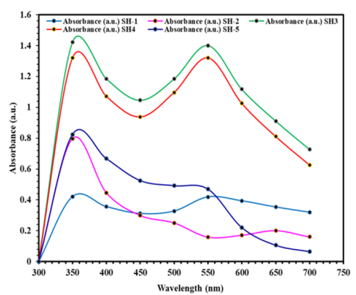
The following UV Vis Spectrometric characterization involves five shell-based extracts, which were further subjected to green synthesis for reduced AuNP synthesis. The following graphical illustration provides an overall indication of the absorbance peaks within the wavelength ranges of 350-700 nm. A lower absorption value indicates a low concentration of gold nanoparticles and as the size of the nanoparticles increases, an increased tendency in absorbance is expected. One of the most serious issues with nanoparticle solutions is their proclivity to agglomeration over time, resulting in an increase in size and a loss of characteristics. Further, UV characterization studies were done by comparing at a different wavelength to quantify the sample’s wavelength-dependent absorbance values of five different AuNP extracts (Figure 3).
Figure 3:Graphical Illustration of UV Characterization Across Different Plant-Derived Extracts of Aunps (SH-1: Pista; SH-2: Badam; SH-3: Walnut; SH-4: Groundnut; SH-5: Tamarind)
Further stability analysis of these extracts up to 37 days indicated better stability with walnut and tamarind AuNPs (Table 1 and 2).
Table 1:Stability Check of Aunps of Walnut and Tamarind on 20th, 28th And 37th Day by UV Spectroscopy Absorbance
Days
Sh-3 (Walnut)
Sh-5 (Tamarind)
20th
Wavelength
Abs
Wavelength
Abs
538.0
1.2
524.0
0.5
28th
470.0
538.0
0.9
1.3
524.0
0.5
37th
534.0
1.4
-
-
Table 2: Stability Check of Five Different Aunps Preparations Up to 11 Days by UV Spectroscopy Absorbance
Days
Sh-1 (Pista)
Sh-2 (Badam)
Sh-3 (Walnut)
Sh-4 (Groundnut)
Sh-5 (Tamarind)
W.L
Abs
W.L
Abs
W.L
Abs
W.L
Abs
W.L
Abs
1st
544.0
560.0
0.4272
0.4425
Nil
Nil
352.0
538.0
1.3457
1.4165
-
-
468.0
534.0
0.4371
0.6328
2nd
544.0
564.0
582.0
0.4554
0.4790
0.4676
Nil
Nil
536.0
1.4205
-
-
532.0
0.5894
5th
-
Nil
Nil
Nil
532.0
540.0
1.3279
1.3318
-
-
530.0
0.5752
5th
-
-
-
-
534.0
1.3675
-
-
532.0
0.6459
6th
-
-
-
-
536.0
1.3680
-
-
532.0
0.6186
8th
-
-
-
-
354.0
538.0
1.3629
1.3871
-
-
532.0
0.6191
11th
-
-
-
-
538.0
1.3131
-
-
482.0
528.0
0.6601
0.7509
Aunp Characterization of Aunp Walnut Extracts via EDX and TEM Analysis
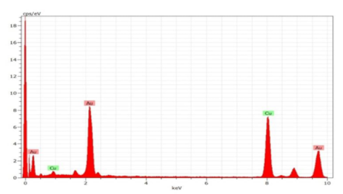
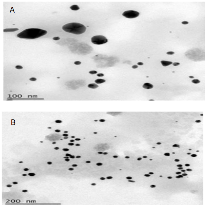
The samples were analyzed to obtain a better understanding of the properties of the silver nanoparticles. AuNP extracts of walnut using TEM and EDAX techniques. The results of TEM showed that the reaction result was made up of high-purity materials, Walnut SH AuNPs (Figure 4).
Figure 4:TEM of Aunps of Walnut SH Extract (A) 100nm and (B) 200 Nm
A similar EDAX spectrum was obtained for each sample analyzed and (Figure 5)shows the walnut SH AuNP. The shape and size features of the silver nanoparticles were studied further using scanning electron microscopy. The proposed bioreduction approach was used to obtain the upcoming depiction of TEM imaging on AuNPs.
Figure 5:Energy-Dispersive X-Ray Spectroscopy (EDAX) Image Showing Aunp Peaks of Walnut SH Extracts
Effects of Various Concentrations of Aunps of Walnut SH in Zebrafish by Weight
From our investigation, zebrafish exposure to gold synthesis that was derived was poured in twice in their controlled environment and was subsequently added in the next day (2nd day). Later on, the gold synthesis was further added to their tank for up to 5 days. Controls, G1(4mL), G2 (8mL), G3 (16mL) and concentrations of AuNPs of walnut SH 0,50,25 and 12.5 mg were treated respectively. Observation in terms of their growth and weight assessment of their masses were determined up to the 5th day (Table 3).
Table 3:Observed Data about the Individual Weight of Zebrafish within a Group for Up to 5 Days
Group
Weight(gms)
Control
1st fish
0.28
2nd fish
1.12
3rd fish
2.39
4th fish
0.87
Group1
1st fish
0.62
2nd fish
2.05
3rd fish
3.06
4th fish
1.78
5 the fish
2.67
Group2
1st fish
1.35
2nd fish
2.19
3rd fish
1.26
4th fish
1.82
5th fish
2.27
Group3
1st fish
0.61
2nd fish
2.06
3rd fish
1.28
4th fish
1.06
5th fish
2.03
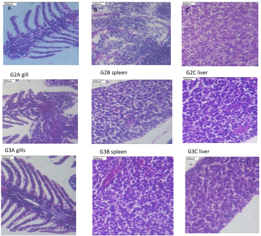
After assessment of the weight up to 5 days, the test organisms were further subjected to biopsy after sacrificing them on 16th day for histological studies. Furthermore, for assessing the cytotoxicity effects of the gold synthesis loaded in the respective groups, it is essential for undergoing a histological examination. The following biopsy investigation about specialized tissue linings was represented in the control and three different groups. The study as shown in (Figure 6) indicated that the sample organisms from all the four groups showed viable, displaying no significant toxicity which results no death. Taking off the primary organs or tissues is the most typical technique of investigating the distribution, in general, involves with liver, kidney, lung, heart, spleen, pancreas, brain, fat, and muscle after animal sacrifice. In our case, we investigated on tissues of gills, spleen and liver, as it predominantly exhibits no signs of toxicity as these organs play a greater degree of metabolic activity in our model organism (Figure 6). Thus from tracking the toxicity levels of AuNPs it could be inferred that there is no significant degree of toxicity impact in terms of visual screening was found up to 16 days. No prominent histological changes were observed between the control and Groups (G1, G2 and G3 respectively).
Figure 6:Histopathological Examination of Zebrafish Organ in Aunp Walnut SH
CONCLUSION
In conclusion, plant extracts of walnut, tamarind, groundnut, almond, and pista were successfully green synthesized into stable, well-characterized Au nanoparticles, and there in vivo cytotoxic impact was studied in zebrafish models. The size and zeta potential of produced nanoparticles were determined to be 300nm to 402nm and 38mV12mV, respectively, by physiochemical analysis of Au NP (data not shown). Our findings revealed a unique green production technique for Au nanoparticles. The discharge of AuNP into the environment can have a significant impact on the aquatic ecosystem's growth and generations. Synthesis and usage of green produced AuNP might be a viable answer in this case, as long as they are employed at the right concentration. Because of these discoveries, metal oxide nanoparticle manufacturers will be looking for green synthesis as an alternative. Furthermore, these findings will open the way for more molecular study into gold nanotoxicity in Zebrafish, as well as demonstrating the relevance of green produced AuNP over commercially available AuNPs.
REFERENCE
Elahi, N. et al. “Recent biomedical applications of gold nanoparticles: A review.” Talanta, vol. 184, 2018, pp. 537–556.
Geetha, R. et al. “Green synthesis of gold nanoparticles and their anticancer activity.” Cancer Nanotechnology, vol. 4, nos. 4–5, 2013, pp. 91–98.
Li, N. et al. “Anisotropic gold nanoparticles: Synthesis, properties, applications, and toxicity.” Angewandte Chemie International Edition, vol. 53, no. 7, 2014, pp. 1756–1789.
Schrand, A.M. et al. “Metal-based nanoparticles and their toxicity assessment.” Wiley Interdisciplinary Reviews: Nanomedicine and Nanobiotechnology, vol. 2, no. 5, 2010, pp. 544–568.
Mao, L. et al. “Design and application of nanoparticles as vaccine adjuvants against human coronavirus infection.” Journal of Inorganic Biochemistry, vol. 219, 2021, pp. 111454.
Luo, Z. et al. “Advances in gold nanoparticles-based colorimetric aptasensors for the detection of antibiotics: An overview of the past decade.” Nanomaterials, vol. 11, no. 4, 2021, pp. 840.
Akintelu, S.A. et al. “Bioremediation and pharmacological applications of gold nanoparticles synthesized from plant materials.” Heliyon, vol. 7, no. 3, 2021, pp. e06591.
Lu, W. et al. “Nanomedicines: Redefining traditional medicine.” Biomedicine and Pharmacotherapy, vol. 134, 2021, pp. 111103.
Khan, A.K. et al. “Gold nanoparticles: Synthesis and applications in drug delivery.” Tropical Journal of Pharmaceutical Research, vol. 13, no. 7, 2014, pp. 1169–1177.
Patra, J.K. et al. “Nano-based drug delivery systems: Recent developments and prospects.” Journal of Nanobiotechnology, vol. 16, no. 1, 2018, pp. 1-33.
D’Amora, M. et al. “Toxicological profile of calcium carbonate nanoparticles for industrial applications.” Colloids and Surfaces B: Biointerfaces, vol. 190, 2020, pp. 110947.
Adewale, O.B. et al. “Toxicological behavior of gold nanoparticles on various models: Influence of physicochemical properties and other factors.” International Journal of Toxicology, vol. 38, no. 5, 2019, pp. 357–384.
Chowdhury, A. et al. “Nanotechnology and nano carrier-based approaches on treatment of degenerative diseases.” International Nano Letters, vol. 7, no. 2, 2017, pp. 91–122.
Howe, K. et al. “The zebrafish reference genome sequence and its relationship to the human genome.” Nature, vol. 496, no. 7446, 2013, pp. 498–503.
Dedeh, A. et al. “Impact of gold nanoparticles on zebrafish exposed to a spiked sediment.” Nanotoxicology, vol. 9, no. 1, 2015, pp. 71–80.
Tedesco, S. et al. “Oxidative stress and toxicity of gold nanoparticles in Mytilus edulis.” Aquatic Toxicology, vol. 100, no. 2, 2010, pp. 178–186.
Bourdineaud, J.P. et al. “Gold and silver nanoparticles effects to the earthworm Eisenia fetida: The importance of tissue over soil concentrations.” Drug and Chemical Toxicology, vol. 44, no. 1, 2021, pp. 12–29.
Hanan, N.A. et al. “Cytotoxicity of plant-mediated synthesis of metallic nanoparticles: A systematic review.” International Journal of Molecular Sciences, vol. 19, no. 6, 2018, pp. 1725.
Libralato, G. et al. “Toxicity effects of functionalized quantum dots, gold and polystyrene nanoparticles on target aquatic biological models: A review.” Molecules, vol. 22, no. 9, 2017, pp. 1439.
Tsyusko, O.V. et al. “Short-term molecular-level effects of silver nanoparticle exposure on the earthworm, Eisenia fetida.” Environmental Pollution, vol. 171, 2012, pp. 249–255.
Shoults-Wilson, W.A. et al. “Evidence for avoidance of Ag nanoparticles by earthworms (Eisenia fetida).” Ecotoxicology, vol. 20, no. 2, 2011, pp. 385–396.
Pacheco, A. et al. “Toxicological interactions induced by chronic exposure to gold nanoparticles and microplastics mixtures in Daphnia magna.” Science of the Total Environment, vol. 628, 2018, pp. 474–483
Recommended Articles
Research Article
Electrochemical Comparative Studies On The Redox Behavior Of Cu(II) And Cr(II) Before And After Interactions With Ciprofloxacin In Solutions
H. M. Naseem Akhtar,
M. Q. Ehsan
Published: 10/08/2021
Download PDF
Research Article
A Rare Case Report of Waugh Syndrome in an Eight Months Old Male
Pielly Hanna,
...
Bassem Abou Merhi
Published: 10/03/2021
Download PDF
Research Article
Study on Prevalence and Etiology of Early Neonatal Deaths in Jasin district, Melaka from 2014-2017
Sujat a Khobragade,
...
Yadneshwar Khobragade
Published: 30/05/2021
Download PDF
Research Article
Clinicopathological Correlation in Cases of Acute Appendicitis: An Overview
Rahul Rai,
...
Tushar
Published: 05/04/2025
Download PDF
PO Box 101, Nakuru
Kenya.
Email: office@iarconsortium.org
Editorial Office:
J.L Bhavan, Near Radison Blu Hotel,
Jalukbari, Guwahati-India
Jeyakumar, M., Deepika, D. & Jeyameenakshi, A. (2023). Stability analysis of gold nanoparticles synthesized using various nutshell extract and it’s in vivo toxicology analysis on adult zebrafish. International Academic Journal of Applied Bio-Medical Sciences, 4(1), 1-6.
MLA
Jeyakumar, Mahalingam, Durai Deepika and Annamalai Jeyameenakshi. "Stability analysis of gold nanoparticles synthesized using various nutshell extract and it’s in vivo toxicology analysis on adult zebrafish." International Academic Journal of Applied Bio-Medical Sciences 4.1 (2023): 1-6.
Chicago
Jeyakumar, Mahalingam, Durai Deepika and Annamalai Jeyameenakshi. "Stability analysis of gold nanoparticles synthesized using various nutshell extract and it’s in vivo toxicology analysis on adult zebrafish." International Academic Journal of Applied Bio-Medical Sciences 4, no. 1 (2023): 1-6.
Harvard
Jeyakumar, M., Deepika, D. and Jeyameenakshi, A. (2023) 'Stability analysis of gold nanoparticles synthesized using various nutshell extract and it’s in vivo toxicology analysis on adult zebrafish' International Academic Journal of Applied Bio-Medical Sciences 4(1), pp. 1-6.
Vancouver
Jeyakumar M, Deepika D, Jeyameenakshi A. Stability analysis of gold nanoparticles synthesized using various nutshell extract and it’s in vivo toxicology analysis on adult zebrafish. International Academic Journal of Applied Bio-Medical Sciences. 2023 Jan;4(1):1-6.


 Download PDF
 Download XML