Comparative Study of Heavy Metals Concentration in Blood and Urine of Chronic Kidney Disease Patients and Healthy Persons in Gashua, Yobe State, Nigeria
H. T. Aminu
1
,
B. Uba
2
,
M. A. Gadaka
3
1
Department of Chemistry Yobe State University Damaturu, Nigeria
2
Desert Reseach Monitoring and Control Center Yobe State University Damaturu, Nigeria
3
Department of Biochemistry Yobe State University Damaturu, Nigeria
Chronic kidney disease (CKD) is a condition that is characterized by a gradual loss of kidney function over time, and when it progresses can lead to kidney failure. The number of chronic kidney patients is increasing annually in Gashua Yobe State. Therefore this research work intend to compare the level of heavy metals in blood and urine of chronic kidney disease patients and healthy persons in Gashua using atomic absorption spectroscopic techniques. The concentration of Zn, Pb, Cd, Fe, Ni and Cr in the blood and urine of males and females CKD patients, healthy persons, and WHO permissible limit were compared. Based on the findings of this research it revealed higher metals concentration in the blood of males CKD patients Pb(0.087mg/l), Ni(0.075mg/l), Fe(0.133mg/l) and Cd(0.108mg/l) but the concentration of Zn(0.108mg/l) and Cr(0.238mg/l) were higher in the blood of male healthy persons. While for females CKD patients, the concentration of Pb(0.04mg/l), Ni(0.039mg/l), Fe(0.223mg/l), Cr(0.112mg/l) were higher than that of females healthy persons but the concentration of Zn(0.062mg/l) and Cd(0.247mg/l) were higher in the blood of female healthy persons as compared with that of female CKD patients. On the other hand the concentration of Zn, Pb, Cd, Fe, Ni and Cr in the urine of males and females CKD patients were found to be higher than that of healthy persons. The concentration of heavy metals in the blood and urine of CKD patients detected in these research were higher than the WHO permissible limit while for healthy persons the concentration of the heavy metals in the blood were above the WHO permissible limit and the heavy metals such as Zn, Ni, and Pb are higher than WHO standard but Cd, Fe and Cr were within the WHO permissible limit in the urine of males while for the urine of females healthy persons it showed that the concentration of Zn, Ni, Cd, Fe and Cr were within the WHO permissible limit except Pd which is higher than WHO standard.
Keywords
Patient
CKD
Comparative
Blood
Urine
Heavy Metals
INTRODUCTION
Chronic kidney disease (CKD), also known as chronic renal disease, is a condition that is characterized by a gradual loss of kidney function over time, and when it progresses can lead to kidney failure [1]. CKD is a quiet disease because it does not have noticeable signs and symptoms that are apparent to the patient at the early stage of the disease. Studies in some parts of Nigeria indicated that 10 people out of every 100 will have CKD, and in some parts, one out of every five people will have CKD at some point in life according to Bamgboye [2]. The rate of CKD prevalence in Nigeria is between 8% and 45%, depending on the region and population that was studied [3]. In addition, the number of environmental risk factors has been recognized globally as probable causes of CKD, namely exposure to heavy metals (Arsenic (As), Cadmium (Cd), chromium (Cr), copper (Cu), and lead (Pb), agrochemicals, and nephrotoxic substances. Chronic arsenic exposure due to presence of arsenic in drinking water has been reported as one of the primary causes of CKD in Bangladesh, Taiwan and India [4]. As, Cd, Cr, Cu, and Pb are some of the heavy metals that occurred naturally in the environment, with the industrial revolution and human activities contributing significantly to their dispersal [5]. Most metals are known to have some toxic effect on human health, with both acute and chronic effects. As the metals are nephrotoxic and accumulate in the kidney, the concentrations in kidney cortex are of particular interest. However, kidney biopsies from human beings are seldom available because of the risks associated with the procedure, and therefore most of the previous knowledge on metal concentrations in the kidney comes from autopsy studies [6].
Chronic kidney disease with unknown etiology is a growing health problem worldwide. The first chronic kidney diseases cases in Gashua town was recognized and announced in early 1990’s according to the hospital information. The underline cause(s) is yet to be identified and disease is proved to be endemic.
Record obtained from General Hospital Gashua had shown that one hundred and sixty one (161) CKD patient of which sixty eight (68) are males and 93 females have been admitted to the hospital from January, 2018 to July, 2021. Hence there is an urgent need to investigate the causes of this problem which is anticipated to be due to higher concentration of heavy metals. In view of the above this study intends to compare the level of heavy metals in blood and urine of chronic kidney disease patients and healthy persons in Gashua town.
Number of chronic kidney failure patients sustained by hemodialysis has been on the increase in Gashua area of Yobe state. The chronic kidney disease among the people living in area of Gashua was first recognized and announced in early 1990’s according to the hospital information and was later found to be endemic to such areas. The underline cause(s) of the CKD have been yet unknown and is yet to be identified. The disease is proved to be endemic, therefore it is highly believed to be an environmentally induced disease.
MATERIALS AND METHODS
Reagents and Chemicals
All reagents and chemicals used for this research were analytical grade purchased from sigma Aldrich and used without further purification. A procedure described by Uba et al., [7] was adopted for sample dilution and rinse of glass wares.
Sample Collection
Blood and urine samples was collected from CKD patients and healthy persons in a sandoz sterile vials at General Hospital Gashua in the department of chemical pathology (urine sample) while a blood samples was collected in 5ml capacity plain plastic bottles in the department of haematology This was done after seeking approval from research and ethics committee of the General Hospital (ethical clearance, 2013) and with the assistance of health personnel at the hospital. A standard method of collection described by [8], was used for this research.
Digestion Procedure of Human Urine Samples
Procedure described by IAEA [9] was adopted with little modification 100 cm3 of each urine samples were evaporated in a hot plate to 20 ml the resulting solution were frozen, lyophilized and stored in a refrigerator .The lyophilized sample of urine was allowed to stand at room temperature and then reconstituted with 20 cm3 distilled water. 5cm3 of concentrated HNO3 was added to the 20 cm3 reconstituted solution of urine and evaporated to 10 cm3 on a water bath and allowed to cool. An equal mole of HNO3 and HClO4 were transferred into a fume chamber. The solution was heated on a hot plate and the heating process continue until the appearance of a dense and white fume and a clear solution. After cooling water was added and the solution was boiled for 10 minute to remove free chloride and oxide of nitrogen. The resulting solution was then filtered through acidified washed Whatmann 540 filter paper into 100 cm3 volumetric flask and fill up to the mark with distilled water.
Preparation of Blood Samples for Heavy Metals Determination
Blood samples was collected in a plain bottles and allow to clot. The clotted samples were centrifuge at 2000 rpm for 10 minute to obtained serum. The serum samples were also stored in a plastic vias at 20oC prior to elemental analysis 1 cm3 of serum was diluted to 10 cm3 with de-ionized water [10].
Determination of Heavy Metals in Blood and Urine
The concentration of Zn, Pb, Cd, Fe, Ni and Cr in blood and urine sample were determine using Atomic Absorption Spectrophometer (Buck Scientific Model 210 VGP).
RESULTS
Zinc
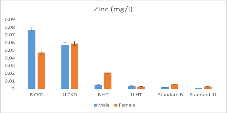
The concentration of zinc in blood and urine of male and female CKD patient were 0.0765mg/l, 0.049mg/l, 0.057mg/l and 0.059mg/l respectively, these values are greater than the one obtained from healthy persons in blood and urine 0.005mg/l, 0.021mg/l, 0.004mg/l, and 0.003mg/l and WHO reported values 0.0021mg/l, 0.006mg/l, 0.001mg/l and 0.003mg/l in both male and female. Zinc concentrations in patients with CKD vary depending on individual diets and medications. Oxidative stress plays an important role in the progression of adverse complications in CKD patients, and many trace elements are involved in the oxidant-antioxidant balance. Zinc deficiency may increase oxidative stress and atherosclerotic complications, especially in late-stage CKD patient [11]. Therefore high level of zinc is an important indicator that is not associated kidney damage (Figure 1).
Figure 1: Mean Concentration of Zinc in Urine and Blood of Male and Female CKD Patient with That of Healthy Persons
Lead
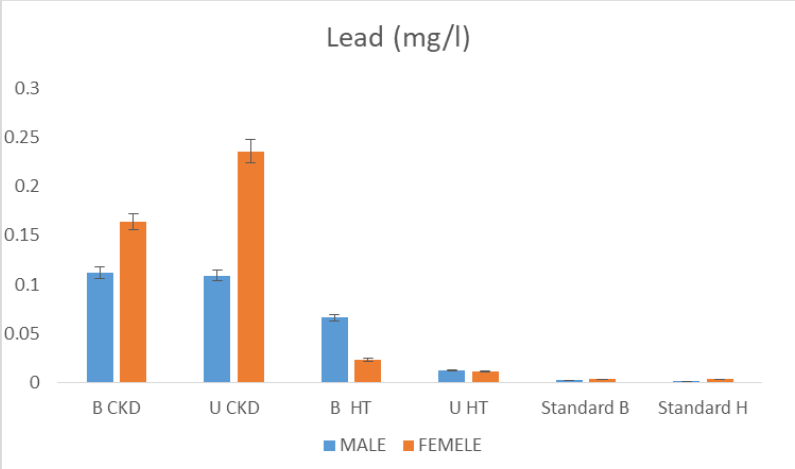
the mean concentration of lead in blood and urine of CKD patients and a healthy persons of both male and female was determined and the results of these findings reveals higher concentration of lead in blood and urine of CKD patients with concentration of 0.112mg/l, 0.164mg/l, 0.109mg/l and 0.236mg/l for male and female respectively. The values obtained for healthy persons are less than the one recorded for CKD patients. The values obtained in both CKD and healthy persons are greater than WHO standard in blood and urine of both male and female. The high values of lead detected could be due to the high level pb in irrigation water and the uptake of these heavy metals through roots of the plants grown in that area can lead to their accumulation in the tissues of organisms that feed on the water or plants grown in that area as reported by salamatu et al. many researchers reported direct relationship between Pb exposure several kidney diseases [12-13]. An exposure to Pb can cause oxidative stress in tubular and glomerular cells, leading to the generation of free radicals, which can contribute to cellular apoptosis and subsequent changes in renal structure and function [14] (Figure 2).
Figure 2: Mean Concentration of Lead in Urine and Blood of Male and Female CKD Patient with That of Healthy Persons
Nickel
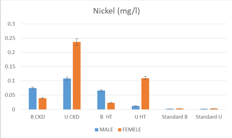
The nickel concentration was found to be greater than WHO standard level in male and female CKD patient with concentration of 0.075mg/l, 0.039 mg/l, 108mg/l and 0.236mg/l blood and urine respectively. The concentration in healthy persons as control were still less than that of CKD patients in urine and blood of both male and females with the concentration of 0.066mg/l, 0.023mg/l 0.012mg/l and 0.110mg/l respectively. It was reported that excess Ni has been shown to trigger an inflammatory response by activating nuclear factor-kB and tubular apoptosis through the phosphoinositide 3-kinase (PI3K)–RAC serine/threonine-protein kinase (AKT) pathway [15-16] (Figure 3).
Figure 3: Mean Concentration of Nickel in Urine and Blood of Male and Female CKD Patient with That of Healthy Persons
Cadmium
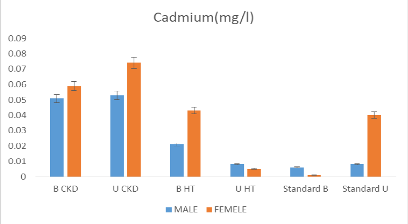
The results of these findings reveals that cadmium concentration in blood and urine of CKD patients is higher than the one obtained from healthy persons with mean concentration of 0.051mg/l, 0.059mg/l, 0.053mg/l and0.074mg/l for CKD patients and 0.021mg/l, 0.043mg/l, 0.008mg/l and 0.005mg/l for healthy persons in blood and urine respectively. All the values obtained were greater than WHO permissible limit. Cd was reported to be a nephrotoxic environmental pollutant [17]. High levels of exposure can result in the accumulation of Cd in the proximal tubules of the kidney, which result to impair tubular function and protein reabsorption [18]. Cd exposure is one of the major causes of proteinuria (26). It was also reveals that chronic Cd exposure has direct relationship to reduced eGFR and an increased risk of CKD [15,13] (Figure 4).
Figure 4: Mean Concentration of Cadmium in Urine and Blood of Male and Female CKD Patient with That of Healthy Persons
Iron
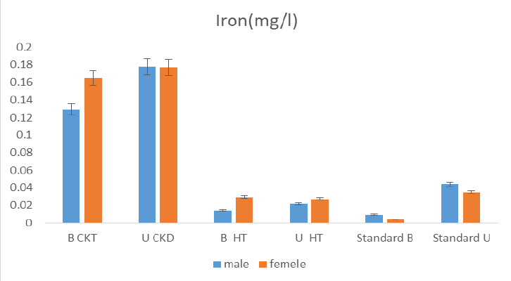
The mean concentration of iron detected in blood and urine of CKD patients were 0.129mg/l, 0.165mg/l, 0.178mg/l and 0.177mg/l in male and female respectively these concentrations are higher than values obtained in healthy persons 0.014mg/l, 0.029mg/l, 0.022mg/l and 0.027mg/l and WHO permissible limit. It was reported that a lower concentration of iron in CKD patient can cause anaemia and also an iron overload represents a condition of increased total body iron content that is possibly associated with a time dependent risk of organ dysfunction. Pathologic iron overload represents a condition of increased body iron content associated with signs of organ dysfunction that are presumably caused by excess iron [19] (Figure 5).
Figure 5: Mean Concentration of Iron in Urine and Blood of Male and Female CKD Patient with That of Healthy Persons
Chromium
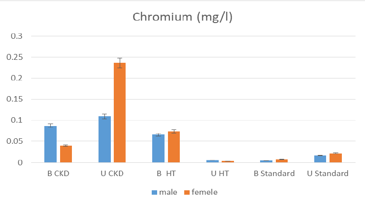
The chromium concentration detected in male and female were 0.087mg/l, 0.040mg/l, 0.109mg/l and 0.236mg/l in blood and urine which is higher than the values obtained for male and female healthy persons with the concentration of 0.066mg/l, 0.074mg/l, 0.005mg/l and 0.003mg/l blood to urine and also less than WHO permissible limit. The kidney is considered to be a critical organ for Cr toxicity, Cr compounds have been reported to accumulate in proximal convoluted tubules [20]. Cr-induced cytotoxicity, DNA damage, and oxidative stress have also been reported in animal kidneys [21-22].
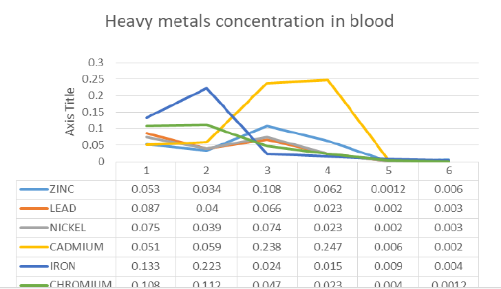
The concentration of heavy metals in human blood serum could serve as indicators for various pathological conditions, determination of certain metals level in the serum provide a very interesting information on diagnosis and treatment of various diseases [23-24]. Blood is responsible for metals transportation to the body tissues, therefore blood test is best for detecting recent heavy metals poisoning and for measuring levels of minerals in the body. The level of heavy metals in blood of male and female CKD patient, healthy persons were compared and the result obtained showed that the concentration of zinc, lead, nickel, cadmium, iron and chromium is metal and sex specific as the concentration of Zn (0.053mg/l), Pb (0.087mg/l) and Ni (0.075mg/l) in the blood of male CKD patient is higher than that of female CKD patient while the concentration of Cd (0.059mg/l), Fe (0.223mg/l) and Cr (0.112mg/l) in female is higher than that of male. In the other hand the results of heavy metal concentration in healthy persons reveals that the concentration of Zn (0.108mg/l), Pb (0.066mg/l), Cd (0.074mg/l), Ni (0.238mg/l), Fe (0.024mg/l) and Cr (0.047mg/l) in the blood of males is higher than that of females. The heavy metals concentration in the blood of males CKD patients and healthy persons showed higher concentration of Pb(0.087mg/l), Ni(0.075mg/l), Fe(0.133mg/l) and Cd(0.108mg/l) while concentration of Zn(0.108mg/l) and Cr(0.238mg/l) is higher in the blood of healthy persons for females CKD patients and healthy persons showed high concentration of Pb(0.04mg/l), Ni(0.039mg/l), Fe(0.223mg/l), Cr(0.112mg/l) while the concentration of Zn(0.062mg/l) and Cd(0.247mg/l)were higher in female healthy persons as compared with that of female CKD patients. The heavy concentration detected in CKD patients and healthy persons were higher than WHO standard (Figure 6).
Figure 6: Mean Concentration of Chromium in Urine and Blood of Male and Female CKD Patient with That of Healthy Persons
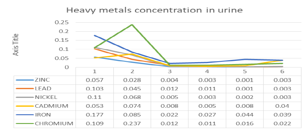
The concentration of zinc, lead, cadmium, iron, nickel and chromium in the urine of males and females CKD patients and healthy persons were compared. the results of the findings reveals that males CKD patient has the highest concentration of Zn(0.057mg/l), Pb(0.103mg/l), Ni(0.11mg/l), and Fe(0.177mg/l) while the concentration of Cd(0.074mg/l) and Cr(0.237mg/l) were found to be higher in females CKD patients. For healthy persons the results showed higher concentration of Zn(0.004mg/l), Pb(0.012mg/l), Cd(0.008mg/l), Ni(0.005mg/l) and Cr(0.012mg/l) but the concentration of Fe(0.022mg/l) is higher in females as compared with that of male healthy persons. The concentration of Zn, Pb, Cd, Fe, Ni and Cr in the urine of males and females CKD patients were found to be higher than that of healthy persons. the concentration of heavy metals in the urine of CKD patients detected in these research are higher than the WHO permissible limit while for healthy persons heavy metals such as zinc, nickel, and lead is higher than WHO standard but cadmium, iron and chromium are within the WHO permissible limit in the urine of males while for the urine of females healthy persons it showed that the concentration of zinc, nickel, cadmium, iron and chromium is within the WHO permissible limit except lead which is higher than WHO standard. High concentration of Cr, Cd and Pb are associated with an additional decline of eGFR [25].
Summary of Major Findings
The concentration of Zn, Pb, Cd, Fe, Ni and Cr in the blood and urine of males and females CKD patients, healthy persons, and WHO permissible limit were compared. Based on the findings of these research it revealed higher metals concentration in the blood of males CKD patients Pb(0.087mg/l), Ni(0.075mg/l), Fe(0.133mg/l) and Cd(0.108mg/l) but the concentration of Zn(0.108mg/l) and Cr(0.238mg/l) is higher in the blood of male healthy persons. While for females CKD patients the concentration of Pb(0.04mg/l), Ni(0.039mg/l), Fe(0.223mg/l), Cr(0.112mg/l) is higher than that of females healthy persons but the concentration of Zn(0.062mg/l) and Cd(0.247mg/l) were higher in the blood of female healthy persons as compared with that of female CKD patients. In the other hand the concentration of Zn, Pb, Cd, Fe, Ni and Cr in the urine of males and females CKD patients were found to be higher than that of healthy persons. The concentration of heavy metals in the blood and urine of CKD patients detected in these research is higher than the WHO permissible limit while for healthy persons the concentration of the heavy metals in the blood are above the WHO permissible limit and the heavy metals such as zinc, nickel, and lead are higher than WHO standard but cadmium, iron and chromium are within the WHO permissible limit in the urine of males while for the urine of females healthy persons it showed that the concentration of zinc, nickel, cadmium, iron and chromium is within the WHO permissible limit except lead which is higher than WHO standard. it was reported that a combine exposure of heavy may lead to nephrotoxicity [26] (Figure 7-8).
Figure 7: Mean Concentration of Heavy Metals in Blood
Figure 8: Mean Concentration of Heavy Metals in Urine
CONCLUSION
The concentration of Zn, Pb, Cd, Fe, Ni and Cr in the urine of males and females CKD patients were found to be higher than that of healthy persons, except for the blood which showed high concentration of zinc in both male and female healthy persons while chromium for male and cadmium for female as compared with male and female CKD patients. It was also revealed that heavy metals concentration are sex specific and the concentration of heavy metals detected in CKD are above WHO permissible limit while for healthy persons are within and above WHO standard.
Acknowledgment
The authors acknowledge Tertiary Education Trust Fund (TET-FUND) Abuja-Nigeria and Management of Yobe State University Damaturu Nigeria for sponsoring the research. We also appreciated the contributions of Chemistry Department and Desert Research Monitoring and Control Center Yobe State University.
REFERENCE
National Kidney Foundation. “About chronic kidney disease.” 2013, Retrieved September.
Bamgboye E. “One out of five Nigerians has kidney disease.” 12 December 2013, http://theeagleonline.com .ng/one-out-of-fivenigerians-has-kidney-deases-nephrolo gist/.
Alebiosu C.O. and Ayodele O.E. “The global burden of chronic kidney disease and the way forward.” Ethnicity and Disease, vol. 15, no. 3, 2005, pp. 418.
Pritchard J.D. Arsenic toxicological overview. CRCE HQ, Health Protection Agency, UK, Version 2, 2007.
United Nations Environment Programme. Final review of scientific information on lead. Version of December, 2010, UNEP Chemicals Branch, http://www.unep.org/chemicalsandwaste /Portals/9/Lead_Cadmium/docs/Interim_re.
Bahemann-Hoffmeister A. et al. “Lead concentrations in human tissues with regard to habits and occupational exposure.” Z Arbeitsmed, vol. 38, 1988, pp. 30-35.
Uba B. et al. “Determination of some heavy metals concentration in muscle and bone of Osteoglossidae, catfish, and tilapia fish of River Yobe.” East African Scholars Multidisciplinary Bulletin, vol. 2, no. 7, 2019, pp. 221-224.
Radojevic M. and Bashkin V.N. Practical environmental analysis. Royal Society of Chemistry, 1999, Cambridge.
International Atomic Energy Agency. Assessments of levels and health effect of airborne particulate matter in mining, metal refining and metal working industries using nuclear and related analytical techniques. IAEA-TECDOC 1576, 2008.
Akan J.C. et al. “Determination of heavy metals in blood, urine and water samples by inductively coupled plasma atomic emission spectrophotometer and fluoride using ion-selective electrode.” Journal of Analytical & Bioanalytical Techniques, vol. 5, no. 9, 2014, pp. 1-7.
Shih C.T. et al. “Changes in levels of copper, iron, zinc, and selenium in patients at different stages of chronic kidney disease.” Genomic Medicine, Biomarkers, and Health Sciences, vol. 4, no. 4, 2012, pp. 128-130.
Evans M. and Elinder C.G. “Chronic renal failure induced by lead.” Kidney International, vol. 79, 2011, pp. 688-689.
Afsar B. et al. “Air pollution and kidney disease: review of current evidence.” Clinical Kidney Journal, vol. 12, no. 1, 2019, pp. 19-32.
Liu G. et al. “Mitochondrial permeability transition and its regulatory components are implicated in apoptosis of primary cultures of rat proximal tubular cells exposed to lead.” Archives of Toxicology, vol. 90, no. 5, 2016, pp. 1193-1209.
Xu X. et al. “Environmental pollution and kidney diseases.” Nature Reviews Nephrology, vol. 14, no. 5, 2018, pp. 313-324.
Guo H. et al. “Modulation of the PI3K/Akt pathway and Bcl-2 family proteins involved in chicken’s tubular apoptosis induced by nickel chloride.” International Journal of Molecular Sciences, vol. 16, no. 9, 2015, pp. 22989-23011.
Soderland P. et al. “Chronic kidney disease associated with environmental toxins and exposures.” Advances in Chronic Kidney Disease, vol. 17, no. 3, 2010, pp. 254-264.
Johri N. et al. “Heavy metal poisoning: the effects of cadmium on the kidney.” Biometals, vol. 23, no. 5, 2010, pp. 783-792.
Macdougall I.C. et al. “Iron management in chronic kidney disease: conclusions from a Kidney Disease: Improving Global Outcomes controversies conference.” Kidney International, vol. 89, no. 1, 2016, pp. 28-39, http://dx.doi. org/10.1016/j.kint.2015.10.002.
Kulathunga M.R.D.L. et al. “Chronic kidney disease of unknown aetiology in Sri Lanka and the exposure to environmental chemicals: a review of literature.” Environmental Geochemistry and Health, vol. 41, no. 5, 2019, pp. 2329-2338.
Eastmond D.A. et al. “Trivalent chromium: assessing the genotoxic risk of an essential trace element and widely used human and animal nutritional supplement.” Critical Reviews in Toxicology, vol. 38, no. 3, 2008, pp. 173-190.
Patlolla A.K. et al. “Oxidative stress, DNA damage, and antioxidant enzyme activity induced by hexavalent chromium in Sprague-Dawley rats.” Environmental Toxicology, vol. 24, 2008, pp. 66-73.
Bárány E. et al. “Trace element levels in whole blood and serum from Swedish adolescents.” Science of the Total Environment, vol. 286, no. 1-3, 2002, pp. 129-141.
Canellas C.G.L. et al. “Trace and major elements in serum of patients with chronic myelogenous leukemia.” Journal of Radioanalytical and Nuclear Chemistry, vol. 269, no. 3, 2006, pp. 631-634.
Tsai T.L. et al. “The decline in kidney function with chromium exposure is exacerbated with co-exposure to lead and cadmium.” Kidney International, vol. 92, no. 3, 2017, pp. 710-720.
Tsai H.J. et al. “Associations among heavy metals and proteinuria and chronic kidney disease.” Diagnostics, vol. 11, no. 2, 2021, pp. 282, https://doi.org/10.3390/diagnost ics11020282.
Recommended Articles
Research Article
Electrochemical Comparative Studies On The Redox Behavior Of Cu(II) And Cr(II) Before And After Interactions With Ciprofloxacin In Solutions
H. M. Naseem Akhtar,
M. Q. Ehsan
Published: 10/08/2021
Download PDF
Research Article
A Rare Case Report of Waugh Syndrome in an Eight Months Old Male
Pielly Hanna,
...
Bassem Abou Merhi
Published: 10/03/2021
Download PDF
Research Article
Study on Prevalence and Etiology of Early Neonatal Deaths in Jasin district, Melaka from 2014-2017
Sujat a Khobragade,
...
Yadneshwar Khobragade
Published: 30/05/2021
Download PDF
Research Article
Clinicopathological Correlation in Cases of Acute Appendicitis: An Overview
Rahul Rai,
...
Tushar
Published: 05/04/2025
Download PDF
PO Box 101, Nakuru
Kenya.
Email: office@iarconsortium.org
Editorial Office:
J.L Bhavan, Near Radison Blu Hotel,
Jalukbari, Guwahati-India
Aminu, H. T., Uba, B. & Gadaka, M. A. (2021). Comparative Study of Heavy Metals Concentration in Blood and Urine of Chronic Kidney Disease Patients and Healthy Persons in Gashua, Yobe State, Nigeria. International Academic Journal of Applied Bio-Medical Sciences, 2(2), 1-7.
MLA
Aminu, H. T., B. Uba and M. A. Gadaka. "Comparative Study of Heavy Metals Concentration in Blood and Urine of Chronic Kidney Disease Patients and Healthy Persons in Gashua, Yobe State, Nigeria." International Academic Journal of Applied Bio-Medical Sciences 2.2 (2021): 1-7.
Chicago
Aminu, H. T., B. Uba and M. A. Gadaka. "Comparative Study of Heavy Metals Concentration in Blood and Urine of Chronic Kidney Disease Patients and Healthy Persons in Gashua, Yobe State, Nigeria." International Academic Journal of Applied Bio-Medical Sciences 2, no. 2 (2021): 1-7.
Harvard
Aminu, H. T., Uba, B. and Gadaka, M. A. (2021) 'Comparative Study of Heavy Metals Concentration in Blood and Urine of Chronic Kidney Disease Patients and Healthy Persons in Gashua, Yobe State, Nigeria' International Academic Journal of Applied Bio-Medical Sciences 2(2), pp. 1-7.
Vancouver
Aminu HT, Uba B, Gadaka MA. Comparative Study of Heavy Metals Concentration in Blood and Urine of Chronic Kidney Disease Patients and Healthy Persons in Gashua, Yobe State, Nigeria. International Academic Journal of Applied Bio-Medical Sciences. 2021 Jul;2(2):1-7.


 Download PDF
 Download XML