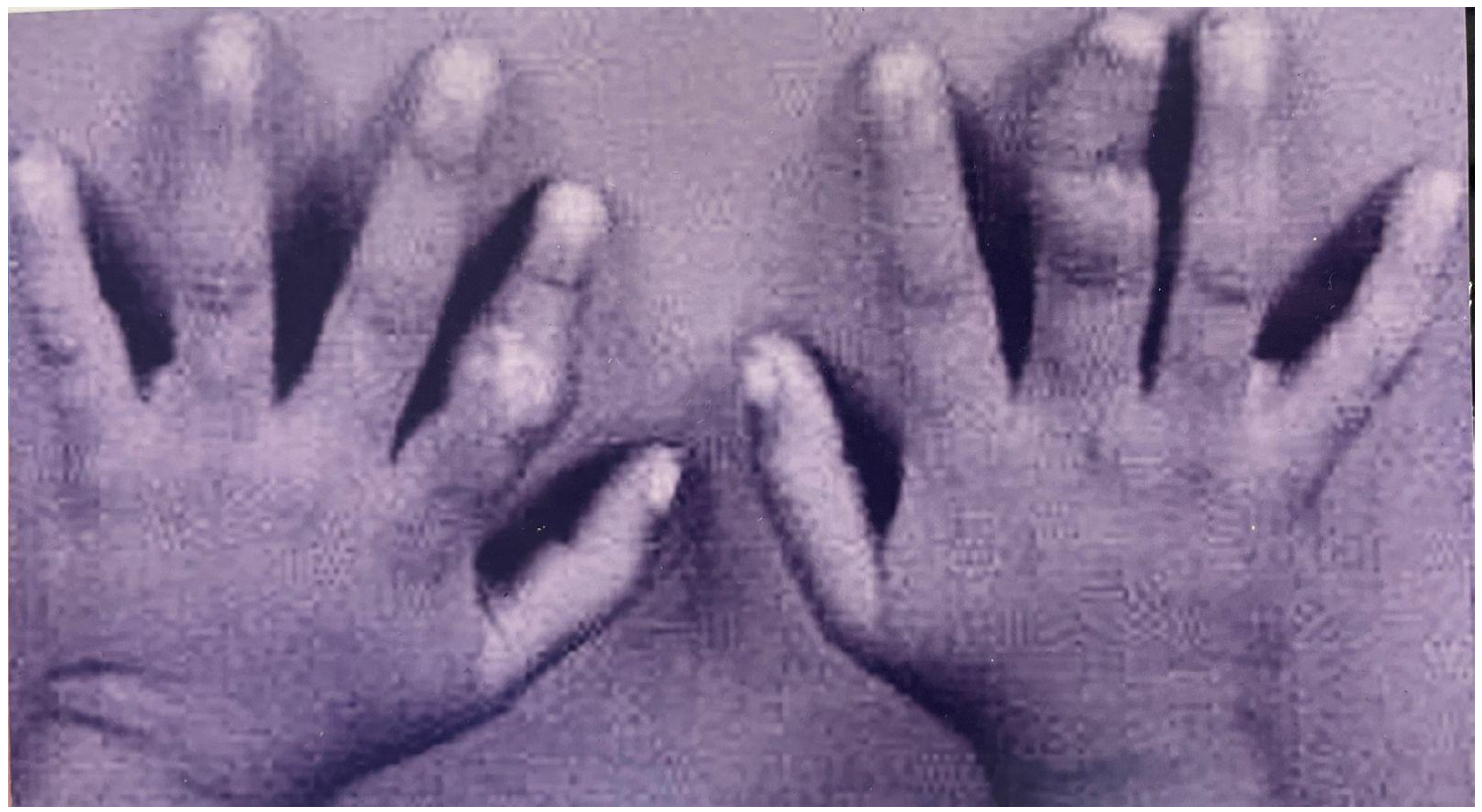
Most of the described large series of tuberculous dactylitis were reported before 1970 (3-5); only isolated cases have been reported in the last two decades(6-8).Tubercular dactylitis is commonly seen in young children; the incidence decreases after five years and rare after ten years. Childhood dactylitis differs from adult dactylitis by increased sequestrum formation, fistula formation, bone expansion, periosteal reaction, multiple bone involvement and positive chest X ray; while rate of pathological fracture is more in adult tubercular dactylitis(9).
Skeletal tuberculosis usually occurs as the outcome of lympho-hematogenous spread of tuberculous bacilli from primary complex. Depending on, quantity of bacilli released and host resistance, active metastatic tuberculous lesion occurs. Skeletal lesion typically begins as an endarteritis in the metaphysis of long bones progresses to form tuberculous follicle and then to granulation tissue.
Depending on host resistance this tuberculous lesion may progresses either to sclerotic or productive form or to destructive or exudative form. The lesion of tuberculous dactylitis appears mainly due to exudative forn of disease with superadded sclerosis. Tuberculous granulation tissue once developed erodes and destroys the trabecular pattern of bone by pressure necrosis and direct bacterial action with progressive demineralization. The periosteal new bone formation is due to increased vascularity with secondary deposition of bone. Gradual destruction with sequestration of the cortical bone then occurs while involucrum thickens.
If much of the involucrum's internal aspects are absorbed, a cyst-like cavity remains, which appears to be ballooned out, or injected with air; typically described as 'Spina Ventosa'. Concomitantly, dense stippling of one or more areas may be seen so that the involved bone, in addition to appearing twice it's normal diameter, may be of greater density and stippled without normal cancellous trabeculae. If the sequestrum suppurates, the new periosteum undergoes necrosis with resultant fistulae through which small fragments are extruded. The disease may advance and involve growth plate and epiphysis resulting in stunted growth and joint destruction. The pull on tendons exerted by contracting granulation tissues adds further deformity.
Dactylitis usually presents within 4-6 weeks after infection as the principal lesion or as a minor manifestation of generalized form. Although a chest roentgenogram positive for tuberculosis is commonly seen in children with tuberculous dactylitis, but chest roentgenogram was unremarkable in the present case.
Although isolated involvement of one bone of hand or foot is the most common manifestation, multiple foci can be identified in 25-30% of cases(10). Tuberculous dactylitis involves hand eight times more common than foot. Simultaneous involvement of the multiple bones of hands and feet, as seen in our patient, is very rare.
Diagnosis is based on high degree of clinical suspicion and must be confirmed by histology and/or bacteriology studies. Pathology examination of a biopsy specimen reveals granulomatous osteitis with typical caseous necrosis.
The radiological features of tuberculous dactylitis on plain X rays include soft tissue swelling, often marked & rusiform; peri-osteitis with bone expansion with cortical and cancellous bone destruction; diffuse uniform infiitration with honey combing of affected phalanx and localized destruction with reactive osteitis.
The differential diagnosis of tubercular dactylitis includes: congenital syphilis (which usually presents in first year, bilaterally symmetrical and not associated with soft tissue swelling or sequestration); sarcoidosis (bone expansion and periosteal new bone formation are absent); multiple enchondromatosis (lesions tend to be larger than tubercular); hemoglobinopathies (cortical thinning and coarse trabecular pattern due to increased cortical activity, affects all bones of hands symmetrically without new bone formation). Other differential diagnoses include sickle cell anemia and other lytic bony lesions caused by slow growing bony tumors, neuroblastoma, Langerhans' cell histiocytosis, or leukemia.
Bone scans and MRI are neither required nor are helpful in differential diagnosis of the lesion. Treatment includes chemotherapy, radical synovectomy and early mobilization. The major role of surgery is to provide tissue for diagnosis and to allow for the removal of caseous avascular tissue to promote distribution of drugs into surrounding tissues. An important aspect in the rehabilitation of these patients is carly motion to prevent joint contracture and subsequent ankylosis. The clinician should be aware that tuberculosis could clinically mimic almost any disease. Only a high degree of suspicion would result in unnecessary investigations with a delay in diagnosis and treatment.



 Download PDF
 Download XML