CIntestinal parasitic infections remain a significant public health concern of global importance. The developed molecular methods demonstrate considerable specificity and sensitivity compared with microscopy. Compared with conventional methods, methods based on detecting intestinal protozoa DNA are faster, have higher sensitivity and specificity, can detect multiple parasites simultaneously and can quantify and genotype parasitic DNA. The objective of the present study was to Molecular diagnosis of intestinal protozoa for patients in Wasit province. A total of 100 stool samples (44 male and 56 female) were randomly collected from patients in Wasit province during January 2023 to April 2023 and diagnosis by Multiplex PCR methods. Of the 100 patients with abdominal pain, they were found to be infected with intestinal parasites upon examination by Nested PCR method. The protozoa included Cryptosporidium parvum (23.96%), Blastocystis hominis (17.71%), Giardia lamblia (20.83%), Entamoeba histolytica (62.50%) and Isospora (7.29%). Our study supports implementation of molecular tools into the parasitology diagnostic laboratory, even in under-resourced settings. Such tools are particularly important for detecting protozoa that are challenging to identify by microscopy. Molecular tools are also essential for distinguishing between morphologically identical species.
Keywords
Cryptosporidium Parvum
Blastocystis Hominis
Giardia Lamblia
Entamoeba Histolytica
Isospora
INTRODUCTION
Intestinal parasitoses are one of the main causes of health-service consultations in developing countries and are an important cause of morbidity worldwide. Among these, infections with some protozoa are recognized as having significant consequences on health, particularly in children [1]. Direct consequences are usually gastrointestinal disorders, such as diarrhea, dysentery, abdominal pain, vomiting and lack of appetite and indirect impacts include synergistic negative effects on nutritional status, stunting and reduced educational achievement. For instance, every year there are more than 200 million symptomatic patients with giardiosis [2] and cryptosporidiosis which cause diarrhea in children younger than 5 years globally, only second after rotaviral enteritis[3]. Infections with Giardia duodenalis (syn. Giardia lamblia or Giardia intestinalis) Cryptosporidium spp. and Entamoeba histolytica are considered to be among the most common and important causes of parasite-related diarrhea in human populations. In contrast, the pathogenicities and symptoms associated with infections with Blastocystis spp. [3]. Intestinal protozoa are major causes of gastrointestinal infections in humans and are diagnosed, traditionally, using microscopy, the diverse concentration protocols usually involving formalin fixation. Microscopic analysis guided by morphological characteristics of parasites remains the gold standard for diagnosing such infections, often caused by members of the genera Giardia, Entamoeba or Cryptosporidium. Species and genotypes within each of these genera may differ in their pathogenesis, host range and epidemiology, which can only be elucidated using molecular methods [4]. However, other methods are available, including detection of parasite antigen in feces by ELISA or immunochromatograpy (e.g., snap tests) and molecular methods [5]. Given the excellent sensitivities and specificities achieved by molecular methods, detection of parasite-specific DNA by PCR is fast gaining popularity. Not only is PCR simple and potentially offers a more rapid turnaround time, but it also allows discrimination between morphologically indistinguishable parasites that are of different clinical relevance (such as Entamoeba dispar and E. histolytica). In addition, multiplex PCR enables samples to be investigated for several pathogens simultaneously and can also be used to determine relative copy numbers, although the potential for quantification is often not used in the diagnostic setting. However, due to the requirement for comparatively costly equipment and reagents, including some for which refrigeration is essential, such diagnostic assays have not been implemented in many diagnostic labs and are particularly underused in labs in less-developed regions. Nevertheless, where facilities and resources are available, multiplexed PCR-based methods that target the most relevant gastrointestinal parasites can provide fast, reliable results and could be implemented in routine clinical diagnostics [6]. However, comparison of commercial multiplex real-time PCR assays has indicated that performance may be variable and the required sensitivities and specificities, along with lab workflow, diagnostic algorithms and population should be considered, as well as the cost, when considering incorporating such assays into the diagnostic lab [7]. Although, in the past few decades molecular diagnostics of parasitic infections have been accepted as first-line diagnostics, laboratories tend to be reluctant of introducing nucleic acid based tests as they dread the validation of laboratory developed tests (LDTs) compliant with ISO1589 [8].
Aim of the Study
This cross-sectional study aims to Molecular detection of intestinal protozoa parasite infection in patients at Wasit province.
MATERIALS AND METHODS
Study Design and Patients
A cross-sectional study was conducted in Wasit province. The study included 100 stool samples (44 male and 56 female) were randomly collected from patients in Wasit province during January 2023 to May 2023. The samples examined were used to detect the presence of intestinal parasitic infestation while the study utilized a pre-tested structure, after collection, each stool sample was divided into two parts; the first used to diagnosed the parasite by microscopic examination of M.Z.N stain, wet mounts were stained with Lugol's iodine and the second was preserved in the laboratory freezer in -20°C for PCR for detection of parasite DNA. Cases were matched based on demographic parameters, such as age, gender, residence, malnutrition and source water.
Collection of Samples
During this study, 100 stool specimens were collected from both gender and different age groups of patients in clean specimen container tube. During the period from January 2023 till the end of May 2023, data were collected simultaneously, each stool sample was divided into two parts; the first used to diagnose the parasite by microscopic examination and the second was preserved in the laboratory freezer in -20°C for PCR for detection of parasite DNA [9].
Molecular Testing
The Multiplex PCR primers for detection Entamoeba histolytica, Giardia intestinalis, Cryptosporidium parvum, Blastocystis spp., Isospora sp., Balantidium coli and Dientamoeba fragilis based on small subunit ribosomal RNA gene were designed in this study using NCBI-Genbank and primer 3 plus design. These primers were provided from Scientific Resercher. Co. Ltd, Iraq.
Multiplex PCR technique was performed for detection Entamoeba histolytica, Giardia intestinalis, Cryptosporidium parvum, Blastocystis sp., Isospora sp., Balantidium coli and Dientamoeba fragilis based on small subunit ribosomal RNA gene from Human stool samples. This method was carried out according to method described as following steps:
DNA Extraction: DNA from water samples were extracted by using Presto™ Stool DNA Extraction Kit and done according to company instructions
Genomic DNA Estimation: The extracted genomic DNA from stool samples was checked by using Nanodrop spectrophotometer (THERMO. USA) that check and measurement the purity of DNA through reading the absorbance in at (260 /280 nm)
Multiplex PCR Master mix Preparation: The mPCR master mix was prepared by using (GoTaq Green PCR Master Mix) and this master mix done according to company instructions
PCR Thermocycler Conditions: Initial Denaturation (95ºC for 5 min), Denaturation (95ºC for 30 sec) Annealing (58ºC for 30 sec) Extension (72ºC for 1 min), Final extension (72ºC for 5 min) Hold (4°C Forever)
Multiplex PCR product analysis: The mPCR products of was analyzed by agarose gel electrophoresis
Statistical Analysis
Statistical analysis was carried out using of ANOVA by the SPSS program [10].
RESULTS
The study included examination of 100 stool samples of patient by Multiplex Polymerase chain reaction PCR. In this section, the results of the Multiplex PCR test were demonstrated. Table 1 shows that 60% of the samples were E. histolytica followed by 23% Cryptosporidium and then 20% for G. lamblia. Regarding Isospora, there were 7 cases (7%) detected Table 1.
Table 2 shows the association of age with the presence of E. histolytica by Multiplex PCR. There was no significant association (p-value = 0.830) between being in a specific age group and testing positive for this parasite. About 60% of those ages 16-30 years and 60% of 31-45 years old appeared to be positive by PCR test. Also, there was 65% with less than 15 years old.
Table 1: Frequency Distribution of Parasites Detected By Multiplex PCR in 100 Samples
Parasites
E. histolytica for PCR
Frequency
Percentage
E. histolytica
Positive
60
60
Negative
40
40
Total
100
100
G. lamblia
Positive
20
20
Negative
80
80
Total
100
100
Cryptosporidium
Positive
23
23
Negative
77
77
Total
100
100
Blastocystis hominis
Positive
17
17
Negative
83
83
Total
100
100
Isospora
Positive
7
7
Negative
93
93
Total
100
100
Balantidium coli
Positive
0
0
Negative
100
100
Total
100
100
Dientamoeba fragilis
Positive
0
0
Negative
100
100
Total
100
100%
Table 2: Association of Age Groups with the Presence of E. Histolytica by Multiplex PCR
Age group (years)
E. histolytica No. (%)
Total
p-value
Positive
Negative
≤15
26 (65.0%)
14 (35.0%)
40(100%)
0.830
16 - 30
15 (60.0%)
10 (40.0%)
25(100%)
31 - 45
9 (60.0%)
6 (40.0%)
15(100%)
46 - 60
6 (46.2%)
7 (53.8%)
13(100%)
61+
4 (57.1%)
3 (42.9%)
7(100%)
Table 3: Association of Age Groups with the Presence of G. Lamblia by Multiplex PCR
Age group (years)
G. lamblia No. (%)
Total
p-value
Positive
Negative
≤15
10 (25.0%)
30 (75.0%)
40 (100%)
0.799
16 - 30
3 (12.0%)
22 (88.0%)
25 (100%)
31 - 45
3 (20.0%)
12 (80.0%)
15 (100%)
46 - 60
3 (23.1%)
10 (76.9%)
13 (100%)
61+
1 (14.3%)
6 (85.7%)
7 (100%)
Table 4: Association of age groups with Presence of Cryptosporidium by Multiplex PCR
Age group (years)
Cryptosporidium No. (%)
Total
p-value
Positive
Negative
≤15
9 (22.5%)
31(77.5%)
40 (100%)
0.455
16 - 30
3 (12.0%)
22 (88.0%)
25 (100%)
31 - 45
5 (33.3%)
10 (66.7%)
15 (100%)
46 - 60
4 (30.8%)
9 (69.2%)
13 (100%)
61+
2 (28.6%)
5 (71.4%)
7 (100%)
Table 5: Association of Age Groups with the Presence of Blastocystis by Multiplex PCR
Age group (years)
Blastocystis No. (%)
Total
p-value
Positive
Negative
≤15
6 (15.0%)
34 (85.0%)
40 (100%)
0.876
16 - 30
5 (20.0%)
20 (80.0%)
25 (100%)
31 - 45
2 (13.3%)
13 (86.7%)
15 (100%)
46 - 60
2 (15.4%)
11 (84.6%)
13 (100%)
61+
2 (28.6%)
5 (71.4%)
7 (100%)
Table 3 found no significant association between age groups and the presence of G. lamblia by Multiplex PCR test with a p-value = 0.799. One-quarter of patients who were less than 15 years old were positive followed by 23% of those aged between 46-60 and 20% of those (31-45) years.
The diagnosis of Cryptosporidium by Multiplex PCR was also not related to age distribution among the patients (p-value = 0.455). Around a third (33.3%) of those belonging to the age group (31-45) years were infected with this parasite and 30.8 % of the age group (46-60) years were also infected followed by 28.6% of those above 61 years Table 4.
Table 5 shows that 28.6% of samples aged above 61 years old had positive Multiplex PCR results of blastocysts followed by 20% for age groups (16-30) years, then 15.4% for (46-60) years. That means no significant difference between positive and negative cases according to age groups (p-value = 0.876).
In Table 6 there is no significant association between those diagnosed with Isospora and age groups (p-value= 0.608).
Table 6: Association of Age Groups with the Presence of Isospora by Multiplex PC
Age group (years)
Isospora No. (%)
Total
p-value
Positive
Negative
≤15
3 (7.5%)
37 (92.5%)
40 (100%)
0.608
16 - 30
2 (8.0%)
23 (92.0%)
25 (100%)
31 - 45
0 (0%)
15 (100%)
15 (100%)
46 - 60
2 (15.4%)
11 (84.6%)
13 (100%)
61+
0 (0%)
7 (100%)
7 (100%)
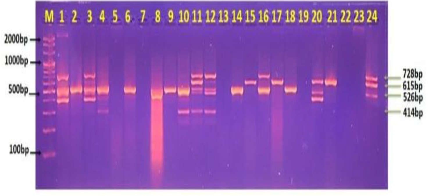
Figure 1: Agarose Gel Electrophoresis Showing Multiplex PCR Analysis of Small Subunit Ribosomal RNA Gene for the Detection of Entamoeba Histolytica, Giardia Intestinalis, Cryptosporidium Parvum, and Blastocystis Sp. from Human Stool Samples. Lane M Represents the DNA Marker (2000–100 Bp), While Lanes 1–24 Show Positive PCR Products for E. Histolytica (526 Bp), G. Intestinalis (615 Bp), C. Parvum (728 Bp), and Blastocystis Sp. (414 Bp), Respectively.
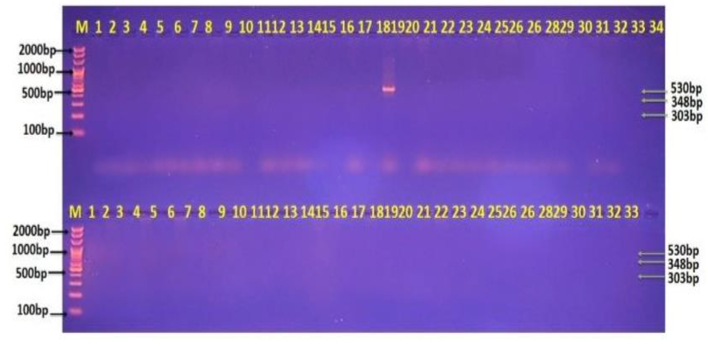
Figure 2: Agarose Gel Electrophoresis Showing Multiplex PCR Analysis Of The Small Subunit Ribosomal RNA Gene For The Detection Of Isospora Sp., Balantidium Coli, And Dientamoeba Fragilis From Human Stool Samples. Lane M Represents the DNA Marker Ladder (2000–100 Bp), While Lanes 1–34 and 1–33 Show Positive PCR Products for Isospora Sp. (530 Bp), Balantidium Coli (348 Bp), and Dientamoeba Fragilis (303 Bp), Respectively.
DISCUSSION
Percentage of Infection for parasites that cause Intestinal parasitic by PCR technique examination results were shown the highest infection with E. histolytca (60%) and followed by C. parvum (23%), G. lamblia (20%) and the lowest infection with Blastocystis hominis (17%), Isospora ( 7%). The current study agrees with Hraija et al. [11] in Wasit, Iraq and Abed et al. [12] in Salah al-Din and Alkhuzaey in Thi-Qar [13] also agree with Yakoob et al. [14] in Iraq and Ngosso and Namkinga [15] in Dar Es Salaam were detected 33.3% as E. histolytica by PCR higher than other parasites (Figure 1-2).
These disagree with Mathurin et al. in Cote d’Ivoire; they saw (36.93%) C. parvum is greater than another parasites and disagrees with, Ngosso and Namkinga in Dar Es Salaam they saw the highest parasites in total positive samples with G. lamblia (35.6%) and followed by E. histolytica, also disagree with study by Akram [16] in Thi-Qar was found the infection with G. lamblia higher than other parasites in total positive samples and followed by E. histolytica and differs with Parčina et al. in Germany they examined of 200 diarrheic stool samples by using RT-PCR and registered G. lamblia (69.0%) the highest infection. and multiplex PCR assay showed sensitivity of 90.91%, for G. intestinalis and Cryptosporidium spp., whereas multiplex PCR assay achieved sensitivity of 86.36% for the detection of E. histolytica by Bairami [17] in Iran. Disagree with Maas et al. [18] in The Netherlands shows the prevalence of intestinal protozoan infection according to patient’s age. The average age of the patients was 35 years, so they divided in to five groups for the purpose of study. The infection with the five parasites was more prominent in ≤15 years old with infection rates estimated as 65.0% for E. histolytica, (25.0%) for G. lamblia and (25.0%) C. parvum, (15.0%) Blastocystis hominis and (7.5%) Isospora. Subjects within 16-30 years age group have the lowest infection rates, 60.0%, 12.0%, 12.0%, 20.0% and 8.0% for E. histolytica, G. lamblia and C. parvum, Blastocystis hominis, Isospora, respectively. There was no association between the infection with the five parasites and age group agrees with Rahi and Majeed [19] in Wasit and disagreeing with Ten Hove et al. [20] in the Netherlands who found that Cryptosporidium infections were strongly associated with the age of the patients and my study also disagreeing with Mahmoud et al. [21] in Taif. We observed rate Blastocystis sp infestations in 11 (73.3%) from 82 samples in age group 1-40 years. And we see through study Masar Hadi [22], in Diyala the highest prevalence rate Blastocystis sp. In patients observed 27.7% in the age group 19-28 years, 22.7% in age group 8-18 years and finally the age groups 29-38 and 39-48 years old (18.1%).
CONCLUSION
Our study supports implementation of molecular tools into the parasitology diagnostic laboratory, even in under-resourced settings. Such tools are particularly important for detecting protozoa that are challenging to identify by microscopy. Molecular tools are also essential for distinguishing between morphologically identical species that are of differing clinical relevance. In addition, molecular and traditional techniques have overlapping, but complementary, roles in diagnosing protozoan infections resulting in clinical manifestations in patients. In conclusion, a multiplex PCR for the detection of intestinal protozoa is a technically feasible tool in a routine microbiological laboratory.
REFERENCE
Hamdy, D. A. et al. “Blastocystis spp. and Giardia intestinalis Co-infection Profile in Children Suffering from Acute Diarrhea.” Journal of Parasitic Diseases, vol. 44, 2020, pp. 88–98.
Mmbaga, B. T. and E. R. Houpt. “Cryptosporidium and Giardia Infections in Children: A Review.” Pediatric Clinics of North America, vol. 64, 2017, pp. 837–850.
Puebla, L. E. J. et al. “Diagnosis of Intestinal Protozoan Infections in Patients in Cuba by Microscopy and Molecular Methods: Advantages and Disadvantages.” Journal of Microbiological Methods, vol. 179, 2020, article 106102.
Lee, M. F. et al. “Successful Extraction and PCR Amplification of Giardia DNA from Formalin-Fixed Stool Samples.” Experimental Parasitology, vol. 198, 2019, pp. 26–30.
Garcia, L. S. et al. “Practical Guidance for Clinical Microbiology Laboratories: Laboratory Diagnosis of Parasites from the Gastrointestinal Tract.” Clinical Microbiology Reviews, vol. 31, no. 1, 2017, e00025-17. https://doi.org/10.1128/CMR.00025-17
Laude, A. et al. “Is Real-Time PCR-Based Diagnosis Similar in Performance to Routine Parasitological Examination for the Identification of Giardia intestinalis, Cryptosporidium parvum/Cryptosporidium hominis and Entamoeba histolytica from Stool Samples?” Clinical Microbiology and Infection, vol. 22, no. 2, 2016, pp. 190.e1–190.e8.
Paulos, S. et al. “Comparative Performance Evaluation of Four Commercial Multiplex Real-Time PCR Assays for the Detection of Diarrhoea-Causing Protozoa.” PLoS One, vol. 14, no. 4, 2019, article e0215068. https://d oi.or g/10.1371/journal.pone.0215068
Verweij, J. J. “Validation and Maintaining Laboratory Developed Molecular Tests Compliant with ISO 15189 for Diagnosis of Intestinal Parasitic Infections.” Expert Review of Molecular Diagnostics, vol. 22, no. 6, 2022, pp. 595–601.
Alkhanaq, M. N. and G. J. Thamer. “Prevalence of Cryptosporidium spp. among Patients with Diarrhea at Wasit Province, Iraq.” International Journal of Forensic Medicine and Toxicology, vol. 16, no. 1, 2022, pp. 771–780.
Al-Gharban, H. A. A. J. “Seroepidemiological Detection and Culture Utilization for Diagnosis of Carrier Horses and Donkeys with Strangles.” Journal of Education College Wasit University, vol. 1, no. 28, 2017, pp. 649–660.
Kareem Kadhim, D. et al. “Molecular-Genotyping Detection of Entamoeba histolytica in Diarrheic Patients.” Advances of Research in Immunology, vol. 78, no. 1, 2023, pp. 337–343.
Habeeb, F. F. et al. “Assessment of the Level of Interleukin-5 and Interleukin-17 in Patients Infected with Some Intestinal Parasites and Helicobacter pylori.” International Journal for Research in Applied Sciences and Biotechnology, vol. 8, no. 6, 2021, pp. 4–9.
Alkhuzaey, A. and B. Aboody. “Co-infection of Intestinal Parasites and Helicobacter pylori in Patients with Chronic Diarrhea.” International Journal of Biology, vol. 18, no. 2, 2019, pp. 18–27.
Yakoob, J. et al. “Association of Helicobacter pylori and Protozoal Parasites in Patients with Chronic Diarrhoea.” British Journal of Biomedical Science, vol. 75, no. 3, 2018, pp. 105–109.
Ngosso and Namkinga. “Identification of Pathogenic Intestinal Parasitic Protozoa Associated with Diarrhea among Under-Five Children in Dar Es Salaam, Tanzania.” International Invention Journal of Medicine and Medical Sciences, vol. 2, no. 4, 2015, pp. 49–55.
Akram, R. S. Molecular Detection and Prevalence of Cryptosporidium parvum, Entamoeba histolytica and Giardia lamblia among Patients with Diarrhea at Al-Rifea City. Thesis, College of Science, University of Thi-Qar, 2018.
Bairami, A. et al. “Synchronous Identification of Entamoeba histolytica, Giardia intestinalis and Cryptosporidium spp. in Stool Samples Using a Multiplex PCR Assay.” Iranian Journal of Parasitology, vol. 13, no. 1, 2018, pp. 24.
Maas, L. et al. “Detection of Intestinal Protozoa in Paediatric Patients with Gastrointestinal Symptoms by Multiplex Real-Time PCR.” Clinical Microbiology and Infection, vol. 20, no. 6, 2014, pp. 545–550.
Rahi, A. A. and L. Majeed. “Epidemiological Study of Intestinal Protozoa at Wasit Province.” European Journal of Parasitology, vol. 1, no. 3, 2019, pp. 26–28.
Ten Hove, R. et al. “Detection of Diarrhea-Causing Protozoa in General Practice Patients in the Netherlands by Multiplex Real-Time PCR.” Clinical Microbiology and Infection, vol. 13, no. 10, 2007, pp. 1001–1007.
Mahmoud, A. M. et al. “Molecular Identification of Blastocystis hominis Isolates in Patients with Autoimmune Diseases.” Applied Microbiology, vol. 3, no. 2, 2023, pp. 417–428.
Recommended Articles
Research Article
Evaluation of Mortality at One Year Follow-Up of Patients Having Early Tumors of Oropharynx Undergoing Trans-Oral Robotic Surgery in a Tertiary Care Hospital
Tapender Singh,
Vinay Gupta
Published: 27/01/2025
Download PDF
Research Article
Molecular Study to Detect Some Virulence Factors in Methicillin – Resistance Staphlococcus Aureus Isolated from Eye Infections in Wasit Province
Nofel Hossin Amhi Alshammari,
Haider Kareem Jassem
Published: 15/12/2023
Download PDF
Research Article
Occurrence of Multi-Drug Resistance of E. coli Isolated from Urinary Tract Infection in Najaf City
Alaa Hamza Jaber Al-Jelehawy,
...
Haneen Ali Kadham
Published: 05/10/2025
Download PDF
Research Article
Estimation Of Hba1c in Non-Diabetic Patients with Myocardial Infarction
Tamarah Riyadh Sabeeh
Published: 19/07/2024
Download PDF
PO Box 101, Nakuru
Kenya.
Email: office@iarconsortium.org
Editorial Office:
J.L Bhavan, Near Radison Blu Hotel,
Jalukbari, Guwahati-India
Ahmed, B. A. A. & Hraija, B. A. (2023). Molecular Diagnosis of Intestinal Protozoa for Patients in Wasit Province, Iraq. Scientific Research Journal of Medical Sciences, 3(1), None-None.
MLA
Ahmed, Bassam A. A. and Baraa A. Hraija. "Molecular Diagnosis of Intestinal Protozoa for Patients in Wasit Province, Iraq." Scientific Research Journal of Medical Sciences 3.1 (2023): None-None.
Chicago
Ahmed, Bassam A. A. and Baraa A. Hraija. "Molecular Diagnosis of Intestinal Protozoa for Patients in Wasit Province, Iraq." Scientific Research Journal of Medical Sciences 3, no. 1 (2023): None-None.
Harvard
Ahmed, B. A. A. and Hraija, B. A. (2023) 'Molecular Diagnosis of Intestinal Protozoa for Patients in Wasit Province, Iraq' Scientific Research Journal of Medical Sciences 3(1), pp. None-None.
Vancouver
Ahmed BAA, Hraija BA. Molecular Diagnosis of Intestinal Protozoa for Patients in Wasit Province, Iraq. Scientific Research Journal of Medical Sciences. 2023 Jan;3(1):None-None.


 Download PDF
 Download XML