Background: Breast cancer is the most common malignant tumor and the leading cause of cancer deaths in women worldwide, including Iraq. Early detection of breast cancer has been recommended by the WHO as one of the major tools to control the disease. The Biofield Diagnostic breast cancer System (BDS) has been recently introduced in Iraq. It is a non-invasive device that measures the level of DC voltage skin surface electropotentials associated with suspicious breast lumps. The main objective of its use is to distinguish between benign and malignant breast lesions detected clinically or radiologically. Aim of Study: To determine the utility of BDS in the diagnosis of breast cancer and to correlate the findings of BDS with: The Triple Assessment Test (which comprises clinical parameters, ultrasonography and mammography and FNAC) of palpable breast lumps, Clinicopathological parameters including staging, grading and type of breast carcinoma and the Results of Ag NOR cytochemical marker. Patients and Methods: This is a prospective study of 50 cases collected from the Main Training Center for Early Detection of Breast Cancer in the Medical City Teaching Hospital. For each patient a full questionnaire was prepared including all demographic and clinical data. All patients were subjected to the Triple Assessment Test and BDS test. Results: In this study, the BDS findings displayed "Probably Benign" in three cases(6%); " Probably Malignant " in 36 cases(72%) and "Malignant" in eleven cases(22%). The clinical level of suspicion was classified as III in 44 cases for the 50 cases studied (88%) and level II in six cases (12%). The ultrasound level of suspicion was recorded as level III in 38 cases (76%) and level II in eleven cases for the 50 cases studied (22%). The mean age for the "Probably Benign" category was (31 years); for the "Probably Malignant" category was (45.5 years) and for the "Malignant" category was (55.5 years). The fine needle aspiration cytology results showed ductal carcinoma in 49 cases out of 50 cases studied (98%) and lobular carcinoma in only one case. The mean Ag-NOR count was (7.86) for "Probably Benign" category, (8.23) for "Probably Malignant" category and (8.5) for "Malignant" category. Histopathological diagnosis was available for only 24 cases out of the 50 cases of this study (48% of total cases examined). Those findings are comparable to those displayed in another previous report which was carried out within the Main Training Centre for Early Detection of Breast Tumors in the Medical City Teaching Hospital [1]. The latter was the only published report from Iraq, which dealt with BDS. Our results were compatible as well with those recorded in other studies from USA [2]. The sensitivity of the BDS test in our study was superior to the sensitivity of clinical breast examination (94% versus 88%). Breast cancer is the most common type of malignancy among Iraqi women accounting for about one third of the registered female cancers ( according to the results of the latest Iraqi Cancer registry). Traditional approaches to the diagnosis of breast cancer include clinical examination, imaging techniques, fine needle aspiration cytology and histopathological examination of the surgical specimens. The detection of a breast mass in an apparently healthy woman before it is palpable is a technique that saves lives and saves breasts. All women are candidates for screening since all women are at risk for breast cancer development. Conclusions: The BDS could provide additional discriminatory information regarding the malignant nature of the disease process in suspicious breast lesions and could serve as a marker of cellular proliferation. We conclude that The BDS test should be an adjunct tool to CBE and not to compete with the other diagnostic utilities of the breast cancer and BDS should reduce the number of unwanted biopsies in cases where CBE, FNAC and BDS show benign changes.
Keywords
Biofield Breast Cancer Diagnostic System
Mammary Carcinoma
Iraq
INTRODUCTION
Breast cancer is the most common malignant tumor and the leading cause of cancer deaths in women worldwide, including Iraq.
Early detection of breast cancer has been recommended by the WHO as one of the major tools to control the disease. The Biofield Diagnostic breast cancer System (BDS) has been recently introduced in Iraq. It is a non-invasive device that measures the level of DC voltage skin surface electropotentials associated with suspicious breast lumps. The main objective of its use is to distinguish between benign and malignant breast lesions detected clinically or radiologically.
The Ag-NOR represents a cytochemical marker of both DNA and the level of its transcription. It is used as a useful tool for the study of variations in the nucleolar activity. Qualitative and quantitative variations in Ag-NOR distribution and number has been studied in breast carcinoma and positively correlated with other markers of cellular proliferation including Ki67 immunoreactivity, hormone receptor contents etc.
Aim of Study
To determine the utility of BDS in the diagnosis of breast cancer
To correlate the findings of BDS with
The Triple Assessment Test (which comprises clinical parameters, ultrasonography and mammography and FNAC) of palpable breast lumps
Clinicopathological parameters including staging, grading and type of breast carcinoma
Results of Ag NOR cytochemical marker
MATERIALS AND METHODS
This is a prospective study of 50 cases collected from the Main Training Center for Early Detection of Breast Cancer in the Medical City Teaching Hospital. For each patient a full questionnaire was prepared including all demographic and clinical data. All patients were subjected to the Triple Assessment Test and BDS test [3-5].
After excision of the breast mass , histopathological examination of the tissue was carried out; recording the type, grading and TNM staging of mammary carcinoma.
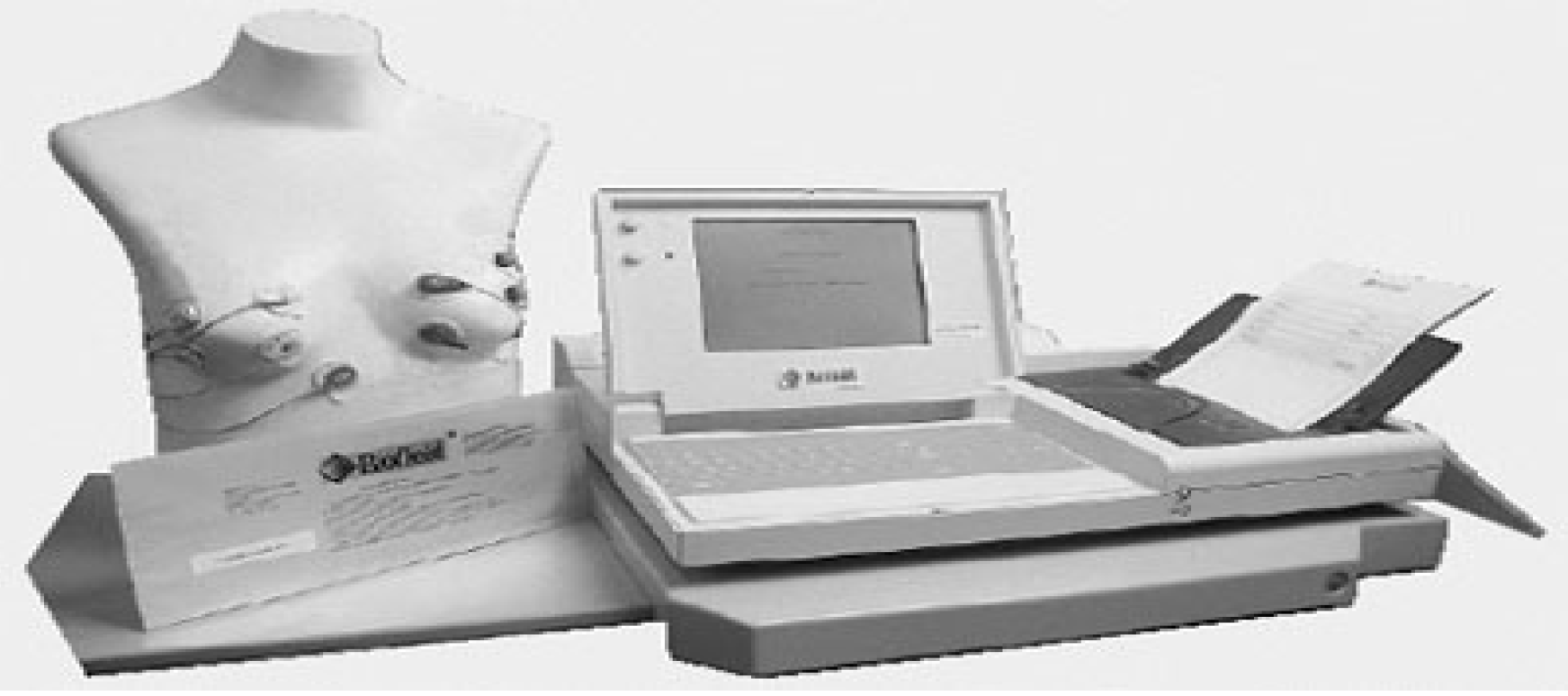
Figure 1: The BDS Equipment
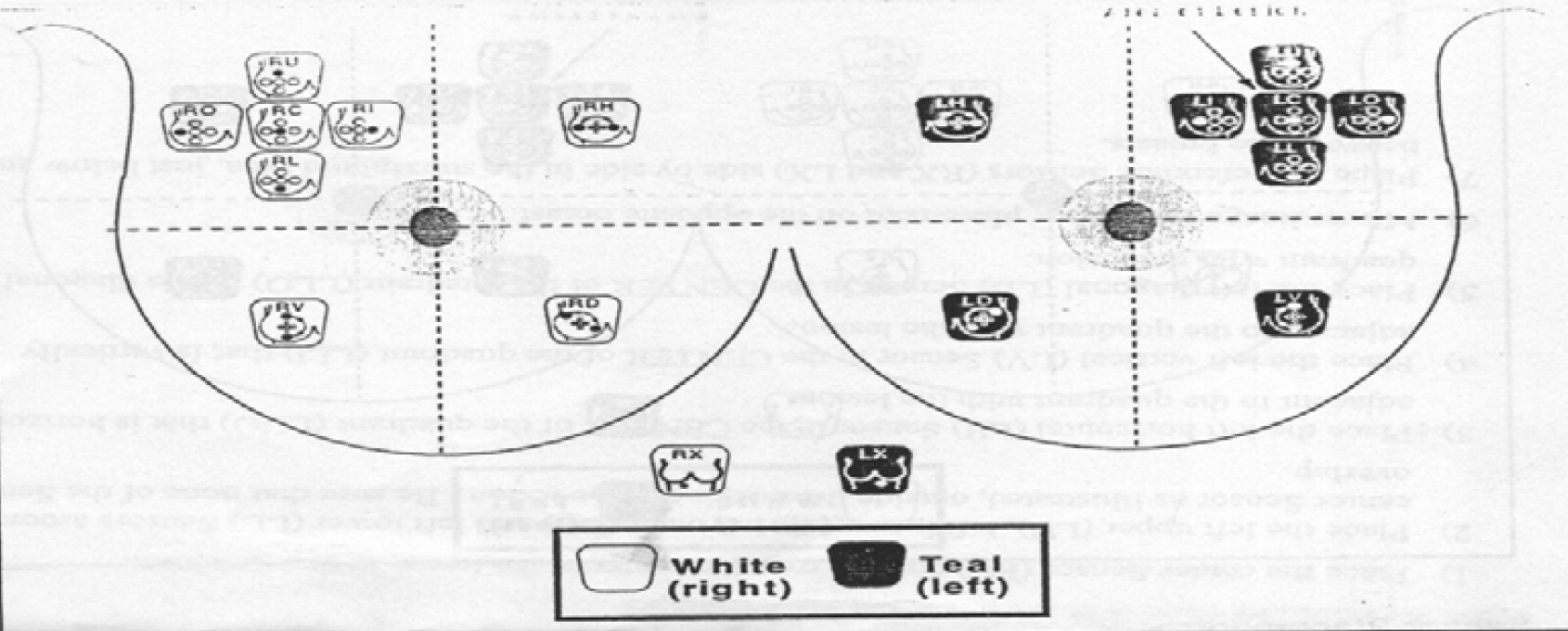
Figure 2: Sensor Placement When the Lesion Is Located In the Upper Outer Quadrant of the Left Breast
Table 1: The Adjusted Level of Suspicion
Level of Suspicion III :Probably Malignant
Level of Suspicion II :Probably Benign
Level of Suspicion I :Normal/ANDI
BDS Index
Probably Benign
Probably Benign
Probably Benign
-5.0 to -1.7
Probably Malignant.
Probably Benign
Probably Benign
-1.6 to +0.2
Malignant.
Probably Malignant.
Probably Benign
+0.3 to +1.5
Malignant.
Probably Malignant.
Probably Malignant.
+1.6 to +5.0
RESULTS
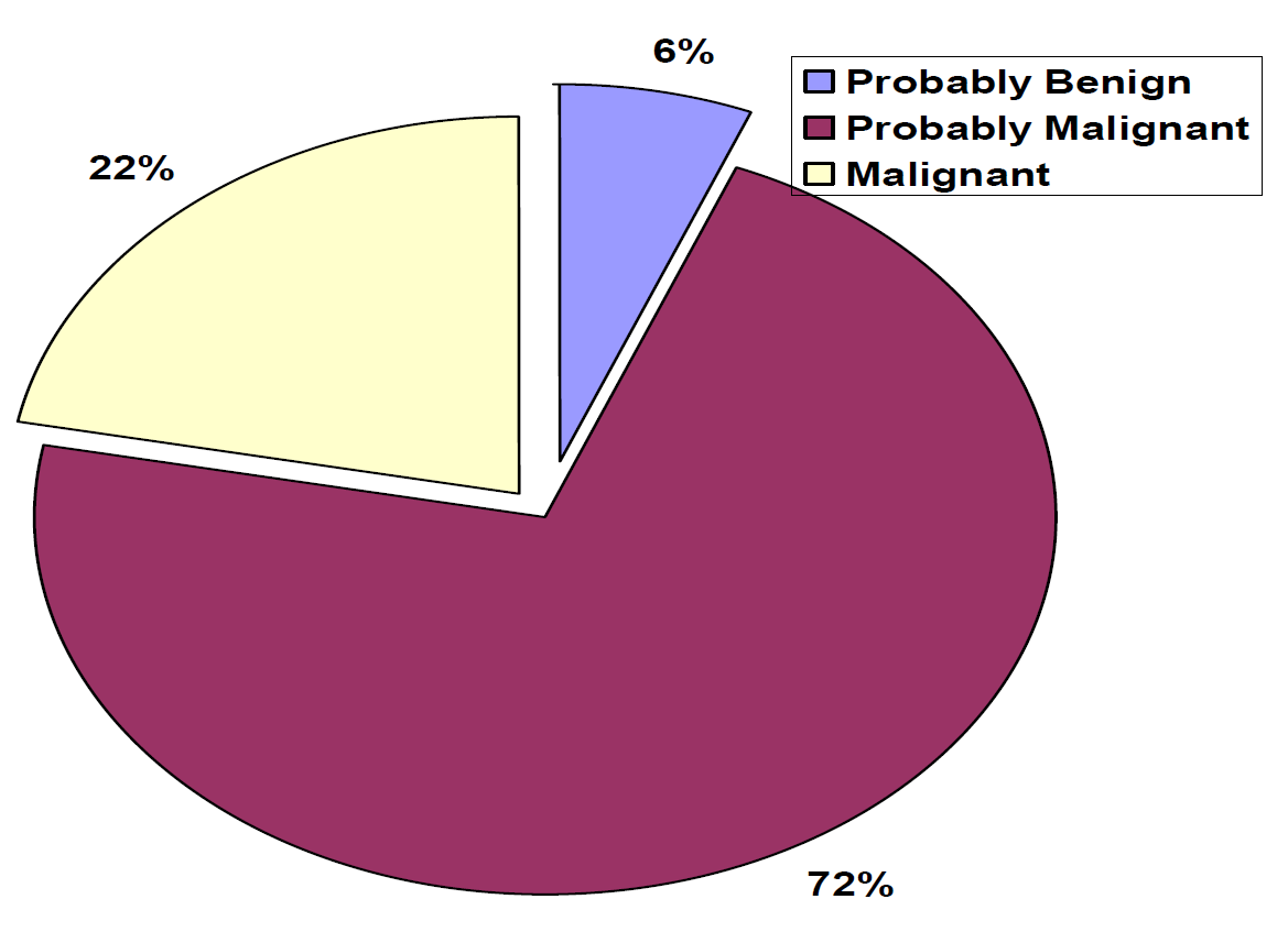
In this study, the BDS findings displayed "Probably Benign" in three cases(6%); " Probably Malignant " in 36 cases(72%) and "Malignant" in eleven cases(22%). The clinical level of suspicion was classified as III in 44 cases for the 50 cases studied (88%) and level II in six cases (12%). The ultrasound level of suspicion was recorded as level III in 38 cases (76%) and level II in eleven cases for the 50 cases studied (22%). The mean age for the "Probably Benign" category was (31 years); for the "Probably Malignant" category was (45.5 years) and for the "Malignant" category was (55.5 years).
The fine needle aspiration cytology results showed ductal carcinoma in 49 cases out of 50 cases studied (98%) and lobular carcinoma in only one case. The mean
Ag-NOR count was (7.86) for "Probably Benign" category, (8.23) for "Probably Malignant" category and (8.5) for "Malignant" category. Histopathological diagnosis was available for only 24 cases out of the 50 cases of this study (48% of total cases examined).
McKee cytolopathological grading showed concordance with the histopathological grading system in 91.7% of ductal carcinomas.
The current study demonstrated as well a significant association between BDS findings, Ag-NOR counts, pathological grading and staging of breast cancer.
The sensitivity of BDS was superior to Physical breast examination (94 % for the former versus 89% for the latter).
Table 2: Clinical and Imaging Levels of Suspicions versus BDS Findings, Ag-NOR Count Results and Other Clinico-Pathological Parameters in 50 Cases of Mammary Carcinomas
Case No.
Age
(years)
Clinical level
U/S level
BDS Index
BDS Result
FNA Result*
Ag-NOR count
Cytological grade
Histologic grade
Stage
1
56
III
III
-0.4
PM
DC
7.6
G II
G III
II
2
32
III
III
-0.7
PM
DC
8.1
G II
G II
II
3
67
III
III
+0.5
M
DC
8.6
G II
-
-
4
60
II
II
+2.0
PM
DC
8.5
G II
-
-
5
39
III
III
+0.3
M
DC
8.7
G III
G III
IV
6
47
III
III
+0.2
PM
DC
7.3
G II
G II
II
7
67
III
III
+0.8
M
DC
8.4
G III
G III
III
8
45
II
III
+2.9
PM
DC
8.4
G II
-
-
9
48
III
II
-0.5
PM
DC
8.8
G II
G II
II
10
38
III
III
-1.6
PM
DC
8.3
G III
G III
IV
11
57
III
III
-0.1
PM
DC
8.6
G III
G III
III
12
73
II
III
+1.8
PM
DC
8.5
G II
-
-
13
35
III
II
-1.3
PM
DC
8.6
G II
-
-
14
47
III
II
-0.0
PM
DC
6.1
G II
G II
III
15
59
III
II
-0.1
PM
DC
8.8
G III
-
-
16
43
III
III
-1.4
PM
DC
7.9
G II
G II
II
17
36
II
I
-1.0
PB
DC
7.0
G II
G I
III
18
70
III
III
-1.0
PM
DC
8.6
G III
G III
IV
19
43
III
III
-1.6
PM
DC
8.8
G III
-
-
20
52
III
III
-0.4
PM
LC
8.3
-
-
--
21
36
III
II
-1.2
PM
DC
7.9
G III
G III
IV
22
65
III
III
-0.1
PM
DC
7.9
G II
G II
II
23
33
II
II
-1.4
PB
DC
8.3
G II
G II
III
24
44
III
III
-1.0
PM
DC
8.1
G III
-
-
25
55
III
III
+0.9
PM
DC
8.9
G III
G III
III
26
50
III
III
-0.3
PM
DC
8.2
G II
G II
III
27
30
II
II
-1.8
PB
DC
8.3
G II
-
-
28
56
III
III
-0.0
PM
DC
8.3
G II
G II
III
29
55
III
III
-1.1
PM
DC
8.7
G II
G II
III
30
48
III
III
-0.4
PM
DC
8.1
G II
-
-
31
37
III
III
-1.6
PM
DC
7.3
G II
G II
III
32
42
III
III
+0.2
M
DC
8.5
G III
G III
IV
33
33
III
III
-1.1
PM
DC
8.6
G III
G III
III
34
33
III
III
-1.1
PM
DC
8.3
G II
-
-
35
57
III
III
-1.0
PM
DC
8.2
G III
-
-
36
62
III
III
+0.7
M
DC
8.3
G III
G III
III
37
57
III
III
+0.5
M
DC
7.9
G II
G II
II
38
46
III
III
-0.2
PM
DC
8.5
G III
-
-
39
32
III
II
+0.0
PM
DC
7.8
G II
-
-
40
40
III
III
+0.5
M
DC
8.8
G II
-
-
41
53
III
III
+0.8
M
DC
7.8
G III
-
-
42
45
III
II
-0.6
PM
DC
8.2
G II
-
-
43
43
III
III
-1.6
PM
DC
8.9
G III
-
-
44
62
III
III
+0.5
M
DC
8.9
G III
-
-
45
49
III
III
-0.2
PM
DC
8.8
G II
-
-
46
47
III
II
-0.2
PM
DC
7.8
G II
-
-
47
42
III
III
-0.6
PM
DC
8.9
G II
-
-
48
70
III
III
+2.7
M
DC
8.9
G II
-
-
49
39
III
III
+0.5
M
DC
8.7
G III
-
-
50
45
III
III
+0.1
PM
DC
8.7
G II
-
-
Table 3: Descriptive Statistics of the Different BDS Results
BDS Results
No. of cases
%
Mean BDS Index
S.D
Min.
Max.
Range
Probably Benign
3
6
-1.4
0.4
-1.8
-1.0
0.8
Probably Malignant
36
72
-0.394
0.989
-1.6
2.9
4.5
Malignant
11
22
+0.791
0.657
0.3
2.7
2.4
Total
50
100
-
-
-
-
-
Figure 3:Percentages of Ddifferent BDS Results
Stastical Analysis
In this study the BDS results were obtained from the application of the technique to 50 patients with suspicious breast lumps. The findings were Probably Benign findings in three cases (6%). Probably Malignant in 36 cases (72%) and Malignant in eleven cases (22%). Those findings are comparable to those displayed in another previous report which was carried out within the Main Training Centre for Early Detection of Breast Tumors in the Medical City Teaching Hospital [1]. The latter was the only published report from Iraq which dealt with BDS. Our results were compatible as well with those recorded in other studies from USA [2].
The Sensitivity of the Bds Test in Our Study Was Superior to the Sensitivity of Clinical Breast Examination (94% Versus 88%)
Regarding the histological grade, it has been reported that the 10-year survival rate for patients harboring Grade I tumors is around 80%, dropping to 45% in Grade III tumors.
In our study there was significant relationship between each of the different BDS category results and the histological and cytological grades in mammary carcinoma cases; indicating that BDS could be used as a marker of cell aggressiveness in breast lesions.
In our study there was significant relationship between each of the different BDS category results and the stages of mammary carcinoma in breast cancer patients.
The results of Ag-NOR staining of cytological smears from suspicious breast masses in our study revealed a mean Ag-NOR count of (8.2+/- 0.3).
CONCLUSION
The BDS test should be an adjunct tool to CBE and not to compete with the other diagnostic utilities of the breast cancer
BDS should reduce the number of unwanted biopsies in cases where CBE, FNAC and BDS show benign changes
This diagnostic tool should be supplied to underserved health centers and women in remote areas where other diagnostic tools such as mammography and/or ultrasound are not available. In these populations, there should be nation wide education for both health staff and women on its benefits
Recommendations
The BDS test should be an adjunct tool to CBE and not to compete with the other diagnostic utilities of the breast cancer
BDS should reduce the number of unwanted biopsies in cases where CBE, FNAC and BDS show benign changes
This diagnostic tool should be supplied to underserved health centers and women in remote areas where other diagnostic tools such as mammography and/or ultrasound are not available. In these populations, there should be nation wide education for both health staff and women on its benefits
REFERENCE
Qadir, A.H. The role of biofield diagnostic system “BDS” in diagnosing breast tumors. Thesis, Council of Arab Board for Surgeons, supervised by N.A.S. Al-Alwan, 2005.
Study of the use of skin surface electropotentials as measured using the biofield breast cancer diagnostic system in the evaluation of suspicious breast lesions. Biofield Clinical Report, TR 005, Alpharetta, GA, USA, 2004.
Rosai, J. and L.V. Ackerman. Rosai and Ackerman’s surgical pathology. 9th ed., vol. 2, Elsevier, 2004, pp. 1763–1839. Chapter 20, Breast.
Koss, L.G. Diagnostic cytology and its histopathologic bases. 4th ed., vol. 2, Lippincott, 1992, pp. 1293–1313.
Bailey, H. Hamilton Bailey’s physical signs: demonstrations of physical signs in clinical surgery. 18th ed., edited by J.S.P. Lumley, Churchill Livingstone, 2001, pp. 230.
Recommended Articles
Research Article
Evaluation of Mortality at One Year Follow-Up of Patients Having Early Tumors of Oropharynx Undergoing Trans-Oral Robotic Surgery in a Tertiary Care Hospital
Tapender Singh,
Vinay Gupta
Published: 27/01/2025
Download PDF
Research Article
Molecular Study to Detect Some Virulence Factors in Methicillin – Resistance Staphlococcus Aureus Isolated from Eye Infections in Wasit Province
Nofel Hossin Amhi Alshammari,
Haider Kareem Jassem
Published: 15/12/2023
Download PDF
Research Article
Occurrence of Multi-Drug Resistance of E. coli Isolated from Urinary Tract Infection in Najaf City
Alaa Hamza Jaber Al-Jelehawy,
...
Haneen Ali Kadham
Published: 05/10/2025
Download PDF
Research Article
Estimation Of Hba1c in Non-Diabetic Patients with Myocardial Infarction
Tamarah Riyadh Sabeeh
Published: 19/07/2024
Download PDF
PO Box 101, Nakuru
Kenya.
Email: office@iarconsortium.org
Editorial Office:
J.L Bhavan, Near Radison Blu Hotel,
Jalukbari, Guwahati-India
Mohammad, S. H. & Salman, R. A. (2022). Correlation of the Findings of Biofield Breast Cancer Diagnostic System (BDS) with Clinicopathological Parameters of Mammary Carcinoma in Iraq. Scientific Research Journal of Medical Sciences, 2(2), 1-4.
MLA
Mohammad, Salam H. and Ruaa A. Salman. "Correlation of the Findings of Biofield Breast Cancer Diagnostic System (BDS) with Clinicopathological Parameters of Mammary Carcinoma in Iraq." Scientific Research Journal of Medical Sciences 2.2 (2022): 1-4.
Chicago
Mohammad, Salam H. and Ruaa A. Salman. "Correlation of the Findings of Biofield Breast Cancer Diagnostic System (BDS) with Clinicopathological Parameters of Mammary Carcinoma in Iraq." Scientific Research Journal of Medical Sciences 2, no. 2 (2022): 1-4.
Harvard
Mohammad, S. H. and Salman, R. A. (2022) 'Correlation of the Findings of Biofield Breast Cancer Diagnostic System (BDS) with Clinicopathological Parameters of Mammary Carcinoma in Iraq' Scientific Research Journal of Medical Sciences 2(2), pp. 1-4.
Vancouver
Mohammad SH, Salman RA. Correlation of the Findings of Biofield Breast Cancer Diagnostic System (BDS) with Clinicopathological Parameters of Mammary Carcinoma in Iraq. Scientific Research Journal of Medical Sciences. 2022 Jul;2(2):1-4.



 Download PDF
 Download XML