Contents
Download PDF
pdf Download XML
294 Views
67 Downloads
Share this article
Case Report | Volume 4 Issue 1 (Jan-June, 2024) | Pages 1 - 8
The Utility of Maternal Serum Amyloid a in the Prediction of Preterm Birth in Symptomatic Women
 ,
1
M.B.Ch.B Kirkuk health directorate, Iraq, 36001
2
M.B.Ch.B. D.G.O- C.A.B.O.G, College of Medicine, Kirkuk University, Iraq, 36001
Under a Creative Commons license
Open Access
Received
March 3, 2024
Revised
April 9, 2024
Accepted
May 19, 2024
Published
June 30, 2024
Abstract

Background: Preterm birth affects about 15 million births around the world and is still a big problem in obstetrics. Maternal serum amyloid A associated with inflammatory response and has an essential role in acute phase reaction. Aim: To determine the maternal serum amyloid A level in women with threatened preterm labor and its rule in prediction of preterm birth.  Patients and methods: A prospective case-control study, conducted at Azadi Teaching Hospital in Kirkuk, Iraq, spanned for nine months from February to October 2023.It included 90 pregnant women aged 17-43 years (with singleton pregnancies between 24-36+6days weeks and intact membranes) presented with symptoms & sign of threatened preterm labor. Participants were categorized according to their fate into the preterm birth group (n:45) consisting of pregnant women who had preterm birth and the term control group (n:45) consisting of preterm pregnant women who continued their pregnancy and delivered at term. Women with multiple pregnancies, uterine or cervical abnormalities and signs of systemic infection were excluded from the study. serum amyloid A levels were measured using ELISA kit. All Pregnant women were admitted for observation and followed up until birth. Results: Mothers of preterm births exhibited a significantly younger age, higher rate of primparity, history of previous preterm delivery and miscarriage compared to the term group. Preterm births have significantly lower Apgar scores, birth weight and higher NICU admission. Serum amyloid A levels were elevated in preterm compared to term group, with cut-off value at 0.69 ng/ml being (77.8%) sensitive and (71.1%) specific for preterm labor prediction. women with preterm birth demonstrated a negative correlation between serum amyloid A levels with both gestational age at delivery and birth weight. Conclusion: Women with preterm birth had elevated serum amyloid a levels which could be a promising biomarker for predicting spontaneous preterm birth.


Keywords
INTRODUCTION

Preterm birth is a substantial global health issue with significant consequences to the newborn, family and society. It affects nearly 15 million births worldwide, is the leading cause of death in children younger than 5 years and remains a prominent issue in obstetrics [1]. Although the burden of this condition is clear, new interventions tested or in use have shown no impact in reducing the rate of preterm birth [2-3]. Moreover, the etiological factors that lead to various pathways of preterm labor are still largely unknown [4-6]. Therefore, it is crucial to develop a better understanding of the underlying pathways to allow earlier prediction and to develop better, more targeted and more effective interventions [8-11]. There is significant variation in the incidence of preterm birth worldwide. The rates of preterm birth in 184 countries in 2010 ranged from 5% in several Northern European countries to 18% in Malawi [12-13]. Rates are highest in low income and lower-middle income countries (11.8% and 11.3% on average, respectively), whereas rates are lower in upper-middle and high-income countries (9.4% and 9.3%, respectively). More than 60% of all preterm births worldwide occur in low-resource, high-fertility fertility countries in sub-Saharan Africa and south Asia [14-16]. Amongst high-resource countries, the United States has the highest incidence of preterm birth and accounts for 42% of all preterm births in high-resource settings (0.5 million per year in the United States out of 1.2 million total per year in high-income countries) [17-19]. Serum amyloid A1 (SAA1) was originally identified as a serum component recognized by antisera against amyloid fibrils known as amyloid A (AA). Investigators have been trying to disclose the role of SAA in pregnancy for some time. Evidence gathered so far indicates that SAA in maternal circulation may function as a non-specific Damage-Associated Molecular Pattern (DAMP) molecule in response to tissue inflammation and damage incurred in parturition and gestational disorders [20-21]. However, SAA synthesized locally in gestational tissues may function as an early host-derived DAMP molecule exerting more specific effects in placentation, membrane rupture and parturition, thereby helping the embryo implant into the endometrium and aiding the fetus to escape from the endangered in-utero environment [22-24]. Stimulation of hepatic production of acute-phase proteins is by proinflammatory cytokines. In PTB, infection or inflammation results in the release of proinflammatory cytokines that stimulate matrix metalloproteinases (MMPs) and prostaglandin synthesis resulting in uterine contractions. The regulation of mSAA gene expression occurs at the level of transcription [25-26]. Although there are various pathways and transcription factors that can activate mSAA expression under different conditions, mSAA gene expression in inflammation is induced by various proinflammatory cytokines [27-28]. Maternal serum amyloid A (mSAA) is regarded as the most prominent representative amongst acute-phase proteins, it can reach plasma levels 500-1000 fold greater than in the non-inflammatory state, thus representing an ideal marker for clinical use [29-30]. In the view of the role of SAA in the inflammatory states of normal and abnormal pregnancies we decided to evaluate SAA level in patients with threatened preterm labor [31-32]. To determine the maternal serum amyloid A level in women with threatened preterm labor and its rule in prediction of preterm birth.

MATERIALS AND METHODS

This study is a prospective case control study that was carried out in the department of obstetrics and gynecology at Azadi Teaching Hospital in Kirkuk-Iraq. The duration of study was nine months throughout the period from 1st February till the end of October 2023. A convenient sample of 90 pregnant women, between 17- 43 years of age with singleton, viable pregnancy at gestational age between (24 – 36+6days) weeks of pregnancy, with symptoms and sign of threatened preterm labour and intact membrane were consecutively recruited in the study. The gestational age was calculated from last menstrual period and confirmed by 1st trimester ultrasound dating.


The pregnant women with threatened preterm labour included in the study were alienated and divided into two groups according to their clinical fate: 


  • The preterm birth group (n:45) pregnant patients who were admitted and had preterm birth

  • The term control group (n:45) pregnant women who continued their pregnancy and delivered at term


The patients were included in study based on the American College of Obstetricians and Gynecologists definition of threatened preterm labor as a regular uterine contraction (at least 1 contraction in 10 minutes) accompanied by progressive cervical changes (dilation, effacement, or both) or as regular contractions and cervical dilation of at least 2 cm at initial presentation. In addition to other symptoms of preterm labor like pelvic pressure, menstrual-like cramps and lower back pain (27,28). 


Women were excluded from the study based on several criteria, ensuring a specific focus on particular cases. These criteria included multiple pregnancies, such as twins or triplets, or conditions like polyhydramnios. Women with known malformations or structural abnormalities of the uterus, those who had a cervical cerclage in the current pregnancy or a history of previous cervical surgery and those with ruptured amniotic membranes were also excluded. Additional exclusion criteria encompassed antepartum hemorrhage, signs and symptoms of systemic infection or chorioamnionitis and various medical diseases including gestational hypertension, preeclampsia, diabetes, asthma, atherosclerosis, rheumatoid arthritis and thyroid dysfunction. Furthermore, women with a history of taking anti-inflammatory drugs or steroids, those with suspected or confirmed fetal growth restrictions, fetal anomalies, intrauterine fetal death and known malignancies such as lung, renal, ovarian, endometrial cancers, or melanoma, were not included in the study. To collect data, a comprehensive questionnaire was administered to all participants. This questionnaire was developed by the researchers after a thorough review of existing literature. It included questions to gather demographic data such as age, occupation and smoking status. Additionally, it covered presenting symptoms, obstetrical history including last menstrual period (LMP), gestational age, gravidity, parity, history of miscarriage and any previous preterm labor. The questionnaire also sought details on past medical history of chronic diseases, history of pelvic and abdominal surgeries and drug history, particularly focusing on the use of progesterone, steroids and anti-inflammatory drugs.


All participants underwent a complete physical examination, which included a general examination with an assessment of body mass index (BMI) and vital signs. Pregnant women presenting with threatened preterm labor were admitted for observation and managed accordingly. Those in the control group whose symptoms subsided were assessed and discharged after 24 hours, with instructions to return if symptoms recurred. Follow-up was conducted via cell phone contact until delivery to confirm term pregnancy. Women who continued to exhibit symptoms were observed until delivery. For both groups, pregnancy outcomes were documented from delivery notes, including mode of delivery and neonatal outcomes such as gestational age at delivery, birth weight, APGAR scores at one and five minutes and the need for admission to the neonatal intensive care unit (NICU). Low birth weight was defined as a birth weight of 2500 grams or less and a low APGAR score was defined as a score of less than seven at five minutes.


Determination of human Serum Amyloid A 

Five ml of venous blood was collected from each patient and control persons by using sterile disposable syringe and transferred to sterile gel tubes, which was then left at room temperature for 30 minutes in order to initiate the clotting process ,the sample was then centrifuged to separate the serum at 3,000xg for 15 minutes and the obtained serum were aspirate using mechanical micropipette and transferred into eppendorf tubes and stored at -20ºC for determination of The levels of serum amyloid A (SAA) was measured in these samples. The content of SAA was determined using an enzyme-linked immunosorbent assay (ELISA) kit (Sunlong Biotech CO., Ltd, China) (37,38). 


Statistical Analysis 

The data of women were analyzed by application of Microsoft Excel program and Statistical Package for Social Sciences (SPSS) version 23. Outcomes of analysis were arranged in scale variables (means & standard deviation) and in categorical variables. Chi square test was used for categorical variables. Independent sample t-test was used to compare between two means. Pearson Correlation has been used to find the correlation between 2 continuous variables and receiver operating characteristic (ROC) curve was used to evaluate the cutoff values of serum amyloid A level as predictor of preterm labor. P value of 0.05 or less was regarded as significant.


Ethical Consideration

Verbal informed consent was obtained from all participants, after discussing with them the nature of the study. Names were removed and replaced by identification codes, data used exclusively for the research purposes. All investigations offered to patients freely. Administrative Approvals were Granted from the Following 


  • The Council of Iraqi Board of Medical Specializations

  • Approval and agreement of the Department of Obstetrics and Gynecology at Azadi Teaching Hospital

RESULTS

In Table 1, the comparison of demographic characteristics between preterm and term control groups reveals distinct patterns. Mothers of preterm births are notably younger, with a mean age of 30.81±4.1 years, compared to a higher mean age of 38.19±5.2 in term births (p = 0.001). Conversely, BMI showed non-significant differences between preterm and term control groups (32.7±1.4 and 32.9±1.3 kg/m²) respectively. Similarly, the prevalence of smoking did not show a significant difference, with 11.11% of the preterm birth group and 8.89% of the term control group reporting smoking habits.


In Table 2, the study showed non-significant differences between preterm and term control groups regarding gestational age and gravidity. A significantly higher rate of primiparity was observed in the preterm group 15(33.33%) compared to the term control group 7(15.56%), (P: 0.041). A higher percentage of mothers in the preterm birth group had a history of previous miscarriage of one or two and more 14(31.11%) and 2(4.44%) Vs. 4 (8.89%) and 1(2.22%) in the term group respectively. A significantly higher rate of the preterm birth group (40%) had a history of previous preterm delivery compared to only 6.67% in the term control group, (P: 0.001). In addition the study showed higher percentage of women in preterm births group delivered by cesarean section (26.7%) in comparison to the term control group (17.8%) however the difference between both groups in relation to delivery mode was non-significant (P: 0.31).


The study showed that the mean gestational age at delivery was significantly lower in the preterm group in comparison to the term control group (33.44±3.87 and 39.11± 4.95 week) respectively. The mean Birth weight was significantly lower in the preterm group (2.06±0.11 kg) compared to the term control group (3.49±0.13 kg), p-value:0.001 with a significant proportion of preterm group (88.89%) exhibiting low birth weight, in comparison with the term control group (31.11%) (p-value: 0.001). In the current study, the Apgar scores were significantly lower in preterm group at both 1 minute and 5 minutes (6.45±1.05, 7.13±0.63) in comparison to term control group (8.58±1.38 and 9.27±0.79) at both 1 minute and 5 minutes respectively, with a significantly higher percentage of preterm neonates requiring NICU admission(84.44%) compared to just 33.33% in the term Control group (P:0.001).


Table 1: Demographic Characteristics of Preterm and Term Groups

Variable

Preterm Birth 

(n: 45)

Term control group 

(n:45)

p-value
Age, yearsMean±SD30.81±4.138.19±5.20.001*
Range 17-3620-42
BMI, kg/m²Mean±SD32.7±1.432.9±1.30.71 *
Range 22.4-36.523.5-35.4
SmokingYes 5(11.11%)4(8.89%)0.72 ** 
No40(88.89%)41(91.11%)

* Independent sample t-test ** chi-square test


Table 2: Obstetric Characteristics of Preterm and Term Control Groups 

Obstetric characteristicsPreterm Birth (n: 45)Term control group (n:45)p-value
Gestational ageMean±SD30.5±3.4130.2±3.550.88*
Range24-<3724- <37
Gravidity(mean, range)2 (1 – 5)3 (1 – 7)0.81**
ParityPrimiparity15(33.33%)7(15.56%)0.041**
Multiparity30(66.67%)38(84.44%)
History of miscarriageNon29(64.44%)40(88.89%)0.022**
one abortion14(31.11%)4(8.89%)
Two and more2(4.44%)1(2.22%)
History of previous preterm deliveryYes18(40%)3(6.67%)0.001**
No27(60%)42(93.33%)
Delivery modesCesarean section12(26.7%)8(17.8%)0.31**
Vaginal delivery33(73.3%)37(82.2%)

* Independent sample t-test ** chi-square test


Table 3: Comparison of Neonatal Outcomes between Preterm and Term Births

Neonatal outcomesPreterm Birth (n: 45)Term control group (n:45)p-value
Gestational age at delivery (week)Mean±SD33.44±3.8739.11±4.950.001*
Birth weight (Kg)Mean±SD2.06± 0.113.49± 0.130.001*
Range1.96-2.852.45-4.43
Low40(88.89%14(31.11%)0.001**
Normal5(11.11%)31(68.89%)
Apgar score1 min6.45±1.058.58 ±1.380.001**
5 min7.13 ±0.639.27±0.790.001**
Need for admission to NICU (n%)Yes38(84.44%)15(33.33%)0.001**
No7(15.56%)30(66.67%)

 * Independent sample t-test ** chi-square test 


Table 4: Serum Amyloid A Levels in Preterm and Term Births

Studied womenNo.Serum amyloid A (mean±SD) ng/mlp-value
Preterm451.05±0.370.001* 
Term birth (control group)450.67±0.12

* Independent sample t-test 


Table 5: Diagnostic Accuracy of Serum Amyloid A Cutoff Point as Biomarker for Preterm Birth

Values Serum amyloid A ng/ml
Cut off value0.69 ng/ml
Sensitivity77.8%
Specificity71.1
Area under the curve (AUC)80.5%
Accuracy75.93%
Negative Predictive Value (NPV)89.79%
Positive Predictive Value (PPV)24.87% 


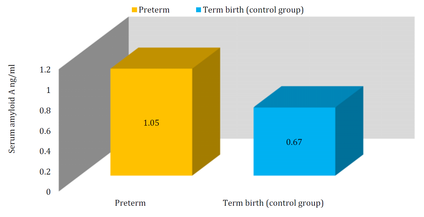
Figure 1: Serum Amyloid A Levels in Preterm and Term Births


The study conducted a comparative analysis of serum amyloid A (SAA) levels in women with preterm births and those in the control group with term births. The findings demonstrate a notable and statistically significant difference in SAA levels between the two groups. Specifically, women who experienced preterm births showed significantly elevated SAA levels (1.05±0.37 ng/ml) in comparison to the term control group (0.67±0.12 ng/ml), with a P-value of 0.001, as illustrated in Table 4 and Figure 1.


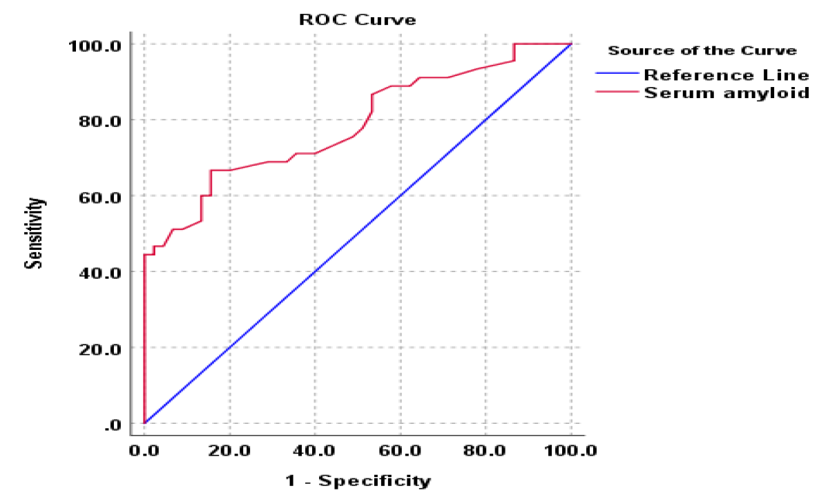
Receiver operating characteristic (RCO) curve analysis was applied for evaluation of serum amyloid A as a diagnostic biomarker for preterm birth.


In the present study the SAA cutoff value was found to be 0.69 ng/ml based on the area under the curve (AUC) reaching 80.5% in the ROC analysis. When the serum amyloid level is above this cut-off point, it is considered indicative of preterm labor with a sensitivity of 77.8% and a specificity of 71.1%. The calculated accuracy of 75.93% reflects a considerable proportion of correct predictions, balancing both true positives and true negatives. Particularly notable is the Negative Predictive Value (NPV) at 89.79%, indicating a high reliability in correctly identifying individuals without preterm delivery as shown in Table 5 and Figure 2.


Table 6 provides a comparison of serum amyloid A (SAA) levels in relation to delivery mode among women who experienced preterm births and the term control group. Among women with preterm births, those who  underwent cesarean sections exhibited significantly higher SAA levels (1.33±0.43 ng/ml) compared to those who had vaginal deliveries (0.95±0.29 ng/ml) with a P-value of 0.014. In contrast, within the control group of women with term births, the SAA levels were lower among those who had cesarean sections (0.16±0.12 ng/ml) compared to those who had vaginal deliveries (0.69±0.11 ng/ml), although the difference did not reach statistical significance (p-value: 0.12).


Table 6: Serum Aamyloid Levels in preterm and Term Births In Relation to Delivery Mode

Studied womenNo.Serum amyloid A (mean±SD) ng/mlp-value
Preterm womenCesarean section121.33±0.430.014*
Vaginal delivery330.95±0.29
Term birth (control group)Cesarean section80.16±0.120.12*
Vaginal delivery370.69±0.11

 * Independent sample t-test 


Figure 2: ROC Curve for the Cutoff Point of Serum Amyloid a In Prediction of Preterm Birth


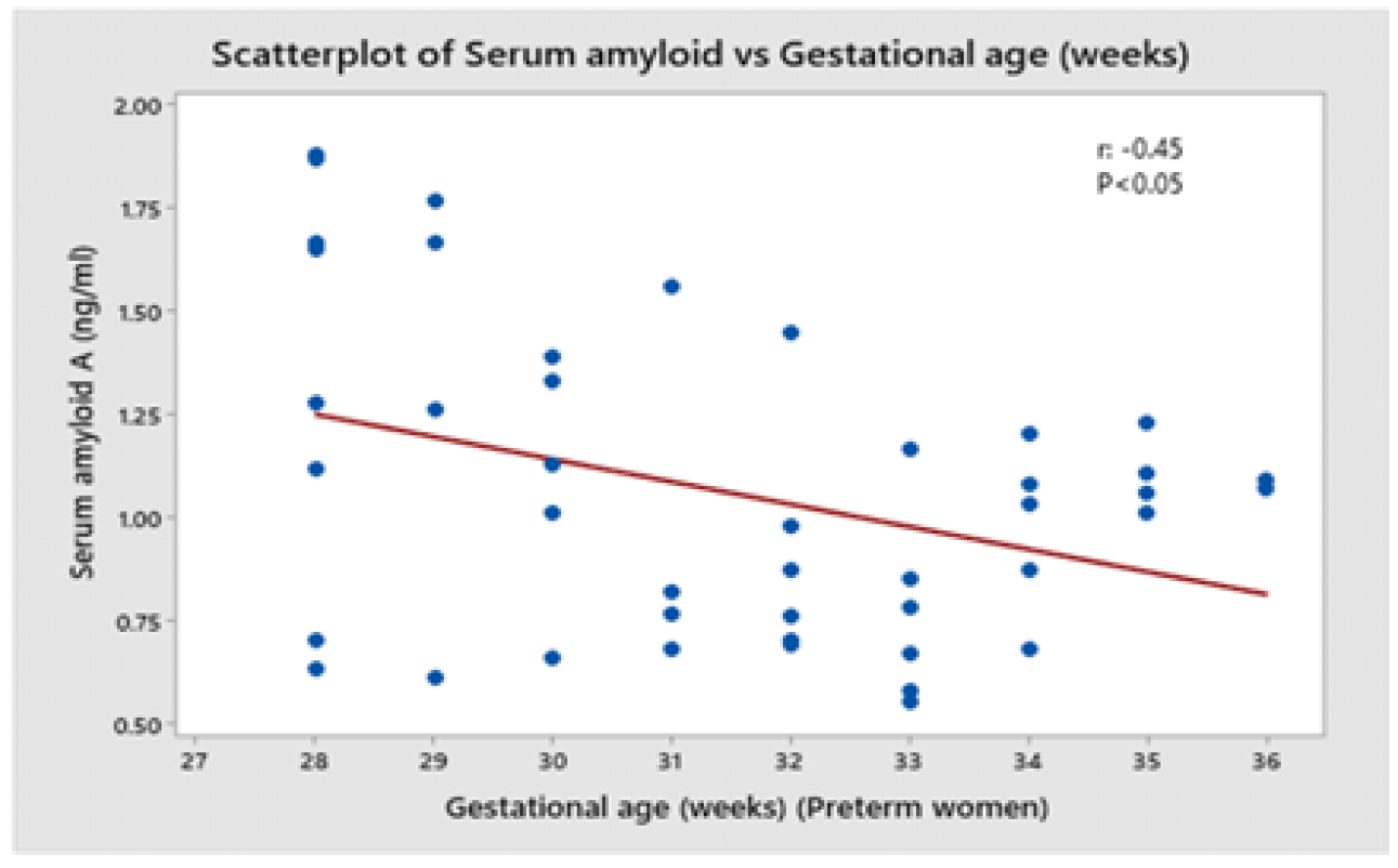
Figure 3: Correlation between Serum Amyloid A Levels And the Gestational Age of Preterm Birth Women


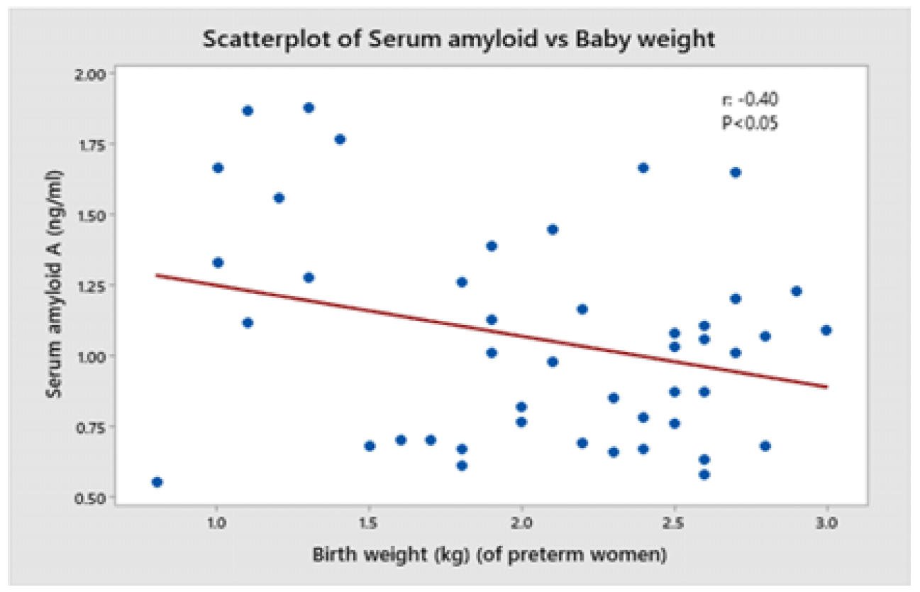
Figure 4: Correlation between Serum Amyloid A Levels And Birth Weight Of Preterm Birth Women

DISCUSSION

Approximately 27% of the 4 million newborn deaths worldwide are due to preterm. Identifying pregnancies at high risk of spontaneous preterm birth represents a challenge that can dramatically improve global health [93-41], however, half of all preterm birth occurs in women with no known clinical risk factors [42-43]. Traditional methods of predicting women at risk for preterm birth such as obstetric history, tocometry, biochemical markers and ultrasonography of the cervix and interventions based on these findings have not lowered the rate of preterm birth. This may be explained by the fact that preterm birth is a complex disease with multiple pathways that culminate in a common terminal pathway [7]. The Recent screening strategies for the preterm delivery focused on early identification of patients at risk, enabling earlier intervention for PTL. In this scope the use of biological markers in predicting PTB and to identify those women at risk has been proposed [4]. Maternal SAA is a member of apolipoproteins associated with high-density lipoproteins in plasma. It is also associated with inflammatory response highly similar to erythrocyte sedimentation rate and CRP [37]. Based on these factors we decided to investigate SAA activity in patients with preterm birth compared to term pregnancies to test its utility as a predictor for the condition. The results revealed a significant association between maternal age and preterm births. Specifically, the average age of mothers in the preterm group tends to be notably younger compared to those in term births, emphasizing the relevance of age in preterm birth occurrence. This aligns with the previous studies by Al-Dabbagh et al. [44] and Sarhan et al. [45] both studies support the notion that the rate of premature deliveries is higher among younger women aged between [20,29] years. Primparity was significantly more prevalent in the preterm group compared to the term control group. While a greater percentage of women with preterm births underwent cesarean section compared to the term control group, the difference in delivery mode between the groups was not statistically significant. These results align with similar finding in study conducted by Aragao et al [40], which identified primiparity as a risk factor for preterm birth, However, it contradicts with the researches done by Al-Dabbagh et al. [44] and Sarhan et al. [45] and Mahapula et al. [39]These studies collectively reported that parity was not identified as a significant risk factor for preterm births. This disparity in findings between these studies may refer to the different family planning programs in different regions and demographical variation between the selected samples in these countries. Additionally, mothers in the preterm birth group exhibited a higher incidence of previous miscarriages in contrast to the term group. Notably, a substantial proportion of the preterm birth group had a history of previous preterm delivery, significantly higher than the term control group. One potential mechanism that could explain an association between previous miscarriage and risk of preterm birth is a weakening of the cervix as a result of damage from the surgical management of miscarriage. Moreover it is thought that inadequate implantation leads to impaired placental function and consequently puts pregnancies at risk for poor fetal growth, placental abruption, preeclampsia, stillbirth and preterm delivery [46], Oliver-Williams et al. [41] the study showed that miscarriage and preterm birth were associated with an increased risk of all-cause preterm birth. These findings show the relevance of primi-parity status, miscarriage history and previous preterm deliveries in elevating the risk of preterm births. The study compared preterm and term births regarding neonatal outcomes, finding that preterm deliveries had a significantly lower gestational age and birth weight compared to term controls. Preterm infants are born before they have fully developed and as a result, their growth and weight may be significantly under what is considered average for a full-term pregnancy [47]. The impact of gestational age on birth weight is well-documented in numerous studies, emphasizing that preterm births are a significant contributor to reduced neonatal weight this trend is further corroborated by findings Gao et al. [42] and Xi et al. [46] illustrating that high rates of preterm births are associated with low birth weight. A higher proportion of preterm infants in our study were recording decreased birth weight, less than 2500 g at birth [48]. The shortened gestation period restricts the time for the fetus to gain weight and complete development, often leading to lower birth weight figures [49]. These babies have been shown to have diminished cognitive development and evidence now suggests that LBW babies are at an increased risk of chronic diseases later in life, including high blood pressure, non-insulin dependent diabetes mellitus, coronary heart disease and stroke [48] .Similarly in a research conducted by de Mendonça et al. [50] preterm infants typically exhibit lower mean birth weights, often below 2.5 kilograms.          In the current study, APGAR scores at 1 and 5 minutes were significantly lower for preterm births than term controls. APGAR scores serve as an essential measure of a newborn's immediate health status, assessing vital signs such as heart rate, respiratory effort, muscle tone, reflex irritability and skin color. Preterm infants, born before completing the full term, often face respiratory and other physiological challenges due to their underdeveloped organ systems. The significant differences observed in APGAR scores between preterm and term births in the current study are in agreement with numerous previous studies. Research by Cnattingius et al. [51] and Abukari et al. [52] showed lower Apgar scores in preterm infants at both 1 and 5 minutes, reflecting the challenges these babies face in adapting to extrauterine life due to their premature birth.


In the current study, a significantly higher percentage of preterm infants required NICU admission compared to term controls. The higher frequency of NICU admission for preterm births aligns with the heightened medical attention and specialized care demanded by these fragile neonates, consistent with observations across various studies [53-54]. The findings demonstrate a notable and statistically significant difference in SAA levels between the two studied groups. Specifically, women who experienced preterm births showed significantly elevated SAA levels (1.05±0.37 ng/ml) in comparison to the term control group (0.67±0.12 ng/ml). The findings of the current study support the role of maternal Serum Amyloid A (mSAA) as a distinguishing and triggering factor for Preterm Birth (PTB). Previous research delved into the association between SAA levels and both normal and abnormal pregnancies, In healthy pregnancies, mSAA levels remained consistent throughout gestation, rising only during intercurrent infections, representing a clinically useful tool to monitor the acute phase response [55]. Interestingly while mSAA concentrations were normal a day before delivery, parturition led to a substantial increase in mSAA levels, surging several hundred-fold [56]. Serum amyloid A released during infection or inflammation stimulates matrix metalloproteinases (MMPs) and prostaglandin production, causing cervical effacement and uterine contractions in PTB [40-41]. In agreement with the current finding, Ibrahim et al. [37] in a recent study highlighted a significant difference in maternal Serum Amyloid a (mSAA) levels between women who experienced preterm birth (PTB) and those who carried pregnancies to term. Specifically, their research demonstrated significantly elevated mSAA levels in women who had preterm births compared to those who completed pregnancies to full term 5.1 (4.5-7.7) mg/l versus 1.2 (0.0–2.5) mg/l, for cases and controls respectively. The current study also agreed with a study done by Kim et al (54) who focused on analyzing the role of mSAA levels in predicting tocolytic failure in preterm delivery, by examining 100 pregnant women who received continuous prenatal care and delivered between 20 and 37 weeks of gestation and conclude that high mSAA measurement might serve as a quick and minimally invasive diagnostic method to predict tocolytic failure in threatened PTL. Furthermore, a study conducted by Çekmez et al. [57] highlighted increased levels of maternal Serum Amyloid A (mSAA) in pregnant women diagnosed with Preterm Premature Rupture of Membranes (PPROM). In addition to the rise of SAA levels in maternal circulation, SAA levels in cord blood were also significantly higher in patients with PPROM) than in patients without PPROM [58]. Ali et al. [59] in their study found that SAA levels were markedly increased in maternal circulation in preterm birth with chorioamnionitis when compared with gestational age-matched iatrogenic preterm birth. In the present study the SAA at a cutoff value 0.69 ng/ml found to be indicative of preterm labor with a sensitivity of 77.8% and a specificity of 71.1%, Ibrahim MI et al. [37] study find mSAA had an excellent predictive value to discriminate between women with and without PTB at a best cut-off value of mSAA >3.0 mg/l with a sensitivity of 96.6% and specificity of 86.2%. The difference in the mean mSAA and cat off value for PTL may be related to the use of different kits and methods for evaluation of mSAA levels between the former study and ours. The current study showed that preterm children who underwent cesarean sections had significantly higher SAA levels compared to those who experienced vaginal deliveries. Conversely, in the control group of women with term births, SAA levels were lower among those who had cesarean sections compared to those who had vaginal deliveries, although this difference did not reach statistical significance. These results suggest that the mode of delivery may play a role in influencing SAA levels, highlighting a potential link between the inflammatory response, delivery mode and gestational age. The observed association between delivery mode and serum amyloid A (SAA) levels may be attributed to the physiological differences in the inflammatory response triggering labor in preterm and term births. In preterm births, the heightened inflammatory state in some cases might lead to increased SAA production and necessitate termination by cesarean section added to the already high level caused by the inflammatory process associated with PTL. On the other hand, term births, particularly in cesarean sections, may involve a different inflammatory profile influenced by factors such as timing of the surgical procedure for different indications, before the onset of spontaneous labor. The study demonstrated a notable negative correlation between serum amyloid a levels and each of the gestational age and birth weight in women who experienced preterm delivery. This may furtherly indicate that SAA had an excellent value to predict PTB. The finding of correlations between (mSAA) with gestational age and birth weight was uncommon in the studies included in our review. Only one study conducted by Ibrahim et al. [37] reported significant negative correlations between mSAA levels and both gestational age and birth weight. Kayabas et al. [58] found that the SAA levels did not correlate with gestational ages. The lack of correlation may be due to the specific study population or the methodological limitations of their study.

CONCLUSION

Based on our study results, we concluded:


  • Serum amyloid A (SAA) at a cut off value (0.69 ng/ml) has a fair sensitivity and specificity for prediction of patient at risk of preterm birth, allowing earlier care, monitoring and intervention for the cases

  • The significant negative correlation found between SAA levels and each of gestational age at birth and birth weight in women who experienced preterm delivery indicates excellent value of the marker for predicting poor neonatal outcome

  • The mode of delivery through caesarean section in the context of preterm births, was associated with higher SAA levels


Recommendations


  • Future studies with different designs including serial measurements of SAA in women at risk of PTL are needed to explore the exact profile of SAA during these abnormal pregnancies

  • The potential of SAA as a predictive biomarker for preterm births needs to confirmed by more expanded studies including multiple centers and larger sample size before its wide application with clinical practice

  • Further study including patients with PROM are recommended to evaluate the role of SAA in the development of this complication

REFERENCE
  1. Cunningham, F.G. et al. “Preterm birth.” Williams obstetrics, edited by Diane M. Twickler, McGraw-Hill Education, 2022, pp. 829–855.

  2. Crum, C.P. et al. Diagnostic gynecologic and obstetric pathology. Elsevier Health Sciences, 2011, pp. 6–22.

  3. Diemert, A. and P.C. Arck. “Preterm birth: Pathogenesis and clinical consequences revisited.” Seminars in Immunopathology, 2020, pp. 375–376.

  4. Blencowe, H. et al. “Born too soon: The global epidemiology of 15 million preterm births.” Reproductive Health, vol. 10, 2013, pp. 1–14.

  5. Walani, S.R. “Global burden of preterm birth.” International Journal of Gynecology & Obstetrics, vol. 150, no. 1, 2020, pp. 31–33.

  6. Durbach, N. “Dead or alive? Stillbirth registration, premature babies and the definition of life in England and Wales, 1836–1960.” Bulletin of the History of Medicine, vol. 94, no. 1, 2020, pp. 64–90.

  7. Purisch, S.E. and C. Gyamfi-Bannerman. “Epidemiology of preterm birth.” Seminars in Perinatology, 2017, pp. 387–391.

  8. Niyibizi, J. et al. “Association between human papillomavirus infection among pregnant women and preterm birth.” JAMA Network Open, vol. 4, no. 9, 2021, p. e2125308.

  9. Noben, L. et al. “The fetal electrocardiogram to detect the effects of betamethasone on fetal heart rate variability.” Early Human Development, vol. 130, 2019, pp. 57–64.

  10. DeCherney, A.H. et al. Current diagnosis and treatment: Obstetrics and gynecology. 2013, pp. 6–11.

  11. Truong, N. et al. “The role of fetal membranes during gestation, at term and preterm labor.” Placenta and Reproductive Medicine, vol. 2, 2023, pp. 2–5.

  12. Letouzey, M. et al. “Cause of preterm birth and late-onset sepsis in very preterm infants: The EPIPAGE-2 cohort study.” Pediatric Research, vol. 90, no. 3, 2021, pp. 584–592.

  13. Nadeau, H.C. et al. “Infection and preterm birth.” Seminars in Fetal and Neonatal Medicine, vol. 21, no. 2, 2016, pp. 100–105.

  14. Maymon, R. et al. “Association between routine ultrasound cervical length measurement at 19–25 weeks’ gestation and prevalence of preterm birth in Israel.” Ultrasound in Obstetrics & Gynecology, 2022, pp. 164–169.

  15. Schmitz, T. et al. “Preterm premature rupture of the membranes: Guidelines for clinical practice from the French College of Gynaecologists and Obstetricians.” European Journal of Obstetrics & Gynecology and Reproductive Biology, vol. 236, 2019, pp. 1–6.

  16. Lomotey, A.Y. et al. “Experiences of mothers with preterm babies at a mother and baby unit of a tertiary hospital.” Nursing Open, vol. 7, no. 1, 2020, pp. 150–159.

  17. Pummara, P. et al. “Association of first-trimester pregnancy-associated plasma protein A levels and idiopathic preterm delivery.” Taiwanese Journal of Obstetrics and Gynecology, vol. 55, no. 1, 2016, pp. 72–75.

  18. Rose, M.S. et al. “Prenatal maternal anxiety as a risk factor for preterm birth.” BioMed Research International, 2016, pp. 110–117.

  19. Morgan, T.K. “Role of the placenta in preterm birth: A review.” American Journal of Perinatology, 2016, pp. 258–266.

  20. Teoh, P. et al. “Gender and preterm birth: Is male fetal gender a clinically important risk factor?” European Journal of Obstetrics & Gynecology and Reproductive Biology, vol. 225, 2018, pp. 155–159.

  21. Konar, H. DC Dutta’s textbook of obstetrics. JP Medical Ltd., 2018, pp. 294–306.

  22. Aregawi, G. et al. “Preterm births and associated factors among mothers in Axum and Adwa Town public hospitals.” BMC Research Notes, vol. 12, 2019, pp. 1–6.

  23. Baker, P.N. and L. Kenny. Obstetrics by ten teachers. CRC Press, 2011, pp. 132–143.

  24. Schleußner, E. “The prevention, diagnosis and treatment of premature labor.” Deutsches Ärzteblatt International, vol. 110, no. 13, 2013, p. 227.

  25. American College of Obstetricians and Gynecologists. “Practice bulletin no. 171: Management of preterm labor.” Obstetrics & Gynecology, vol. 128, no. 4, 2016, pp. e155–e164.

  26. Bitzer, E.M. et al. “Self-testing of vaginal pH to prevent preterm delivery: A controlled trial.” Deutsches Ärzteblatt International, vol. 108, no. 6, 2011, p. 81.

  27. Gotsch, F. et al. “The fetal inflammatory response syndrome.” Clinical Obstetrics and Gynecology, vol. 50, no. 3, 2007, pp. 652–683.

  28. Gabbe, S. et al. Obstetrics: Normal and problem pregnancies. 1st South Asia ed., 2017, pp. 5–11.

  29. Crane, J. and D. Hutchens. “Transvaginal sonographic measurement of cervical length to predict preterm birth.” Ultrasound in Obstetrics and Gynecology, vol. 31, no. 5, 2008, pp. 579–587.

  30. Greco, E. et al. “First-trimester screening for spontaneous preterm delivery.” Fetal Diagnosis and Therapy, vol. 31, no. 3, 2012, pp. 154–161.

  31. Shah, P.E. et al. “Neurodevelopmental outcomes of very low birth weight infants.” Clinics in Perinatology, vol. 43, no. 2, 2016, pp. 347–363.

  32. Saigal, S. and L.W. Doyle. “An overview of mortality and sequelae of preterm birth from infancy to adulthood.” The Lancet, vol. 371, no. 9608, 2008, pp. 261–269.

  33. Behrman, R.E. and A.S. Butler. Preterm birth: Causes, consequences and prevention. National Academies Press, 2007, pp. 45–67.

  34. Blencowe, H. et al. “National, regional and worldwide estimates of preterm birth.” The Lancet, vol. 379, no. 9832, 2012, pp. 2162–2172.

  35. Goldenberg, R.L. et al. “Epidemiology and causes of preterm birth.” The Lancet, vol. 371, no. 9606, 2008, pp. 75–84.

  36. March of Dimes. Premature birth report card. March of Dimes Foundation, 2019.

  37. Beck, S. et al. “The worldwide incidence of preterm birth.” Bulletin of the World Health Organization, vol. 88, 2010, pp. 31–38.

  38. Kramer, M.S. et al. “Determinants of preterm birth.” American Journal of Epidemiology, vol. 158, no. 2, 2003, pp. 115–124.

  39. Lawn, J.E. et al. “Global report on preterm birth and stillbirth.” BMC Pregnancy and Childbirth, vol. 10, suppl. 1, 2010, pp. S1–S24.

  40. Steer, P. “The epidemiology of preterm labour.” BJOG: An International Journal of Obstetrics & Gynaecology, vol. 112, suppl. 1, 2005, pp. 1–3.

  41. Slattery, M.M. and J.J. Morrison. “Preterm delivery.” The Lancet, vol. 360, no. 9344, 2002, pp. 1489–1497.

  42. Mercer, B.M. “Preterm premature rupture of the membranes.” Obstetrics & Gynecology, vol. 101, no. 1, 2003, pp. 178–193.

  43. Hack, M. et al. “Outcomes in young adulthood for very low birth weight infants.” New England Journal of Medicine, vol. 346, no. 3, 2002, pp. 149–157.

  44. Saigal, S. et al. “School-age outcomes in children who were extremely low birth weight.” Pediatrics, vol. 105, no. 2, 2000, pp. 325–331.

  45. Bhutta, A.T. et al. “Cognitive and behavioral outcomes of school-aged children who were born preterm.” JAMA, vol. 288, no. 6, 2002, pp. 728–737.

  46. Hack, M. and A.A. Fanaroff. “Outcomes of children of extremely low birth weight.” Seminars in Neonatology, vol. 5, no. 2, 2000, pp. 89–106.

  47. Moore, T. et al. “Neurological and developmental outcomes in extremely preterm children.” New England Journal of Medicine, vol. 358, no. 14, 2008, pp. 1470–1471.

  48. Doyle, L.W. et al. “Outcome at 2 years of age of infants born extremely preterm.” New England Journal of Medicine, vol. 357, no. 21, 2007, pp. 2148–2159.

  49. Wood, N.S. et al. “Neurologic and developmental disability after extremely preterm birth.” New England Journal of Medicine, vol. 343, no. 6, 2000, pp. 378–384.

  50. Marlow, N. et al. “Neurologic and developmental disability at six years of age after extremely preterm birth.” New England Journal of Medicine, vol. 352, no. 1, 2005, pp. 9–19.

  51. Aylward, G.P. “Neurodevelopmental outcomes of infants born prematurely.” Journal of Developmental and Behavioral Pediatrics, vol. 26, no. 6, 2005, pp. 427–440.

  52. Allen, M.C. “Neurodevelopmental outcomes of preterm infants.” Current Opinion in Neurology, vol. 21, no. 2, 2008, pp. 123–128.

  53. Johnson, S. and N. Marlow. “Early and long-term outcome of infants born extremely preterm.” Archives of Disease in Childhood, vol. 102, no. 1, 2017, pp. 97–102.

  54. Mwaniki, M.K. et al. “Long-term neurodevelopmental outcomes after intrauterine and neonatal insults.” The Lancet, vol. 379, no. 9814, 2012, pp. 445–452.

  55. Saigal, S. and L.W. Doyle. “Preterm birth 1: An overview of mortality and sequelae.” The Lancet, vol. 371, no. 9608, 2008, pp. 261–269.

  56. Blencowe, H. et al. “Born too soon: The global epidemiology of 15 million preterm births.” Reproductive Health, vol. 10, suppl. 1, 2013, pp. S2–S14.

  57. Howson, C.P. et al. Born too soon: The global action report on preterm birth. World Health Organization, 2012.

  58. World Health Organization. Preterm birth fact sheet. WHO, 2023.

  59. March of Dimes. Global report on preterm birth and stillbirth. March of Dimes Foundation, 2012.
Recommended Articles
Case Report
OROFACIAL GRANULOMATOSIS: A CASE REPORT
...
Published: 19/11/2024
Download PDF
Research Article
The Study of Suicide in Adolescents in The Context of Cognitive Brain Change
...
Published: 29/07/2024
Download PDF
Research Article
Role of First Trimester HbA1c in Screening of Gestational Diabetes Mellitus
...
Published: 28/12/2025
Download PDF
Research Article
Antibiotics and their use in preventing Coronavirus Covid -19
Published: 26/08/2024
Download PDF
Chat on WhatsApp
Flowbite Logo
PO Box 101, Nakuru
Kenya.
Email: office@iarconsortium.org

Editorial Office:
J.L Bhavan, Near Radison Blu Hotel,
Jalukbari, Guwahati-India
Useful Links
Order Hard Copy
Privacy policy
Terms and Conditions
Refund Policy
Shipping Policy
Others
About Us
Team Members
Contact Us
Online Payments
Join as Editor
Join as Reviewer
Subscribe to our Newsletter
+91 60029-93949
Follow us
MOST SEARCHED KEYWORDS
Copyright © iARCON International LLP . All Rights Reserved.