Multinodular goiter is a common disease whose prevalence increases in subsaharan Africa. Compressive goiter, heart failure (HF) and chronic obstructive pulmonary disease (COPD) are both common causes of breathlessness. Clinical presentation of those diseases can be very similar, but the treatment and prognostic implications are very different. In the evidence of compressive goiter, making diagnosis of the coexisting HF and COPD can be difficult especialy in a developing country. This case discusses the diagnostic challenge in a 26 years’ woman with compressive giant goiter, COPD and HF. We highlight the clinical relevance, and therapeutics aspects.
Keywords
Diagnosis
Compressive Goiter
Heart Failure
COPD
INTRODUCTION
Coexisting of compressive giant goiter , HF and COPD are rare. Dyspnea is a common symptom affecting both of those patients seen in the ambulatory setting [1]. According to Wakeley and Mulvany classification in1940, 15% of goiters are “partial” intrathoracic (in which the major part of the mass is situated within the thorax) [2]and can be responsable for tracheal compression. Diagnosing COPD and HF in patient presenting compressive giant goiter is challenging because clinical features overlap [3]. Therefore, it remains a clinical diagnosis requiring a history, physical examination, and laboratory testing. We report herein the diagnosis challenge of coexisting compressive geant goiter, COPD and HF highlighting the clinical relevance and therapeutics aspects.
Case Report
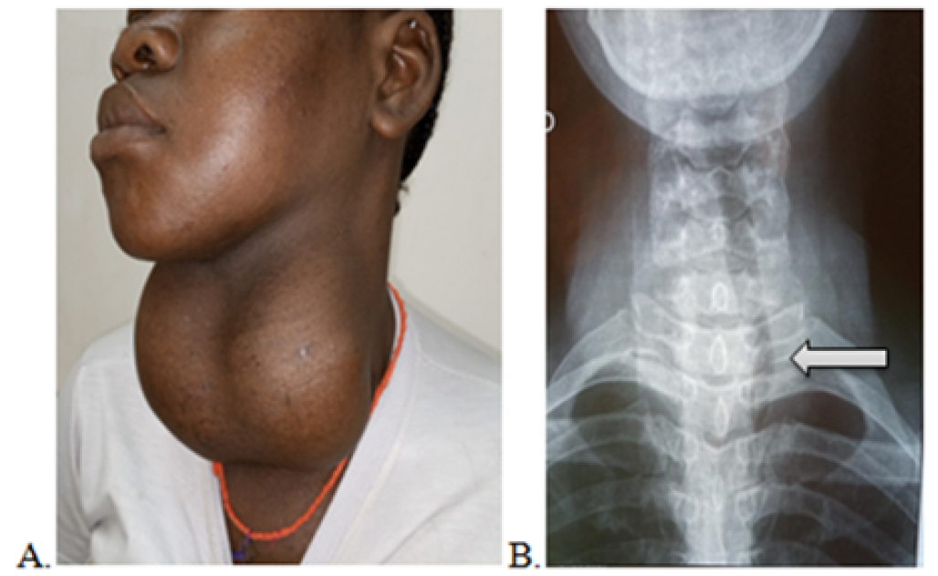
A young adult hypertensive woman of 26-year-old, from an iodine insufficient area, was admitted to the emergency department of our hospital for dyspnea presenting an acute upper airway obstruction (wheezing and stridor) as results of an important progressive tracheal compression, which progressively aggravated by an acute respiratory infection. She had history of thyroid enlargement for 5 years. Physical examination revealed a cervical mass that moved on swallowing (Figure 1A).
The Heart rate was 58 bpm, Blood pressure was 170/100 mmHg. Oxygen saturation on room air was 82%. The patient treatment was provide byasocial assistance program supported by the hospital. The standard cervical X-ray revealed a cervico-mediastinal opacity and tracheal compression with left tracheal deviation (Figure 1B). Laryngoscopy showed a normal mobility of both vocal cords. Cardiac evaluation revealed presence of hypertension, systolic murmur, myocardial hypertrophy and dyspnea. Preanesthetic evaluation revealed a high anesthetic and surgical risk in a patient with respiratory failure and cardiac associated comorbidity. The initial management in this case was oxygen therapy, antihypertensive drugs (captopril 25 mg/ day associated with furosemide 40 mg/day) and vasodilator therapy (isosorbide dinitrate 20 mg/ day). Biological findings of thyroid gland function were within normal limits. Eletrocardiography revealed arythmia.
Figure 1: Cervical Compressive Goiter. (A: Huge Cervical Mass, B: Cervical Radiology with Left Tracheal Deviation)
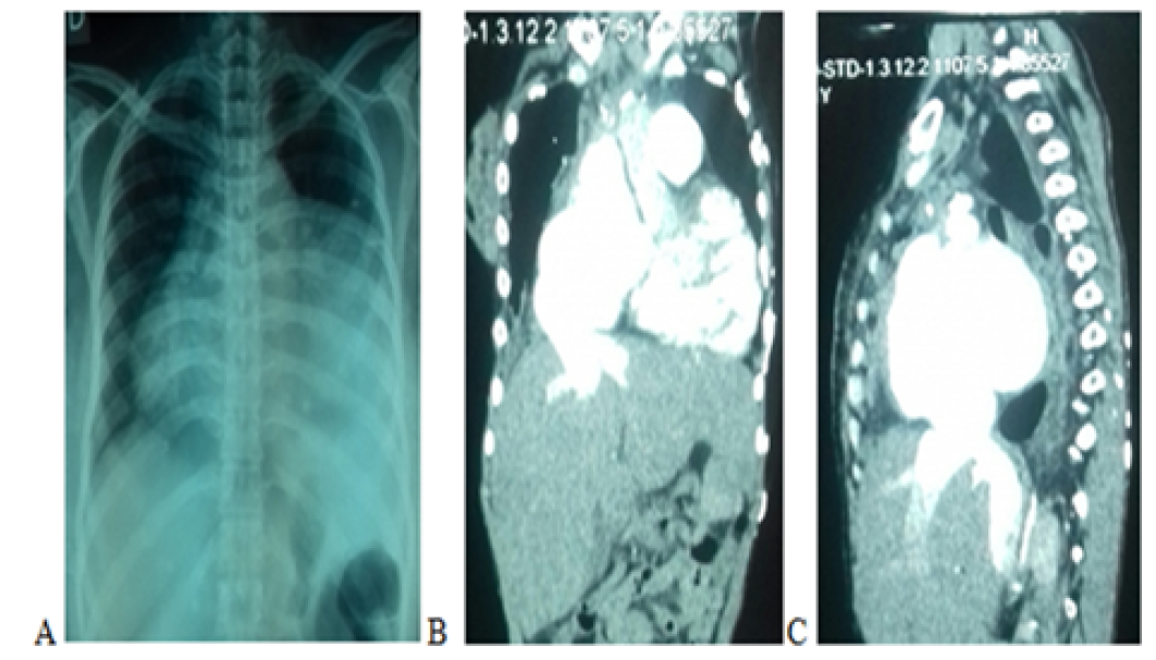
Finally, the patient was taken to the operating theater to achieve the goal of the emergency surgery by complete removal of the goiter and decompression of the trachea. Postoperatively in ICU, they was absence of wheezing and stridor. There was no dysphonia, tracheomalacia and the calcaemia was in normal levels. Postoperative care included substitutive medication levothyroxine 50 mcg/ day. The histologic examination of the thyroid mass revealed a multinodular goiter with epithelium hyperplasia. After 5 days in ICU, the patient present again episode of moderate symptoms of respiratory distress. The Chest X-ray showed mediastinal enlargement (Figure2A) .The patient was taken to the external radiographic center to perform the thoracic CT and the cardiac echography.
CT scan showed myocardial hypertrophy , a huge right atrial enlargement , veina cava dilation (Figure 2 B,C ) and reticular patterns of pulmonary oedema. Transthoracic echocardiography revealed the presence of concentric left ventricular hypertrophy with an ejection fraction of 45%, with relevant valvulopathies affecting both the mitral valve (mild steno-insufficiency), tricuspid valve (mild-to-moderate regurgitation).
Figure 2: Postoperative Chest Imaging. (A: Chest Radiology Showing Cardiomegaly, B: Thoracic CT Showing Dilation of the Right Atrium and the Vena Cava, C: Dilation of Intrahepatic Veins)
On spirometry, forced expiratory volume in the first second (FEV1) was 63% of the predicted value, with a FEV1/forced vital capacity (FVC) ratio of 53%; the COPD Assessment Test (CAT) was performed and the final score was 16. Brain Natriuretic Peptid (BNP) was 150, pg/mL. The patient was discharged after one month on the following medications: ramipril 10 mg/day, bisoprolol 5 mg/day, digoxin 0.25 mg/day, furosemide 50 mg/day, : acénocoumarol 4 mg/day, salbutamol 2 mg tablet 6mg/day, and levothyroxine 50 mcg/day. The patient was re-hospitalized twice in the same year for pneumonia and signs of heart failure (EF of 40%). In particular, the patient presented New York Heart Association (NYHA) functional class III symptoms. She was excluded from valve replacement surgery due to anesthesiological risk. Spirometry was unchanged, with a CAT score of 16. We have added the bronchodilator combination indacaterol/glycopyrronium (85/43 μg/day) to the patient’s therapy, and after 16 months with this therapy, his FEV1 improved to 80% of the predicted value with a FEV1/FVC ratio of 70% and a CAT score of 8. The ejection fraction was 50%, BPN was 100 pg/mL and the blood pressure was 150/90 mmHg (table 1).
Table 1: Patient Respiratory, Laboratory and Clinical Parameters
Parameters
Initially
16 Months
FEV1 (% )
61
80
FEV1/FVC (%)
53
70
CAT score
16
8
Systolic BP ( mm Hg )
170
150
Diastolic BP ( mm Hg )
100
90
Heart rate ( bpm )
58
65
EF ( %)
45
50
BNP ( pg/mL )
150
100
DISCUSSION
The incidence of multinodular goiter is more prevalent in women, has intrathoracic extension, It can be more frequently, acquired, which originates in the cervical thyroid gland and descends through the fascial plane to the mediastinum [4]. Usually, it is located in the upper mediastinum, producing different symptoms [5]. Approximately 50% accounts for multinodular goiter, as in our patient, where iodine deficiency and altered thyroxine synthesis favor tumor growth [6]. Clinical presentation is very diverse and nonspecific, therefore diagnostic images are required. Cervico-thoracic radiography assesses the location of the mass, the displacement or compression of adjacent structures and the presence of calcifications [7]; chest CT is better to characterize limits with intrathoracic structures, it is useful to identify the etiology and plan the best surgical approach. In developing countries, medical equipment is often shut down due to lack of maintenance. In our case there was no possibility to perform a chest CT, or cardiac echography in emergency.Whenever obstructive symptoms or compression of cervical and thoracic structures occur, surgical management is recommended [8]. Complications associated with surgical management are dysphonia, infections, tracheomalacia, hypoparathyroidism, and airway injuries [4]. The postoperative results were very satisfactory, with the absence of those complications. The large size of the tumor along with compressive symptoms of the trachea and the coexisting of HF and COPD make this particular case striking.
Also the poverty of the population contributes to the diagnostic delay. The patient was a hypertensive woman without any medical follow-up. Luckily she received social assistance in the hospital. In this part of the word, expensive examinations are not accessible for the majority of patients and standard radiography and echography are preferred in diagnosis research of compressive goiter. They are less expensive and most available. Usualy the exams are requested gradually depending on cost and availability. CT scan, Echocardiography, and pulmonary function tests along should be performed and carefully interpreted. Despite the difficulties in accessing exams, the CT scan was the examination needed in order to allow a very good exploration in this emergency.
The emergency of this case did not allow us to perform these examinations without any risks.
As reported by Warren and other authors, acute airway obstruction can lead to sudden death [9]. In this case, the approach was the immediate surgical resection of a thyroid mass, thus decompressing the trachea to avoid sudden death. Coexinting COPD and HF can remains undetected in the patient especialy when compressive symptoms of airway was evident. Closer collaboration between cardiologists and pulmonologists and surgeon is required for better identification and management of concurrent compressive goiter, COPD and HF [10].COPD induces several cardiovascular comorbidities and exerts a negative prognostic effect on the outcomes of heart failure patients [11]. Patients with concomitant COPD and heart failure treated with the combination of indacaterol and glycopyrronium results in reciprocal potentiation with a maximal bronchodilatory effect [12] Appropriate diagnosis and therapy for heart failure are important given the poor prognosis. Survival is 89.6 % at one month from diagnosis, 78 % at one year, and only 57.7 % at five years [13]. The prognosis of patients with both disorders is worse than those with only one of the diseases. In the 16-month follow-up our patient condition improved with incidents of exertional dyspnea. Multidisciplinary perioperative management is key for a successful recovery. Selective screening of compressive goiter in patients when necessary for HF and COPD should be considered.
Acknowledgements
To the Mali hospital medical team and the scientific committee for their encouragement and participation.
Funding
There is no financial support for this manuscript.
Lee, D.S. et al. “Relation of disease pathogenesis and risk factors to heart failure with preserved or reduced ejection fraction.” Circulation, vol. 119, no. 24, 2009, pp. 3070–3077.
Katlic, M.R., Grillo H.C. and Wang C.A. “Substernal goiter: analysis of 80 patients from Massachusetts General Hospital.” American Journal of Surgery, vol. 149, 1985, pp. 283–287.
Foroulis, C.N. et al. “Primary intrathoracic goiter: a rare and potentially serious entity.” Thyroid, vol. 19, 2009, pp. 213–218.
Schlumberger, M.J. et al. “Nontoxic diffuse goiter, nodular thyroid disorders, and thyroid malignancies.” In: Melmed S. et al., eds. Williams Textbook of Endocrinology, 13th ed. Philadelphia: Elsevier, 2016, pp. 449–488.
Huang, W.C. et al. “Intrathoracic goiter in elderly patients.” International Journal of Gerontology, vol. 7, 2013, pp. 8–12.
Chen, A.Y. et al. “American Thyroid Association statement on optimal surgical management of goiter.” Thyroid, vol. 24, 2014, pp. 181–189.
Warren, C.P.W. “Acute respiratory failure and tracheal obstruction in the elderly with benign goiters.” Canadian Medical Association Journal, vol. 121, 1979, pp. 191.
Lee, D.S. et al. “Relation of disease pathogenesis and risk factors to heart failure with preserved or reduced ejection fraction.” Circulation, vol. 119, no. 24, 2009, pp. 3070–3077.
Čelutkienė, J. et al. “Challenges of treating acute heart failure in patients with chronic obstructive pulmonary disease.” Cardiac Failure Review, vol. 3, 2017, pp. 56–61.
Cohen, J.S. et al. “Dual therapy strategies for COPD: the scientific rationale for LAMA + LABA.” International Journal of Chronic Obstructive Pulmonary Disease, vol. 11, 2016, pp. 785–797.
Michael, K. and Joe K. “Diagnosis and evaluation of heart failure.” American Family Physician, vol. 85, no. 12, 2012, pp. 1161–1168.
Recommended Articles
Case Report
OROFACIAL GRANULOMATOSIS: A CASE REPORT
Diksha Thakur,
...
Mandeep Kaur
Published: 19/11/2024
Download PDF
Research Article
The Study of Suicide in Adolescents in The Context of Cognitive Brain Change
Zhang Wen,
...
Amiya Bhaumik
Published: 29/07/2024
Download PDF
Research Article
Role of First Trimester HbA1c in Screening of Gestational Diabetes Mellitus
Anu Berwal,
...
Geetika Gupta Syal
Published: 28/12/2025
Download PDF
Case Report
The Utility of Maternal Serum Amyloid a in the Prediction of Preterm Birth in Symptomatic Women
Enas Hussein Ahmed,
Aseel Ghazi Rifat
Published: 30/06/2024
Download PDF
PO Box 101, Nakuru
Kenya.
Email: office@iarconsortium.org
Editorial Office:
J.L Bhavan, Near Radison Blu Hotel,
Jalukbari, Guwahati-India
Togo, S., Bazongo, M., Ouattara, M. A., Cissé, M. A. C., Ouologuem, N., Sidibé, A., Ombotimbé, A., Maïga, I. B., Maïga, A. A., Coulibaly, S., Koita, M. S., Traoré, M. M., Dakouo, J. & Sadio, Y. (2021). Emergency Surgical Tracheal Decompression For Giant Goiter Revealing Co-Existence Of Heart Failure And Copd. Scientific Research Journal of Medical Case Reports, 1(1), 1-3.
MLA
Togo, S., et al. "Emergency Surgical Tracheal Decompression For Giant Goiter Revealing Co-Existence Of Heart Failure And Copd." Scientific Research Journal of Medical Case Reports 1.1 (2021): 1-3.
Chicago
Togo, S., M. Bazongo, M. A. Ouattara, M. A. C. Cissé, N. Ouologuem, A. Sidibé, A. Ombotimbé, I. B. Maïga, A. A. Maïga, S. Coulibaly, M. S. Koita, M. M. Traoré, J. Dakouo and Y. Sadio. "Emergency Surgical Tracheal Decompression For Giant Goiter Revealing Co-Existence Of Heart Failure And Copd." Scientific Research Journal of Medical Case Reports 1, no. 1 (2021): 1-3.
Harvard
Togo, S., Bazongo, M., Ouattara, M. A., Cissé, M. A. C., Ouologuem, N., Sidibé, A., Ombotimbé, A., Maïga, I. B., Maïga, A. A., Coulibaly, S., Koita, M. S., Traoré, M. M., Dakouo, J. and Sadio, Y. (2021) 'Emergency Surgical Tracheal Decompression For Giant Goiter Revealing Co-Existence Of Heart Failure And Copd' Scientific Research Journal of Medical Case Reports 1(1), pp. 1-3.
Vancouver
Togo S, Bazongo M, Ouattara MA, Cissé MAC, Ouologuem N, Sidibé A, Ombotimbé A, Maïga IB, Maïga AA, Coulibaly S, Koita MS, Traoré MM, Dakouo J, Sadio Y. Emergency Surgical Tracheal Decompression For Giant Goiter Revealing Co-Existence Of Heart Failure And Copd. Scientific Research Journal of Medical Case Reports. 2021 Jul;1(1):1-3.


 Download PDF
 Download XML