Annotation of the chest X-ray (CXR) images is costly and unlabeled images are plentiful by comparison. This paper tests a label-efficient semi-supervised design for three-class COVID-19, NORMAL and PNEUMONIA image classification. Following perceptual-hash deduplication, 3,710 unique CXRs were split into training (2,207), validation (473), internal test (473) and an image-disjoint holdout (557). An ImageNet-pretrained MobileNetV3-Small backbone was trained in Fix Match and a matched supervised baseline at 1%, 5%, 10%, 20% and 50% labels per seed (five seeds). For unlabeled images, weak-augmentation prediction was retained where confidence was greater than τ = 0.90 and was optimized under strong augmentation with unsupervised-loss weight set at 1.0. At the primary 10% budget, Fix Match reached 94.29±1.72% accuracy and 94.49±1.69% Macro-F1 on the internal test set, compared to 86.22% accuracy and 86.14% Macro-F1 for supervised training alone. The image-disjoint holdout gave 93.68% accuracy and 94.03±1.23% Macro-F1, while the fully supervised upper bound was 98.01% accuracy and 98.09% Macro-F1. The largest gain occurred at 5% labels (+33.30 percentage points Macro-F1) and the margin decreased with increasing labelled budget. Cross-split auditing found no duplicate-image overlap. Fix Match is supported as a practical research line for annotation economy, while multi-centre validation, calibration and explainability are required before clinical use.
Keywords
Semi-Supervised Learning
Fix Match
Chest X-ray
Mobile NetV3-Small
Label Efficiency
Macro-F1
INTRODUCTION
Chest X-ray is one of the most convenient ways of estimating pulmonary disease as it is inexpensive, fast and available [1-4]. The automated interpretation using CNNs will aid research in pneumonia and screening of COVID-19, however the model enhancement is usually dependent on huge datasets of expert labeled images [5-9]. There are institutions that have massive unlabeled archives, which are greater in number than the curated, labeled datasets and this creates a data-annotation capacity mismatch.
Semi-Supervised Learning (SSL) addresses this by learning from a small labeled set as well as a large unlabeled set and Fix Match combines confidence-based pseudo-labeling with consistency regularization where the augmentation strong [10]. The consistency is applied only when the model is confident and the model is regularized to produce the same prediction from the augmentation. It is computationally appealing as it requires one student network and a very simple confidence gate. Res Net, Efficient Net, attention mechanisms and lightweight CNNs have reported good results in previous CXR studies [11-15]. Many studies focus on fully supervised accuracy, little evidence about label efficiency or do not audit duplicate-image leakage which is a known risk in medical-imaging evaluation [16]. Unsupervised Data Augmentation and Mean Teacher are related SSL approaches [17,18], but their gain is less often quantified under matched CXR label budgets.
There are four practical contributions. The image-level perceptual-hash deduplication, before stratified partitioning, is the first. The complete label-efficiency curve, from 1% to 50% budget, is the second. The comparison between Fix Match and a matched supervised MobileNetV3-Small baseline, over five random seeds, is the third. The internal-test, class-wise and image-disjoint holdout results are the fourth. This is a reproducible classification benchmark and not a demonstration of autonomous clinical diagnosis (Table 1).
Table 1: Comparison of Representative CXR Studies and the Present Work
There are four practical contributions. The image-level perceptual-hash deduplication, before stratified partitioning, is the first. The complete label-efficiency curve, from 1% to 50% budget, is the second. The comparison between Fix Match and a matched supervised MobileNetV3-Small baseline, over five random seeds, is the third. The internal-test, class-wise and image-disjoint holdout results are the fourth. This is a reproducible classification benchmark and not a demonstration of autonomous clinical diagnosis (Table 1).
MATERIALS AND METHOD
Dataset, Deduplication and Partitioning
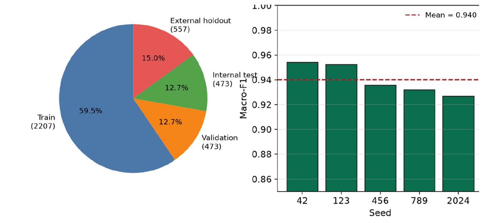
The experiments used the COVID-19 Radiography Database [19] and retained the COVID, NORMAL and PNEUMONIA folders. Perceptual hashes were computed to identify duplicate-image candidates before any train-validation-test allocation was made and duplicate groups were resolved so that a single visual image could not be included in more than one subset. The resulting corpus contains 3,710 unique images. The audit reported in this paper is an image-level duplicate audit. Patient-level independence cannot be inferred from it unless patient identifiers are available (Table 2).
Table 2. Dataset Partitioning After Image-Level Deduplication
Subset
Images
Approx. share
Role
Training
2,207
59.5%
Labeled and unlabeled optimization
Validation
473
12.7%
Checkpoint selection by Macro-F1
Internal test
473
12.7%
Primary performance evaluation
Image-disjoint holdout
557
15.0%
Additional same-database-family check
Total
3,710
100%
Unique audited CXRs
The audited images were further divided by stratified sampling into training, validation, internal test and an image-disjoint holdout, the latter of which is drawn from the same database family and is therefore treated as an additional image-disjoint check rather than as a multi-hospital external validation. The validation set was used only for checkpoint selection and the internal test and holdout sets were not used for optimization (Figure 1).
Figure 1: End-to-end Workflow for the Proposed Label-Efficient CXR Classification Study
Image Preprocessing and Augmentation
All images were resized to 160×160. The pixel values were scaled and standardized with the ImageNet channel statistics of the pretrained backbone. Grayscale radiographs were converted to the three-channel input format of the Mobile Net V3-Small. Weak augmentation consisted of horizontal flipping and mild rotation. Strong augmentation combined affine transformation, color jitter, Gaussian blur and random erasing. The weak view was used to create a stable pseudo-label and the strong view enforced invariance to larger appearance changes.
FixMatch Framework
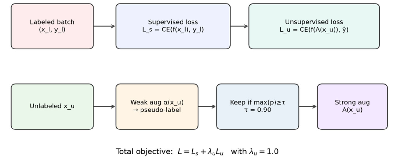
MobileNetV3-Small pretrained on ImageNet was chosen as the backbone because it has a good accuracy-to-computation trade-off [20]. Its final classifier was replaced by a three-class output layer. For a labeled mini-batch the supervised loss was the cross-entropy between the model prediction and the ground-truth label. For each unlabeled image a pseudo-label was computed from the weakly augmented view. The unsupervised loss was only applied when the largest class probability was at least τ = 0.90. The total objective combines the supervised and unsupervised terms, with λu=1.0 (Figure 2).
Figure 2. FixMatch Consistency-Regularization Mechanism Used in this Study
RESULTS
Label-Efficiency Results
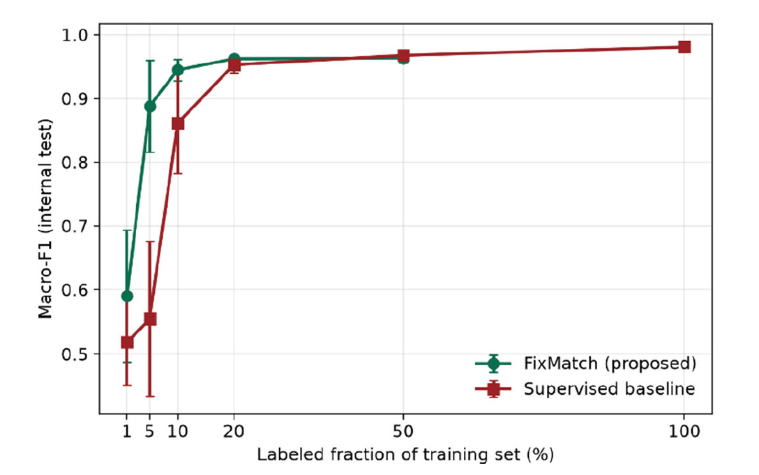
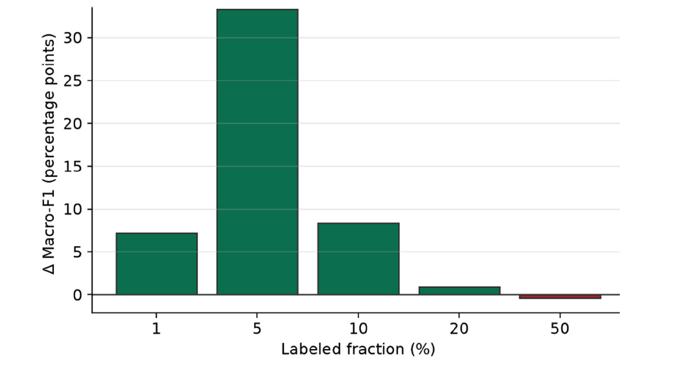
The internal-test means of the five seeds are summarised in Table 4. FixMatch reached 94.29% and 94.49% Macro-F1 at the primary hyperparameters (10% labelled), 8.35 percentage points below the matched supervised baseline on Macro-F1. The biggest gain came at 5%, with 33.30 percentage points and methods were close at 20% and slightly above at 50%. This is the same as what SSL is meant to do: when the labelled set is small the unlabeled images carry a lot of information and when it is large they carry only a little more (Figure 3,4).
Table 4: Mean Internal-Test Accuracy and Macro-F1 Over Five Seeds
Labeled
Sup. Acc.
Sup. Macro-F1
FixMatch Acc.
FixMatch Macro-F1
Δ Macro-F1
1%
60.97%
51.85%
65.88%
59.04%
+7.19 pp
5%
61.73%
55.50%
88.63%
88.80%
+33.30 pp
10%
86.22%
86.14%
94.29%
94.49%
+8.35 pp
20%
95.26%
95.32%
96.11%
96.23%
+0.91 pp
50%
96.62%
96.77%
96.19%
96.35%
−0.42 pp
100%
98.01%
98.09%
—
—
—
Figure 3: Internal-Test Macro-F1 Versus Labeled-Data Fraction (mean ± SD over five seeds)
Figure 4: Mean Macro-F1 Improvement of FixMatch Over Supervised Learning at Each Shared Label Budget
Training Behavior and Representative Error Analysis
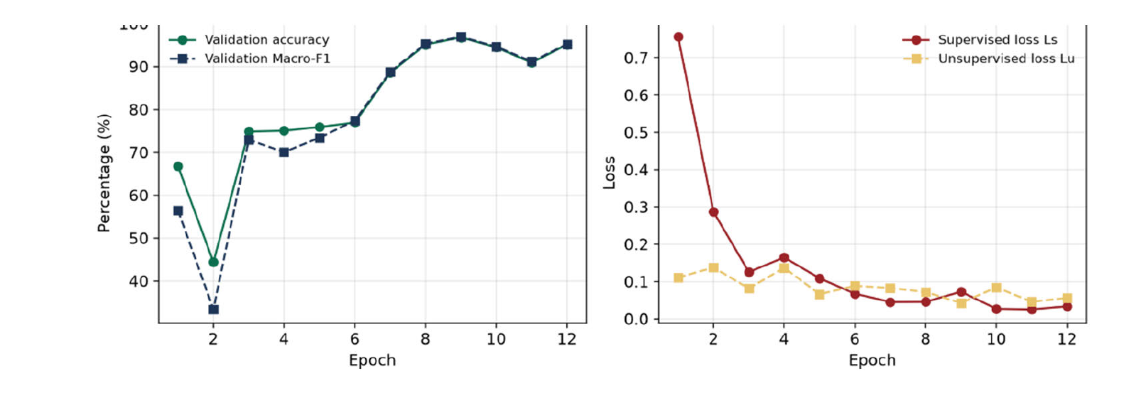
In the representative case (seed #42) the validation accuracy and Macro-F1 improved after the initial epochs and settled in the mid-high 90s. The total loss is a weighted sum of the supervised loss and the unsupervised loss and both have decreased, so the supervised part and the pseudo-labels are optimized jointly. The small changes in Figure 5 are what one should expect with a small set that is labelled temporarily and they reinforce the need to choose a checkpoint on a separate validation set, rather than on the final epoch (Table 5).
Figure 5: Validation Metrics and Training Losses for Fix Match at 10% Labels (seed 42)
Table 5: Class-Wise Performance for Fix Match at 10% Labels (seed 42, internal test)
Class
Precision
Recall
F1-score
Support
COVID
98.50%
99.24%
98.87%
132
NORMAL
92.27%
97.66%
94.89%
171
PNEUMONIA
98.11%
91.76%
94.83%
170
Macro average
96.29%
96.22%
96.20%
473
Overall accuracy
—
—
95.98%
473
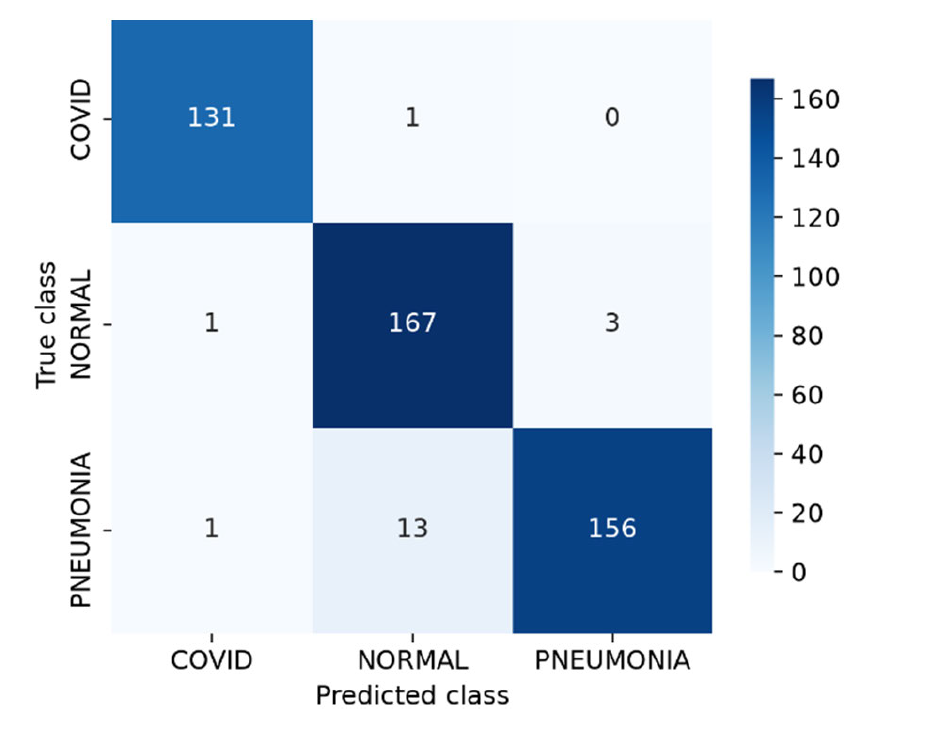
Only 454 images were correctly predicted (95.98% accuracy of the representative seed as demonstrated in the confusion matrix in Figure 6). The recognition result of COVID was the best (F1-score of 98.87%). The majority of residual errors were for the PNEUMONIA images predicted as NORMAL; therefore, the pneumonia recall dropped to 91.76%. It should be noted that the class-wise Macro-F1 value is reported as 96.20% (this is the corrected class-wise Macro-F1 for this run and not the class-wise Accuracy).
Figure 6: Confusion Matrix for Fix Match - 10% Labels (internal test set seed 42)
Image-Disjoint Holdout Evaluation
The image-disjoint holdout gave 93.68% mean accuracy and 94.03±1.23% mean Macro-F1 over the five 10%-label FixMatch checkpoints. There is a small spread across the seeds, which supports the main finding. The internal test and the holdout were based on the same database family. The holdout therefore defends against image-disjoint testing, but not against different populations of patients, acquisition protocols, manufacturers of scanners, or hospitals (Figure 7).
Figure 7: Audited Split Composition and Holdout Macro-F1 for Fix Match at 10% Labels by Random Seed
DISCUSSION
The key outcome is not just a high score, but a reduction in labeled-data requirements. Fix Match achieved most of the fully supervised upper-bound performance and greatly outperformed the supervised training at the same budget, because only 10% of the training images were labeled. The gain at 5% was large, which showed the ability of confidence-gated consistency learning to turn otherwise uninformative images into a useful training signal. MobileNetV3-Small is also designed to be suitable to low computation environments.
Among its strengths, the evaluation design contains: duplicate-image auditing before splitting, a matched supervised comparison, several label budgets, five seeds, a stated primary metric and an image-disjoint holdout. As a result, it is less likely that the ending will depend on a lucky single split or on the split itself. The representative confusion matrix also separates the clinically important error pattern (NORMAL vs. PNEUMONIA) and only summarises the metrics.
Important limitations remain. First, the holdout is not a genuinely independent hospital group. Second, the paper does not incorporate prospective readers, calibrated uncertainty, decision-curve analysis or evidence for lesion localization. Third, the experiment used 12 CPU-heavy epochs and a lightweight backbone; further architecture and hyperparameter searches could establish whether this was a generic result. Fourth, the duplicate audit is image rather than patient level and cannot substitute patient level grouping. Multi-center data, patient-disjoint partitions, calibration, reject-option triage, Grad-CAM or other explanations and comparison with other SSL methods are all worth following up.
CONCLUSION
A FixMatch-based framework with MobileNetV3-Small gave strong label efficiency for a three-class CXR classification. The mean accuracy and mean Macro-F1 on the internal test set using 10% of the labeled training data were 94.29% and 94.49% compared with 86.22% and 86.14% for the matched supervised baseline. The same was observed for an image-disjoint holdout drawn from the same database family. The advantage was greatest under severe label scarcity and diminished when 20-50% of the training set was labeled. This evidence supports semi-supervised learning as a practical annotation-reduction strategy in medical-image research. It does not, by itself, establish clinical safety or cross-hospital generalization.
Acknowledgment
Authors acknowledge the College of Engineering, University of Mosul and the COVID-19 Radiography Database for enabling this reproducible medical-image research.
Data Availability and Reproducibility
Source database. The COVID-19 Radiography Database [19] is publicly available. Final submission requirements. The split manifests, random seeds, duplicate-audit output and training configuration needed to reproduce the reported experiments should be supplied. Raw image pixels are not redistributed.
Author Contributions
Author contributions should be supplied before submission with the CRediT taxonomy: conceptualization, methodology, software, validation, formal analysis, writing, visualization and supervision.
REFERENCES
Ismael, A.M. and A. Şengür. “Deep learning approaches for covid-19 detection based on chest x-ray images.” Expert Systems with Applications, vol. 164, 2021, article 114054. https://doi.org/10.1016/j.eswa.2020.114054.
Narne, V. et al. “Evaluating deep learning models for accurate pneumonia diagnosis from chest x-rays.” Proceedings of the 2024 First International Conference on Innovations in Communications, Electrical and Computer Engineering (ICICEC), 2024, pp. 1–8. https://doi.org/10.1109/ICICEC62498.2024.10808495.
Kudagammana, S.T. et al. “Childhood mortality due to pneumonia: evidence from a tertiary paediatric referral center in Sri Lanka.” BMC Public Health, vol. 24, 2024, article 3351. https://doi.org/10.1186/s12889-024-20883-1.
El-Ghandour, M. and M.I. Obayya. “Pneumonia detection in chest x-ray images using an optimized ensemble with XGboost classifier.” Multimedia Tools and Applications, vol. 84, no. 9, 2025, pp. 5491–5521. https://doi.org/10.1007/s11042-024-18975-6.
Padash, S. et al. “Pediatric chest radiograph interpretation: how far has artificial intelligence come? A systematic literature review.” Pediatric Radiology, vol. 52, no. 8, 2022, pp. 1568–1580. https://doi.org/10.1007/s00247-022-05368-w.
Ali, M. et al. “Pneumonia detection using chest radiographs with novel efficientnetv2l model.” IEEE Access, vol. 12, 2024, pp. 34691–34707. https://doi.org/10.1109/ACCESS.2024.3372588.
Asnake, N.W. et al. “X-ray image-based pneumonia detection and classification using deep learning.” Multimedia Tools and Applications, vol. 83, 2024, pp. 60789–60807. https://doi.org/10.1007/s11042-023-17965-4.
Chen, J. et al. “Machine learning techniques for CT imaging diagnosis of novel coronavirus pneumonia: A review.” Neural Computing and Applications, vol. 36, no. 1, 2024, pp. 181–199. https://doi.org/10.1007/s00521-022-07709-0.
Sohn, K. et al. “FixMatch: Simplifying semi-supervised learning with consistency and confidence.” Advances in Neural Information Processing Systems 33, 2020, pp. 596–608.
Showkat, S. and S. Qureshi. “Efficacy of transfer learning-based resnet models in chest X-ray image classification for detecting covid-19 pneumonia.” Chemometrics and Intelligent Laboratory Systems, vol. 224, 2022, article 104534. https://doi.org/10.1016/j.chemolab.2022.104534.
Li, D. “Attention-enhanced architecture for improved pneumonia detection in chest x-ray images.” BMC Medical Imaging, vol. 24, 2024, article 6. https://doi.org/10.1186/s12880-023-01177-1.
Sajed, S. et al. “The effectiveness of deep learning vs. traditional methods for lung disease diagnosis using chest x-ray images: A systematic review.” Applied Soft Computing, vol. 147, 2023, article 110817. https://doi.org/10.1016/j.asoc.2023.110817.
Bahatheq, T.A.S. et al. “Detection of pneumonia from chest x-ray images using transfer learning and ensemble learning.” Academic Journal of Research and Scientific Publishing, vol. 5, no. 54, 2023, pp. 75–89. https://doi.org/10.52132/Ajrsp.e.2023.54.3.
Sanida, T. and M. Dasygenis. “A novel lightweight CNN for chest x-ray-based lung disease identification on heterogeneous embedded system.” Applied Intelligence, vol. 54, 2024, pp. 4756–4780. https://doi.org/10.1007/s10489-024-05420-2.
Roberts, M. et al. “Common pitfalls and recommendations for using machine learning to detect and prognosticate for covid-19 using chest radiographs and CT scans.” Nature Machine Intelligence, vol. 3, no. 3, 2021, pp. 199–217. https://doi.org/10.1038/s42256-021-00307-0.
Xie, Q. et al. “Unsupervised data augmentation for consistency training.” Advances in Neural Information Processing Systems 33, 2020, pp. 6256–6268.
Tarvainen, A. and H. Valpola. “Mean teachers are better role models: weight-averaged consistency targets improve semi-supervised deep learning results.” Advances in Neural Information Processing Systems 30, 2017, pp. 1195–1204.
Howard, A. et al. “Searching for mobilenetv3.” Proceedings of the IEEE/CVF International Conference on Computer Vision (ICCV), 2019, pp. 1314–1324. https://doi.org/10.1109/ICCV.2019.00140.
Chiu, M.-C. and C.-J. Wei. “Integrating deblurGAN and CNN to improve the accuracy of motion blur X-ray image classification.” Journal of Nuclear Science and Technology, vol. 61, no. 3, 2024, pp. 403–416. https://doi.org/10.1080/00223131.2023.2236106.
M.-C. Chiu and C.-J. Wei, “Integrating deblurGAN and CNN to improve the accuracy of motion blur x-ray image classification,” Journal of Nuclear Science and Technology, vol. 61, no. 3, pp. 403–416, 2024, doi: 10.1080/00223131.2023.2236106.
Recommended Articles
Research Article
Beyond Conventional Crimping: The Case for Cordless Thermal Jointing for Wires and Cables in Power Distribution and Construction Environment
Prashobh Karunakaran,
...
Tonny Ling
Published: 15/09/2026
Download PDF
Research Article
Harnessing Nanotechnology to Conserve Energy in Buildings
Nada S. Abdulmajeed
Published: 30/10/2022
Download PDF
Research Article
Some Properties of Fuzzy g-Regular Spaces
Hassan Abid Al-Hadi Ahmed,
Noor Al-Huda A. Haider
Published: 10/12/2025
Download PDF
Research Article
Selecting The Most Appropriate Type of Connection to Transmit the Necessary Amount of Flight Information on the Air to Ground Path
Al Kadhimi Ali Noori Mohammed,
Zaidoon Khalaf Mahmood
Published: 30/12/2023
Download PDF
PO Box 101, Nakuru
Kenya.
Email: office@iarconsortium.org
Editorial Office:
J.L Bhavan, Near Radison Blu Hotel,
Jalukbari, Guwahati-India
Abdulghani, M. M. (2026). Improvement of Medical Image Classification Based on Semi-Supervised Framework. Scientific Research Journal of Engineering and Computer Sciences, 6(2), 1-7.
MLA
Abdulghani, Mohammed Moath. "Improvement of Medical Image Classification Based on Semi-Supervised Framework." Scientific Research Journal of Engineering and Computer Sciences 6.2 (2026): 1-7.
Chicago
Abdulghani, Mohammed Moath. "Improvement of Medical Image Classification Based on Semi-Supervised Framework." Scientific Research Journal of Engineering and Computer Sciences 6, no. 2 (2026): 1-7.
Harvard
Abdulghani, M. M. (2026) 'Improvement of Medical Image Classification Based on Semi-Supervised Framework' Scientific Research Journal of Engineering and Computer Sciences 6(2), pp. 1-7.
Vancouver
Abdulghani MM. Improvement of Medical Image Classification Based on Semi-Supervised Framework. Scientific Research Journal of Engineering and Computer Sciences. 2026 ;6(2):1-7.



 Download PDF
 Download XML