Background: Pseudomonas aeruginosa is a main pathogens as a cause of healthcare acquired infection. This organism causes severe disaster of general health producing a great burden of mortality and morbidity in growing counties. AmpC-type enzymes are generally resistant to cephalosporins, but lower efficacy toward carbapenems. Nevertheless, carbapenem resistance is noted when AmpC enzymes combine with reduced carbapenem cell penetration due to a loss of porin protein. Overproduction of AmpC β-lactamases combinations occurring when associated with ESBLs and modified membrane channels leads to increasing carbapenem resistance of P. aeruginosa clinical isolates. Study Aim: This thesis is designed to identify and assess the frequency of P. aeruginosa clinical isolates, to examine the occurrence of extensively drug resistant P. aeruginosa in hospitals and to examine the presence of common AmpC β-Lactamases genes in XDR isolates in Najaf City-Iraq. Materials and Method: Basic test including stain of Gram, morphology of the colony and conventional biochemical test were used to isolate and identify clinical isolates of P.aeruginosa. Upon receipt, swab specimens were immediately cultured onto MacConkey agar and the plate were incubated for 24 hours at 37°C. Results: During 3-months, an overall of 1070 clinical specimens were collected from six main hospitals and Burn Center in Najaf province. Present results showed that 66 isolates of P. aeruginosa were obtained from clinical sources. The clinical sample type’s collected during the study period included 821 (76.6%) urine specimens from patients suspected with UTIs, 85 (7.9%) wound exudate, 71 (6.6%) ear swab, 56 (5.2 %) burn exudate and 37 (3.4%) CSF aspiratesas shown in table (3-2). This study showed that 13 isolates from 66 isolates recognized as XDR P. aeruginosa isolates. This study showed that 13 (19.6%) isolates from 66 isolates recognized as XDR P. aeruginosa isolates. Among the 13 XDR isolates, PCR assay showed that all of these isolates harbored blaAmpC and blaCMY AmpC beta lactamase genes by using specific primers for each one.
Keywords
Pseudomonas Aeruginosa
Nosocomial Infection
Cerebrospinal Fluid
Burn Exudate
Wound Exudate
Ear Discharge and Urine
XDR
AmpC
INTRODUCTION
Pseudomonas aeruginosa is a main pathogens as a cause of healthcare acquired infection [1]. This organism causes severe disaster of general health producing a great burden of mortality and morbidity in growing countries [2]. The most worrisome feature of this pathogen is the low sensitivity to antibiotics, which is attributable to low permeability of action of multidrug efflux and bacterial cellular envelopes. P. aeruginosa has a special predilection for water and as result, it has been obtained from disinfectants, solutions of contact lens, hot tubes and cosmetics, all of which have been considered as infection sources [3]. However, it is most commonly present in small numbers in the gastrointestinal tract. It may transiently colonize various surfaces of moist skin including on the perineum and under the arm. The common sites that have been colonized with P. aeruginosa are nose and throat as well [3]. Pseudomonas aeruginosa is often the cause of urinary tract, wound and respiratory tract infections. In a previous study, Al-Muhannak [4] reported that P. aeruginosa was the most frequently isolated species (30.5%) in Najaf hospitals. In IWK Health Centre, Canada, P. aeruginosa accounts for approximately 15% of all hospital-acquired infections [5]. Pseudomonas aeruginosa is a significant bacteria in nosocomial infections, the variety of mucoid being of special interest since it is responsible for the most of death among patients’ of Cystic Fibrosis (CF) in spite treatment of intensive antibiotics [6]. Recurrent infections lead to altars in the epithelium of airways, which in turn permit in most patients, the strain persists for a long period and the infection becomes extensive [7].
AmpC-type enzymes are generally resistant to cephalosporins, but lower efficacy toward carbapenems [8]. Nevertheless, carbapenem resistance is noted when AmpC enzymes combine with reduced carbapenem cell penetration due to a loss of porin protein [9]. Overproduction of AmpC β-lactamases combinations occurring when associated with ESBLs and modified membrane channels leads to increasing carbapenem resistance of P. aeruginosa clinical isolates [10]. AmpC β-lactamase has been found as chromosomal mediated in P. aeruginosa [11].
A cephamycin-resistant strain of P. aeruginosa that produced chromosome mediated AmpC β-lactamase (CMY-1), was initially recorded in Korea in 1989 [12]. These enzymes make the bacteria resistant to connections of penicillin/β-lactamase inhibitors, penicillin, cephalosporins ensuring aztreonam and cephamycins [13].
MATERIALS AND METHODS
Collection of Samples
The study happened through a 3-month period from September to December 2023. The samples were collected from seven main hospitals (Medical City of Al-Sadder, General Hospital of Al-Hakeem, Al-Forat General Hospital, Al-Zahra Teaching Hospital for Children, Al-Sajad Hospital, Al-Manathera Hospital and Burn Center) located in Najaf city.
Demographic details including age, gender, location and site of infection and duration of hospitalization were collected from the medical and laboratory records of each patient recorded in the present thesis. All specimens were identified and analyzed anonymously and all the patients were assigned code numbers.
Clinical specimens of various infection sites were obtained during the study period from patients showing signs and symptoms suggestive of infection. The samples were distributed as burn exudate, wound exudate, ear discharge, Cerebrospinal Fluid (CSF) and urine. One sample was collected from each registered patient who had attained the inclusion criteria. Pus samples were aseptically collected from clinically deep areas of ear, burn and wound using a sterile cotton swab moistened with saline. The swab was placed straight into the transport medium and labeled. The CSF is aspirated by needles, collected into sterile screw-cap tubes and transported to the laboratory within 1 hour for culture and analysis. Clean catch midstream samples of urine were obtained utilizing wide, sterile glass bottles with tops of screw caps. All collected specimens were carried to the Department of Microbiology lab and processed immediately for identification by standard microbiological tests.
Isolation and Identification of P. aeruginosa
Basic tests including stain of Gram, morphology of the colony and conventional biochemical test were used to isolate and identify clinical symptoms of P. aeruginosa. Upon receipt, swab specimens were immediately cultured onto MacConkey agar and the plate were incubated for 24 hours at 37°C. Samples of urine were inoculated utilizing a calibrated 0.01 mL urine plastic loop on agar of MacConkey and BA plates. The plates were incubated for 24 h at 37°C. Specimens of urine were considered positive for urinary tract infections if the colony numbers exceeded 105 Colony-Forming Units (CFU)/mL [1]. The initial detection of known isolates of bacteria was made according to pigmentation and colony appearance. Then, colonies of isolates were primarily stained of Gram by obtaining smear from isolated colonies. Stained smear was investigated microscopically to view microscopic features. Based on the Bergey’s Manual of Determinative Bacteriology guidelines [15] and as described by MacFaddin [16], the isolates were further biochemically characterized and detected. All isolates were confirmed by the VITEK2 system utilizing the GN AST 222 card.
AmpC β-Lactamases Genes Molecular Screening
This thesis was carried out to discover the dissemination of the AmpC β-Lactamases genes in the XDR isolates of P. aeruginosa collected from sixth hospitals of Najaf through a period of 3 months. The early detection of AmpC β-Lactamases genes may inactivate the dissemination of these XDR isolates in the future.
RESULTS
Sample Sources
During the 3-month period of cross-sectional thesis, an overall of 1070 clinical specimens were collected from six main hospitals and Burn Center in Najaf province. The clinical sample type’s collected during the study period included 821 (76.7%) urine specimens from patients suspected with UTIs, 85 (7.9%) wound exudate, 71 (6.6%) ear swab, 56 (5.2%) burn exudate and 37 (3.4%) CSF aspiratesas shown in Table 2.
Overall, these clinical samples were more frequently obtained from Medical City of Al-Sadder (334, 31.2%) followed by General Hospital of Al-Hakeem (271, 25.3%), Al-Forat General Hospital (144, 13.4%), Al-Zahra Teaching Hospital for Maternity and Children (100, 9.3%), Al-Sajad Hospital (85, 7.9%), Al-Manathera Hospital (74, 6.9%) and Burn Center (62, 5.7%).
Prevalence and Sources of P. aeruginosa Isolates
The present study included a total of 1070 clinical specimens. Out of them, 66 (6.2%) non-duplicate P. aeruginosa isolates were identified in Table 1. However, the isolates were primarily detected by standard culture and biochemical tests and finally reconfirmed using the VITEK2 compact automated system.
Pseudomonas aeruginosa isolates sources (n = 66) based on clinical sample type as shown in Table 2.
Table 1: Pseudomonas aeruginosa Isolates Distribution according to Sample Sources
Sample source
Samples No.
No. of isolates of P. aeruginosa
%
Clinical
1070
66
6.1
Table 2: Pseudomonas aeruginosa Clinical Isolates Depending on Clinical Sample and Hospital Origin
Hospital origin
No of P. aeruginosa isolated from
Total (%)
Urine (n = 821
Wound (n = 85)
Ear (n = 71)
Burn (n = 56)
CSF (n = 37)
Al-Sadder (n = 334)
2
2
1
4
1
10 (3)
Al- Hakeem (n = 271)
4
4
1
4
1
14 (5.1)
Al-Forat (n = 144)
3
3
2
2
1
11 (7.6)
Al-Zahra (n = 100)
3
-
-
-
1
4 (4)
Al-Sajad (n = 85)
2
1
2
4
1
10 (11.8)
Al-Manathera (n = 74)
1
1
-
1
-
3 (4)
Burn Center (n = 62)
4
3
1
6
-
14 (22.6)
Total (%)
19 (2.3%)
14 (16.5%)
7 (9.9%)
21 (37.5%)
5 (13.5 %)
66
Table 3: Demographic Features of the Patients Infected with P. aeruginosa (n = 66)
Patients profile
Status
No. (%) of P. aeruginosa
Age group (in years)
1-10
5 (7.6)
11-30
35 (53)
31-50
19 (28.8)
51-70
7 (10.6)
Gender
Male
39 (59)
Female
27 (41)
Most of the clinical isolates were recovered from burn exudate (21, 37.5%) and wound exudate (14, 16.5%), followed by CSF aspirate (5, 13.5%), ear discharge (7, 9.9%) and urine (19, 2.3%). Though, the occurrence of P. aeruginosa isolates in the condition of burned patients was more important than other patients.
The hospital-wise distribution of clinical isolates of P. aeruginosa is observed in Table 2. The most common site from which P. aeruginosa isolates were obtained was Burn Center (14/62, 22.6%), followed by Al-Sajaad Hospital (10/85, 11.8%), Al-Forat General Hospital (11/144, 7.6%), Al-Hakeem General Hospital (14/271, 5.1%), Al-Manathera Hospital (3/74, 4%), Al-Zahra Teaching Hospital for Maternity and Children (4/100, 4%) and Al-Sadder Medical City (10/334, 3%). However, the occurrence of P. aeruginosa isolates in the Burn Center was significantly higher than other hospitals.
Pseudomonas aeruginosa isolates sources depending on the age, hospitalization and gender are presented in Table 3. In case of gender-wise occurrence, Pseudomonas aeruginosa was more common in male patients (39, 59%) as compared to female patients (27, 41). Looking over the age group wise frequency of P. aeruginosa isolates, the patients age ranged from (1-70) years. The high number of infected cases in the age group of (11-30) years (n = 35; 53%), followed by 19 (28.8%), 7 (10.6%) and 5 (7.6%) infected patients, in the age group 31-50 years, 51-70 years and 1-10 years, respectively.
Prevalence of AmpC Beta Lactamase Genes among XDR P. aeruginosa Isolates.
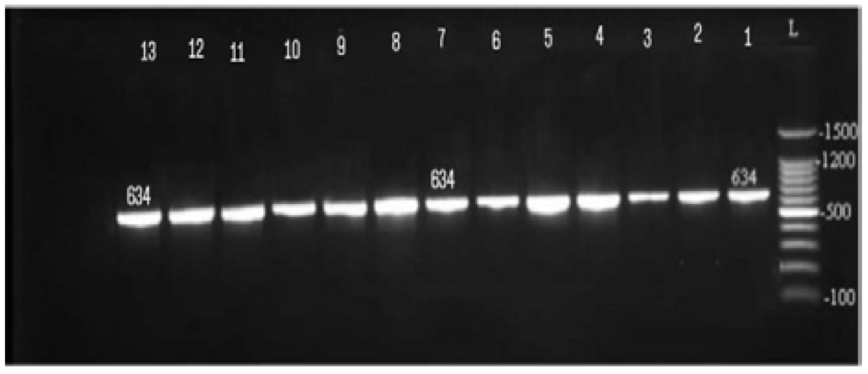
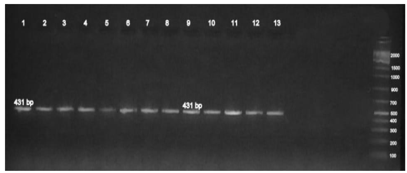
This study showed that 13 (19.6%) isolates from 66 isolates recognized as XDR P. aeruginosa isolates (isolates exhibiting resistance to at least 1 antibiotic in all but one or two classes of antibiotic [18]. About 9 isolates are from burns and 4 are from urine. Among the XDR isolates, PCR assay showed that all of these isolates harbored blaAmpC and blaCMY AmpC beta lactamase genes by using specific primers for each one (Table 4 and Figure 1,2).
Table 4: Dissemination of AmpC β-Lactamase Genes among the 13 P.aeruginosa Isolates
Type of gene
No. (%) of isolates
Isolate code No.
blaAmpC
13 (100%)
All isolates
blaCMY
13 (100%)
Figure 1: Gel of Agarose with Red Save Stain of Mono-Plex PCR Amplified Product Isolates DNA of Isolates with blaAmpC Primer. The Electrophoresis is used for 120 min at 65 Volts. Lane (L) is a Marker of DNA Molecular Size (1500–bp ladder). Lanes (1-13) Show Positive Results with blaAmpC gene (634 bp)
Figure 2: Gel of Agarose with Red Save Stain of Mono-Plex PCR Amplified Product Isolates DNA of Isolates with blaCMY Primer. The Electrophoresis is used for 120 min at 65 volts. Lane (L) is a Marker of DNA Molecular Size (2000–bp ladder). Lanes (1-13) show Positive Results with blaCMY Gene (431 bp)
DISCUSSION
Distribution of Hospital Pseudomonas Aeruginosa Isolates
In Najaf province, P. aeruginosa has been reported as the most commonly isolated organism in hospitals [17,19]. One objective of the current cross-sectional thesis is to evaluate the occurrence of clinical isolates of P. aeruginosa in Najaf hospitals and to find out whether P. aeruginosa has shown up in clinical samples.
The present study showed that 66 (6.2%) P. aeruginosa isolates were obtained and identified from several clinical specimens in Najaf hospitals (Table 1). When compared to previous results in Iraq, higher frequency rates were recorded in other similar studies in Hilla [20] and Al-Nasiriya [21], in which 28.2 and 18.3% P. aeruginosa prevalence rates were reported, respectively. This difference in rate of prevalence among above results due to differences in sample types, geographical location and hygienic strategies.
Regarding clinical samples, 1070 of various body site samples were collected from patients exposed to bacterial infection during a span of four months. Results show that of all the samples screened, only 66 (6.2%) were positive for P. aeruginosa (Table 1). The occurrence observed in this study was in line to that obtained in a study conducted in Najaf hospitals where they observed a prevalence rate of 6% [19].
The current result reported that the main clinical sample from which isolates of Pseudomonas aeruginosa were collected was burn exudate (37.5%). This is higher than the recent result reported by Al-Janahi [19], who showed Pseudomonas aeruginosa in infection of burn with a percent of 28.5% and may be due to inappropriate infection control measures to burn patients. The elevated incidence of P. aeruginosa infections in burn patients in this thesis and the widespread use of multiple antibiotics may be one of the most significant effects on the rate of mortality in patients.
Present results revealed that wound exudate samples were the second most common source of isolation of P. aeruginosa (16.5%).This result was in considerable concinnity with the findings of Al-Janahi [19] who reported a percentage of 15%. Furthermore, the observations on the frequency of P. aeruginosa isolates in wound exudate samples were similar to those of other authors noting that the isolation rate of this bacterium from wound exudates was high among clinical samples [22].
In the current work, the third most common source of isolation of P. aeruginosa was the CSF aspirate (13.5%), which showed that it may be important in causing meningitis among patients. However, in meningitis, P. aeruginosa infection is linked with 40 to 80% mortality [23].
Present result indicates that the distribution of the P. aeruginosa isolates is significantly affected by the type of hospital from which they have been isolated (Table 3-2). The highest number of isolates was from Burn Center (22.6%), most of which originated from burn exudate samples. on the other hand, Pseudomonas aeruginosa isolates were at a less occurrence average as in hospital of Al-Sajad were incidence of Pseudomonas aeruginosa of (11.8)% followed by Al-Forat General Hospital (7.6%), Al-Hakeem General Hospital (5.1%), Al-Manathera Hospital (4%), Al-Zahra Hospital for Children (4%), Al-Sadder Medical City (3%). This variation among hospitals may be associated with the specification of each hospital for admitting patients suffering from specific diseases correlated to those hospitals. As well, high rate detected in Burn Center, may be correlated with that most patients suffering from burns admit in the Burn Center for a long period. In addition, the contamination of isolates in hospitals play an important role in causing nosocomial infections [24].
According to gender, a higher proportion of males (59%) were detected positive for P. aeruginosa than their females (41%) indicating that males show greater vulnerability for infections (Table 3). While, previous study reported prevalence of P. aeruginosa in females (54.1%) than in males (45.9%) in this locality [19]. Another important parameter in this study is age. A high incidence (53%) of P. aeruginosa infections was found in the 11-30 years age group followed by 31-50 years (28.8%). Very young and very old patients had overall lower rates of infection than did other age groups. This study disagreed with several studies which showed the elevated rate of infection of P. aeruginosa among age groups more than 50 years [25].
Over the last decades, the occurrence of P. aeruginosa producing AmpC β-lactamases is increasing globally. Therapeutic options for infections with this bacterium are limited not only by the production of β-lactamases, which confer resistance to all β-lactams, but also by frequent co- resistance to other antimicrobial agents even including colistin and tigecycline [26,27]. One objective of this thesis was to obtain a better understanding about the dissemination of AmpC β-lactamases among 13 XDR P. aeruginosa isolates.
AmpC β-lactamases (such as CMY) confer resistance to penicillins, cephalosporins, aztreonam as well as to cephamycins (cefoxitin) and are poorly inhibited by clavulanic acid [28]. Cefoxitin resistance is supposed to be a discriminative indicator for the identification of AmpC producing isolates [29]. In this study, all the 13 XDR isolates were phenotypic tested for the production of AmpC β-lactamases using the cefoxitin disk method. This thesis showed that all isolates exhibited resistance to cefoxitin and all of which harbored blaAmpC and blaCMY genes with PCR method. The main finding of this thesis was the report of the prevalence of CMY β-lactamase for the initial time in Iraq. However, resistance caused by ESBL production is more common than resistance caused by AmpC β-lactamase in most parts of the world [30]. In addition, AmpC β-lactamases are capable of conferring carbapenem resistance when combined with the mutation of porins [31,32].
CONCLUSION
The current study showed that the most common clinical sample from which P. aeruginosa isolates were obtained was burn exudate. The elevated incidence of P. aeruginosa infections in burn patients in the present study and widespread multiple antibiotics resistance may be one of the major effects on the rate of mortality in patients. The high XDR P. aeruginosa isolates were documented and all these isolates show resistance to most available antibiotics. Present findings indicated the high occurrence of AmpC β-lactamases among XDR P. aeruginosa isolates.
Conflict of Interest
This thesis showed there is no interest collision.
Ethical Approval
Ethics committee show there is no wrong study and there is no plagiarism in this result.
REFERENCES
Lambert, M.L. et al.Clinical Outcomes of Health-Care-Associated Infections and Antimicrobial Resistance in Patients Admitted to European Intensive-Care Units: A Cohort Study.The Lancet Infectious Diseases, vol. 11, no. 1, 2011, pp. 30–38.
Rashid, A. et al. “Clinical Features and Epidemiological Patterns of Infections by Multidrug Resistance Staphylococcus aureus and Pseudomonas aeruginosa in Patients with Burns.” Biomedical Journal of Scientific and Technical Research, vol. 25, no. 4, 2020, pp. 19272–19278.
Moore, N.M. and M.L. Flaws. “Antimicrobial Resistance Mechanisms in Pseudomonas aeruginosa.” Clinical Laboratory Science, vol. 24, 2011, pp. 47–51.
Al-Muhannak, F.H. Spread of Some Extended Spectrum Beta Lactamases in Clinical Isolates of Gram-Negative Bacilli in Najaf. M.Sc. thesis, College of Medicine, University of Kufa, 2010.
Meletis, G. et al. “Double and Multi Carbapenemase Producers: The Excessively Armored Bacilli of the Current Decade.” European Journal of Clinical Microbiology and Infectious Diseases, vol. 34, no. 8, 2015, pp. 1487–1493.
Sadikot, R.T. et al. “Pathogen-Host Interactions in Pseudomonas aeruginosa Pneumonia.” American Journal of Respiratory and Critical Care Medicine, vol. 171, no. 11, 2005, pp. 1209–1230.
Ciofi degli Atti, M.L. et al. “Trend of Healthcare-Associated Infections in Children: Annual Prevalence Surveys in a Research Hospital in Italy, 2007–2010.” Journal of Hospital Infection, vol. 80, 2012, pp. 6–12.
Poirel, L. et al.. “Emergence of NDM-1-Producing Klebsiella pneumoniae in Morocco.” Journal of Antimicrobial Chemotherapy, vol. 66, no. 12, 2011, pp. 2781–2783.
Qin, X. et al. “Prevalence and Mechanisms of Broad-Spectrum Beta-Lactam Resistance in Enterobacteriaceae: A Children’s Hospital Experience.” Antimicrobial Agents and Chemotherapy, vol. 52, 2008, pp. 3909–3914.
Neyestanaki, D.K. et al.. “Characterization of AmpC, CTX-M and MBLs Types of β-Lactamases in Clinical Isolates of Klebsiella pneumoniae and Escherichia coli.” Jundishapur Journal of Microbiology, vol. 7, no. 2, 2014, pp. 31–36.
Denton, M. “Enterobacteriaceae.” International Journal of Antimicrobial Agents, vol. 29, 2007, pp. 9–22.
Wassef, M. et al. “Genotypic Identification of AmpC β-Lactamases Production in Gram-Negative Bacilli Isolates.” Jundishapur Journal of Microbiology, vol. 7, no. 1, 2014, pp. 55–61.
Paltansing, S. et al. “Characterization of Fluoroquinolone and Cephalosporin Resistance Mechanisms in Enterobacteriaceae Isolated in a Dutch Teaching Hospital.” Journal of Antimicrobial Chemotherapy, vol. 68, 2013, pp. 40–45.
Collee, J.G. et al.Mackie and McCartney’s Practical Medical Microbiology. 4th ed., Churchill Livingstone, 1996.
Parte, A. Bergey’s Manual of Systematic Bacteriology: Actinobacteria. Springer, 2012.
MacFaddin, J.F. Biochemical Tests for Identification of Medical Bacteria. 3rd Edn., Lippincott Williams and Wilkins, 2000.
Al-Shara, J.M. Phenotypic and Molecular Detection of Carbapenem-Resistant Pseudomonas aeruginosa in Najaf Hospitals. PhD thesis, University of Kufa, 2013.
Magiorakos, A.P. et al. “Multidrug-Resistant, Extensively Drug-Resistant and Pandrug-Resistant Bacteria.” Clinical Microbiology and Infection, vol. 18, 2012, pp. 268–281.
Al-Janahi, H.C. Occurrence and Molecular Characterization of Metallo-β-Lactamase-Producing Pseudomonas aeruginosa in Najaf Hospitals. M.Sc. thesis, University of Kufa, 2020.
Saleh, R.H. Immunological and Molecular Study on Pseudomonas aeruginosa Isolated from Clinical Samples in Babylon Province. Babylon University, 2012.
Abdul-Wahid, A.A. Dissemination of Aminoglycosides Resistance in Pseudomonas aeruginosa Isolates in Al-Nasiriya Hospitals. M.Sc. thesis, University of Kufa, 2014.
Alharbi, S.A. and M.E. Zayed. “Antibacterial Susceptibility of Bacteria Isolated from Burns and Wounds of Cancer Patients.” Journal of the Saudi Chemical Society, vol. 18, no. 1, 2014, pp. 3–11.
Juhi, T. et al. “Pseudomonas aeruginosa Meningitis in Post-Neurosurgical Patients.” Neurology Asia, vol. 14, no. 2, 2009, pp. 95–100.
Hammami, S. et al. “Diversity in VIM-2-Encoding Class 1 Integrons.” Clinical Microbiology and Infection, vol. 16, 2010, pp. 189–193.
Ahmed, M.S. et al.“Emergence of Multidrug and Pandrug Resistant Pseudomonas aeruginosa from Five Hospitals in Qatar.” Infection Prevention in Practice, vol. 1, nos. 3–4, 2019, article 100027.
Gomez, J. et al. “Clinical Relevance of Bacterial Resistance: A Historical Approach (1982–2007).” Revista Española de Quimioterapia, vol. 21, 2008, pp. 115–122.
Rubio-Perez, I. et al. “Extended Spectrum Beta-Lactamase-Producing Bacteria in a Tertiary Care Hospital in Madrid.” Emerging Health Threats Journal, vol. 5, 2012, pp. 11589–11594.
Jeon, J. et al. “Structural Basis for Carbapenem-Hydrolyzing Mechanisms of Carbapenemases.” International Journal of Molecular Sciences, vol. 16, 2015, pp. 9654–9692.
Peter-Getzlaff, S. et al. “Detection of AmpC Beta-Lactamase in Escherichia coli.” Journal of Clinical Microbiology, vol. 49, no. 8, 2011, pp. 2924–2932.
Fam, N. et al. “Prevalence of Plasmid-Mediated AmpC Genes in Clinical Isolates of Enterobacteriaceae.” British Microbiology Research Journal, vol. 3, no. 4, 2013, pp. 525–537.
Sobia, F. et al. “Occurrence of blaAmpC in Cefoxitin-Resistant Escherichia coli and Klebsiella pneumoniae.” New Zealand Journal of Medical Laboratory Science, vol. 65, 2011, pp. 5–9.
Shahid, M. et al. “Molecular Epidemiology of Carbapenem-Resistant Enterobacteriaceae.” Medical Laboratory Science Journal, vol. 66, 2012, pp. 5–7.
Recommended Articles
Research Article
Knowledge of the Patient's Family about the Level of Emergency Based on Triage with the Satisfaction of the Patient's Family in the Emergency Department of the Sumedang Area General Hospital
Roganda Situmorang,
...
Raida Rihadatul Aisyi
Published: 30/03/2024
Download PDF
Research Article
The Influence of Murottal Al Qur'an Surat Ar Rahman Irama Hijaz Therapy on the Pain Scale of Post-Explorative Laparatomy Patients in the Icu Room of Bandung City Hospital
Andri Nurmansyah,
...
Fifi Siti Fauziah Yani
Published: 30/03/2024
Download PDF
Research Article
The role of IFN-g gene polymorphism (+874 T/A) as predisposing factor for pulmonary tuberculosis
Download PDF
Research Article
Caesarean Scar Pregnancy
Komal Sharma,
...
Jyotshna Sharma
Published: 02/02/2023
Download PDF
PO Box 101, Nakuru
Kenya.
Email: office@iarconsortium.org
Editorial Office:
J.L Bhavan, Near Radison Blu Hotel,
Jalukbari, Guwahati-India
Al-Jelehawy, A. H. J. & Al-Faham, Q. M. H. (2024). Incidence of AmpC β-Lactamases Genes among Extensively Drug Resistant Pseudomonas Aeruginosa Isolated from Clinical Sources in Al-Najaf City-Iraq. Scientific Research Journal of Clinical and Medical Sciences, 4(1), 1-5.
MLA
Al-Jelehawy, Alaa H. J. and Qassim M. H. Al-Faham. "Incidence of AmpC β-Lactamases Genes among Extensively Drug Resistant Pseudomonas Aeruginosa Isolated from Clinical Sources in Al-Najaf City-Iraq." Scientific Research Journal of Clinical and Medical Sciences 4.1 (2024): 1-5.
Chicago
Al-Jelehawy, Alaa H. J. and Qassim M. H. Al-Faham. "Incidence of AmpC β-Lactamases Genes among Extensively Drug Resistant Pseudomonas Aeruginosa Isolated from Clinical Sources in Al-Najaf City-Iraq." Scientific Research Journal of Clinical and Medical Sciences 4, no. 1 (2024): 1-5.
Harvard
Al-Jelehawy, A. H. J. and Al-Faham, Q. M. H. (2024) 'Incidence of AmpC β-Lactamases Genes among Extensively Drug Resistant Pseudomonas Aeruginosa Isolated from Clinical Sources in Al-Najaf City-Iraq' Scientific Research Journal of Clinical and Medical Sciences 4(1), pp. 1-5.
Vancouver
Al-Jelehawy AHJ, Al-Faham QMH. Incidence of AmpC β-Lactamases Genes among Extensively Drug Resistant Pseudomonas Aeruginosa Isolated from Clinical Sources in Al-Najaf City-Iraq. Scientific Research Journal of Clinical and Medical Sciences. 2024 Jan;4(1):1-5.


 Download PDF
 Download XML