Fungal infections have emerged as a worldwide healthcare problem in recent years due to extensive use of broad spectrum antibiotics, long term use of immunosuppressive agents, increased use of indwelling devices and increasing population of terminally ill, debilitated and immune-compromised patients. The local patterns of fungal isolates from clinical specimen may change with time and geographical area and it is important to be familiar with recent local trends in order to improve patient management, hence the need for surveillance. Aim of Study: To study the spectrum of fungal isolates from clinical Specimens in a Tertiary Care Hospital in Sub-Himalayan Region. Material and Methods: Retrospective study was done at Indira Gandhi Medical College, Shimla from July 2021-June 2022. Samples received in mycology laboratory were processed using direct microscopy with 10%, 20% and 40% potassium hydroxide (KOH), India ink and Gram staining to visualize the presence of fungal elements. All the samples were inoculated on Sabouraud’s dextrose agar (SDA) one with chloramphenicol and other with cycloheximide, then further incubated at 250 and 370 C. Result: Of the 752 clinical samples 273 (36%) had fungal growth, 300 (40%) were contaminants and 179 (24%) showed no growth. Among the respiratory samples (369), 121 samples (33%) were positive in fungal culture. The other major samples received were skin scrapings (81), skin biopsy (56) and oral mucosal scrapings (25) in which fungal growth was observed in 28 (35%), 21 (38%) and 17 (68%) respectively. Most common isolate observed among dermatophytes was T.mentagrophytes 15 (71%) followed by T.rubrum 3 (14%) and T.violaceum 2 (10%). Sporothrix schenckii 16 (57%) was predominant in patients suspected of subcutaneous mycosis.Candida albicans 41 (87%) was isolated in reproductive age group females followed by Candida glabrata 4 (9%) and Candida tropicalis 2 (4%).In corneal scrapings, growth of Fusarium 4 (27%), Cladosporium 4 (27%) and Aspergillus fumigatus 3 (20%) was obtained. Conclusion: Predominant fungal isolate incriminated in superficial fungal infections was T.mentagrophytes followed by T.rubrum and T.violaceum.Sporothrix schenckii was the major isolate in patients suspected of subcutaneous mycosis.Candida albicans was the most common isolate in reproductive age group females followed by Candida glabrata and Candida tropicalis. In corneal scrapings, different fungal pathogens were Fusarium , Cladosporium and Aspergillus fumigatus.
Keywords
Spectrum
Fungal isolates
Clinical Specimens
Tertiary Care Hospital
Sub-Himalayan Region
INTRODUCTION
Fungal infections have emerged as a worldwide healthcare problem in recent years [1] due to extensive use of broad spectrum antibiotics [2], long term use of immunosuppressive agents, increased use of indwelling devices [3], increasing population of terminally ill, debilitated and immunocompromised patients [4]. Environmental contaminants have now emerged as significant human pathogens.The local patterns of fungal isolates from clinical specimen may change with time and geographical area hence the need for fungal surveillance to improve patient management.
There is an emergence and re-emergence of different types of fungal diseases of paramount significance which are caused by saccharomyces cerevisiae, candida dubliensis, scedosporium, apiosporum, penicillium marneffei and fusarium sps. Some of fungi, which have been reported as merely environmental contaminants have now emerged as significant human pathogens e.g. apophysomyces elegans and Saksenaea vasiformis may lead to fatal consequences even in immunocompetent individuals [5].
Candida and Aspergillus species are the most common causes of fungal infection but other yeasts and filamentous fungi are emerging as pathogens. Among the filamentous fungi, apart from Aspergillus spp, others like Fusarium spp., Scedosporium spp., Penicillium spp. and Zygomycetes are becoming increasingly common [6,7]. Although C. albicans is the most prevalent species involved in causing fungal infections, the incidence of infections due to non-albicans species is increasing particularly in patients treated in the intensive care unit [8]. Though several reports on fungal profile are available from different parts of country.
The local patterns of fungal isolates from clinical specimen may change with time and geographical area and it is important to be familiar with recent local trends in order to improve diagnosis. The present study was undertaken with a view to find local patterns of fungal isolates from various clinical samples received at our tertiary care hospital.
MATERIALS AND METHODS
All samples received from the patients, suspected of fungal infections (1st July 2021 to 30th June, 2022) in the department of Microbiology, Indira Gandhi Medical College, Shimla were included in the study.Samples included blood, urine, body fluids, skin scrapings, corneal scrapings, respiratory samples, penile and vaginal discharge. Relevant demographic and clinical data was collected.All clinical specimens were collected under appropriate clinical guidelines and proper criteria were maintained during the transportation of specimen.
All samples were analyzed by direct microscopy and culture as per standard microbiological procedures. For direct microscopy, 10%, 20% and 40% potassium hydroxide (KOH), Nigrosin and Gram staining was done to visualize presence of fungal elements.For fungal culture, all samples were inoculated on two isolation media: one in Sabouraud’s dextrose agar (SDA) with chloramphenicol and other with chlorhexidine (0.05mg/ml, 0.5mg/ml). The culture tubes were incubated at 25ºC and 37ºC and examined for six weeks. The identification of fungi was done by macroscopic and microscopic evaluation of the fungal morphology. Macroscopic identification was done by observing colony morphology.Microscopic identification was done by LCB mount/ slide culture/ Gram’s staining/ germ tube test/ Dalmau method and by using CHROM agar and biochemical tests.Candida speciation was done by BD Phoenix TM automated system.
RESULTS
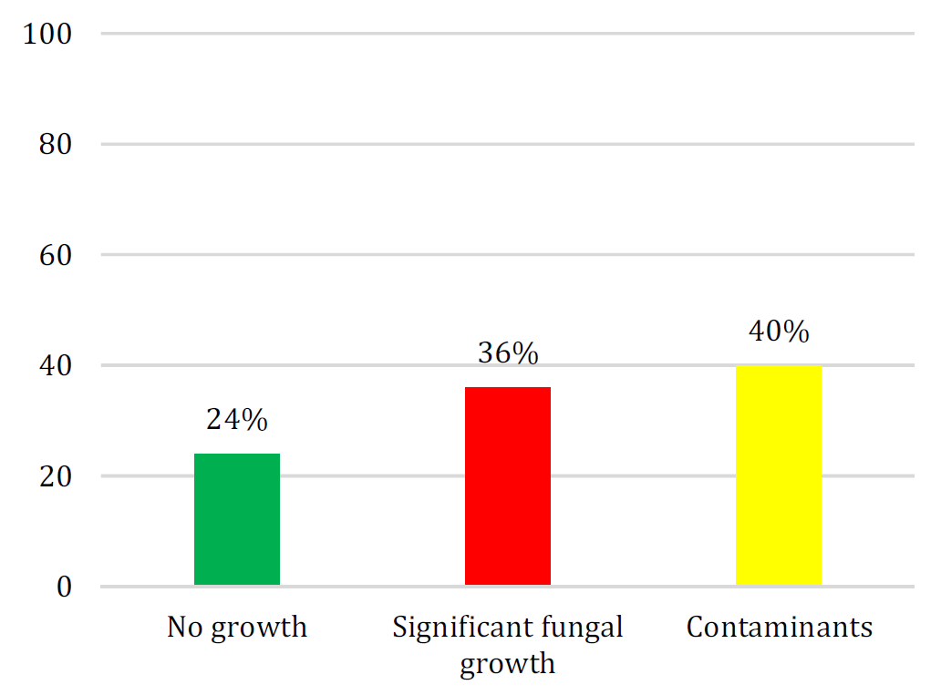
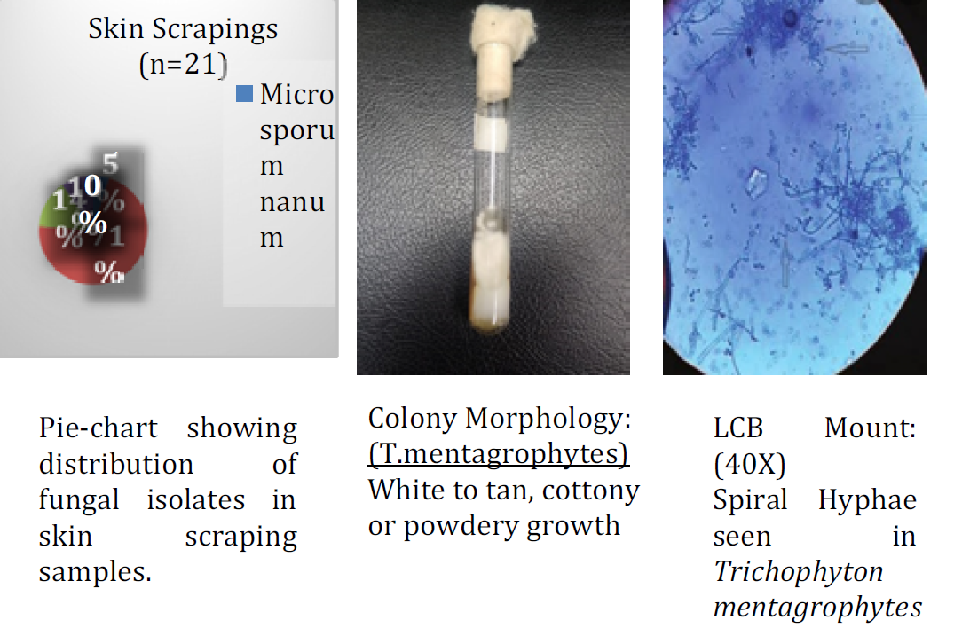
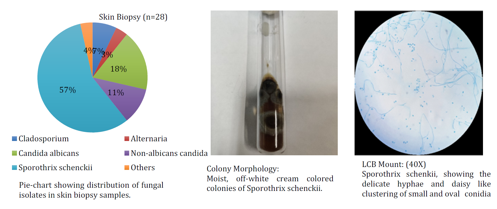
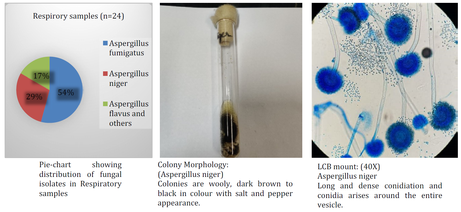
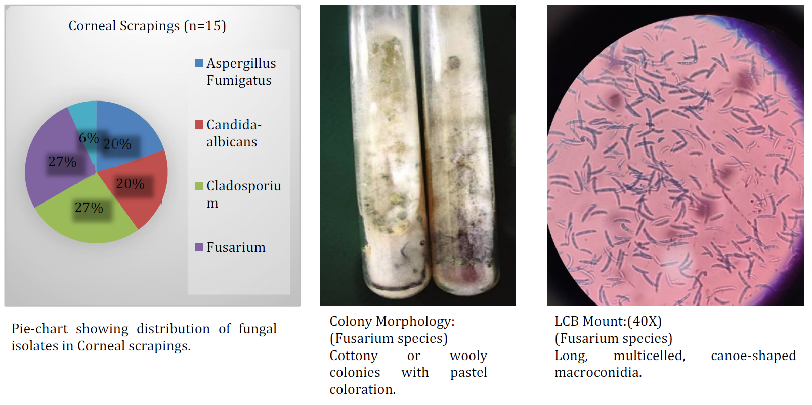
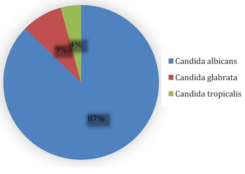
Various clinical samples were received in the department of Microbiology in our hospital. Total 752 samples were included in the study. Of the 752 clinical samples 273 (36%) had fungal growth, 300 (40%) were contaminants and 179 (24%) showed no growth. Among the respiratory samples (369), 121 samples (33%) were positive in fungal culture. The other major samples received were skin scrapings (81), skin biopsy (56) and oral mucosal scrapings (25) in which fungal growth was observed in 28 (35%), 21 (38%) and 17 (68%) respectively. Most common isolate observed among dermatophytes was T.mentagrophytes 15 (71%) followed by T.rubrum 3 (14%) and T.violaceum 2 (10%). Sporothrix schenckii 16 (57%) was predominant in patients suspected of subcutaneous mycosis.Candida albicans 41 (87%) was isolated in reproductive age group females followed by Candida glabrata 4 (9%) and Candida tropicalis 2 (4%).In corneal scrapings, growth of Fusarium 4 (27%) , Cladosporium 4 (27%) and Aspergillus fumigatus 3 (20%) was obtained.
Figure 1: Distribution of Significant Fungal Isolates in Different Samples
Figure 2: Distribution of Fungal Isolates in Skin Scrapings Samples
Figure 3: Distribution of Fungal Isolates in Skin Biopsy Samples
Figure 4: Distribution of Fungal Isolates in Respiratory Samples
Figure 5: Distribution of Fungal Isolates in Corneal Scrapings
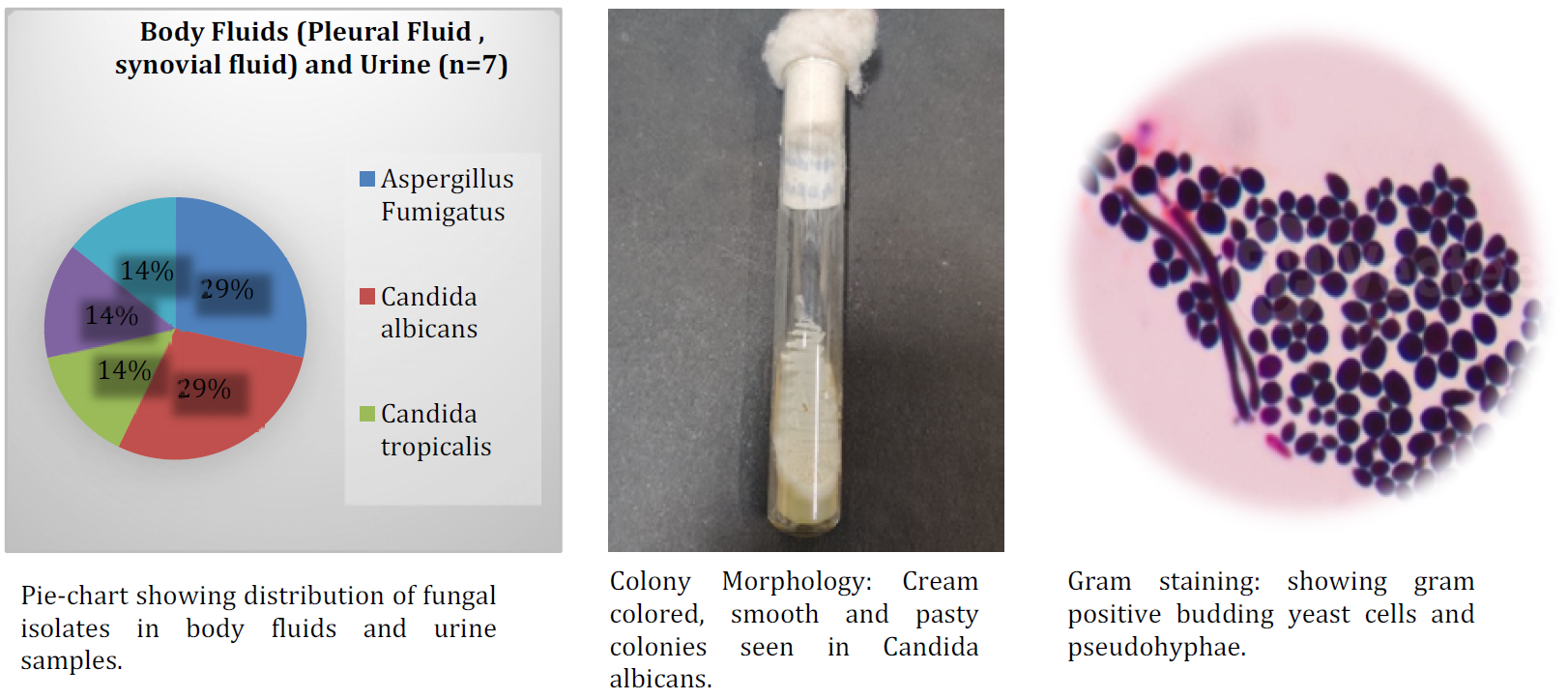
Figure 6: Distribution of Fungal Isolates in Body Fluids (Synovial Fluid, Pleural fluid) and in Urine Samples
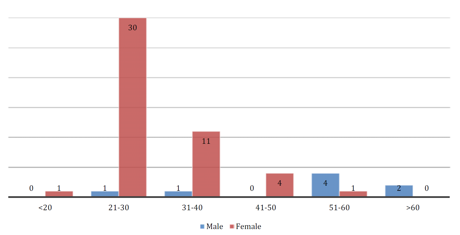
Figure 7: Distribution of Fungal Isolates in Samples Suspected of Vulvo-Vaginitis and Balanoposthitis according to Age Group
Figure 8: Distribution of Candida Species in Reproductive Age Group Females Suspected of Vulvovaginitis
Table 1: Distribution of Significant Fungal Isolates in Different Samples
Type of Specimen
Significant Fungal Growth n(%)
Respiratory Samples (n = 369)
121(33)
Skin Biopsy (n = 81)
28(35)
Vaginal Discharge (n = 66)
47(71)
Skin Scrapings (n = 56) and Nail scrapings (n = 13)
21 (38) and 4 (31)
Corneal Scrapings (n = 45)
15(33)
Pus Samples (n = 26)
1(4)
Oral Mucosal Scrapings (n = 25)
17(68)
Body Fluids (n = 24)
4(17)
Nasal Crusting (n = 18)
3(16)
Penile Discharge (n = 12)
9(69)
Urine (n = 9)
3(33)
Blood and Bone Marrow (n = 8)
0
Total (752)
273
DISCUSSION
Total 752 samples were included in our study. Outof these 752 clinical samples 36% had fungal growth, 40% were contaminants and 24% showed no growth. In our study, among dermatophytes, T.mentagrophytes 15(71%) was the commonest etiological agent followed by T.rubrum 3 (14%) in patients suspected of Tinea corporis, Tinea cruris, and Tinea pedis and 2 (10%) were T.violaceumin patients with clinical suspicion of Tinea capitis.Sporothrix schenckii 16(57%) was predominant in patients suspected ofsubcutaneous mycosis, out of these 11 (69%) were of fixed-cutaneous type and 4 (31%) were of lympho-cutaneous type.Candida albicans 41 (87%) was predominant in reproductive age group females suspected of vulvovaginitis, followed by Candida glabrata 4 (9%) and Candida tropicalis 2 (4%).In Corneal scrapings, Fusarium species 4 (27%) and Cladosporium species4 (27%) were predominant isolates followed by Aspergillus fumigatus 3 (20%).
In a study conducted by Ahuja S et al, various isolateswere Trichophyton species 35 (43.75%), followed by Candida species 18 (22.5%), Aspergillus species 16 (20%),Curvularia and Epidermatophyton species 4 (5%),Alternaria, Fusarium and Penicillium species one (1.25%) 9. The major isolates from another study were Aspergillus niger [8] (34.78%), followed by Rhizopus species [3] (13.04%), Aspergillus flavus, Aspergillus nidulans and Epidermatophyton floccosum 2 (8.70%), Aspergillus terreus, Aspergillus species, Mucor species, Penicillium species, Curvularia and candida species [10].
CONCLUSION
There is distinct pattern of regional variation in the etiology of fungal infections. The role of diagnostic mycology laboratory is important in the management of fungal infections. An understanding of fungal infections in each set up will help greatly in improving clinical diagnostic approaches. Culture and identification of mycotic infections is essential for commencement of antifungal therapy. The clinician-microbiologist collaboration will help in improving patient care.
REFERENCE
Chen, K.Y. et al. “Pulmonary fungal infection: emphasis on microbiological spectra, patient outcome and prognostic factors.” Chest Journal, vol. 120, 2001, pp. 177–184.
Samonis, G. et al. “Prospective study of the impact of broad spectrum antibiotics on the yeast flora of the human gut.” European Journal of Clinical Microbiology and Infectious Disease, vol. 13, 1994, pp. 665–667.
Shrihari, N. et al. “The onychomycosis in a tertiary care hospital.” International Journal of Medical and Health Science, vol. 1, no. 3, 2012, pp. 25–30.
Davies, S.F. “Fungal pneumonia.” Medical Clinics of North America, vol. 78, no. 5, 1994, pp. 1049–1065.
Chander, J. Medical Mycology. 4th ed., Mehta Publishers, 2018.
Marr, K.A. et al. “Epidemiology and outcome of mould infections in hematopoietic stem cell transplant recipients.” Clinical Infectious Diseases, vol. 34, 2002, pp. 909–917.
Husain, S. et al. “Opportunistic mycelia fungal infections in organ transplant recipients: emerging importance of non-Aspergillus mycelia fungi.” Clinical Infectious Diseases, vol. 37, 2003, pp. 221–229.
Sardi, J.C.O. et al. “Candida species: current epidemiology, pathogenicity, biofilm formation, natural antifungal products and new therapeutic options.” Journal of Medical Microbiology, vol. 62, 2013, pp. 10–24.
Ahuja, S. et al. “Etiological agents of onychomycosis from a tertiary care hospital in central Delhi, India.” Indian Journal of Fundamental and Applied Life Sciences, vol. 1, no. 2, 2011, pp. 11–14.
Shrihari, N. et al. “Mycological profile of fungal infections from various clinical samples in a tertiary care hospital.” Journal of Pharmaceutical and Biomedical Sciences, vol. 17, 2017, pp. 22–25.
Recommended Articles
Research Article
Knowledge of the Patient's Family about the Level of Emergency Based on Triage with the Satisfaction of the Patient's Family in the Emergency Department of the Sumedang Area General Hospital
Roganda Situmorang,
...
Raida Rihadatul Aisyi
Published: 30/03/2024
Download PDF
Research Article
The Influence of Murottal Al Qur'an Surat Ar Rahman Irama Hijaz Therapy on the Pain Scale of Post-Explorative Laparatomy Patients in the Icu Room of Bandung City Hospital
Andri Nurmansyah,
...
Fifi Siti Fauziah Yani
Published: 30/03/2024
Download PDF
Research Article
The role of IFN-g gene polymorphism (+874 T/A) as predisposing factor for pulmonary tuberculosis
Download PDF
Research Article
Assessment of Knowledge on Common Pediatric Infections and Their Prevention Among Parents in Chamba, Himachal Pradesh
Isha Bhatia,
Rupika
Published: 20/12/2024
Download PDF
PO Box 101, Nakuru
Kenya.
Email: office@iarconsortium.org
Editorial Office:
J.L Bhavan, Near Radison Blu Hotel,
Jalukbari, Guwahati-India
Sharma, S., Thakur, V., Verma, S., Sharma, V., Mehta, V. & Bhagra, S. (2023). Spectrum of Fungal isolates from Clinical Specimens in a Tertiary Care Hospital in Sub-Himalayan Region. Scientific Research Journal of Clinical and Medical Sciences, 3(1), 1-5.
MLA
Sharma, Swati, et al. "Spectrum of Fungal isolates from Clinical Specimens in a Tertiary Care Hospital in Sub-Himalayan Region." Scientific Research Journal of Clinical and Medical Sciences 3.1 (2023): 1-5.
Chicago
Sharma, Swati, Vibha Thakur, Santwana Verma, Vineeta Sharma, Vinod Mehta and Suruchi Bhagra. "Spectrum of Fungal isolates from Clinical Specimens in a Tertiary Care Hospital in Sub-Himalayan Region." Scientific Research Journal of Clinical and Medical Sciences 3, no. 1 (2023): 1-5.
Harvard
Sharma, S., Thakur, V., Verma, S., Sharma, V., Mehta, V. and Bhagra, S. (2023) 'Spectrum of Fungal isolates from Clinical Specimens in a Tertiary Care Hospital in Sub-Himalayan Region' Scientific Research Journal of Clinical and Medical Sciences 3(1), pp. 1-5.
Vancouver
Sharma S, Thakur V, Verma S, Sharma V, Mehta V, Bhagra S. Spectrum of Fungal isolates from Clinical Specimens in a Tertiary Care Hospital in Sub-Himalayan Region. Scientific Research Journal of Clinical and Medical Sciences. 2023 Jan;3(1):1-5.


 Download PDF
 Download XML