Background: Mucormycosis is an angio-invasive infection, caused by ubiquitous environmental fungal species of the order Mucorales including Rhizopus, Lichtheimia, Mucor, and Rhizomucor species. The clinical presentation of mucormycosis varies according to the anatomical site of involvement. The incidence rate of mucormycosis globally varies from 0.005 to 1.7 per million population. India’s yearly prevalence of CAM is around 0.02-9.5 cases per 100,000 people. COVID -19 associated mucormycosis was mostly isolated during the second wave with mortality of 31-50%. Objective: To identify the outcome of early diagnosis and initiation of antifungal treatment in CAM-associated mortality and morbidity. Material& Methods: A study was done at IGMC Shimla over a period of 8 months (May 2021 to December 2022). All hospitalized patients diagnosed with COVID-19 and with suspected mucormycosis were included in the study. Biopsy/scrapings of affected area were collected and received at Microbiology department. Samples were subjected to direct microscopy using 10%/20% KOH for fungal elements and cultured on Sabouraud’s dextrose agar with chloramphenicol and cycloheximide at 25o and 370C. The fungal diagnosis was done by LCB mount. Results: A total of 913 cases of covid-19 were admitted in the isolation unit during this period out of which 62(6.8%) were suspected of mucormycosis. Samples were sent for processing. KOH positive in 14% and 18% were culture positive. Rhizopus (84%) and Lichtheimia (7%) species were found. Patients affected were mainly in the 5th (15%) and 6th (38%) decade of life. Results identified diabetes (76%) as the primary predisposing factor. Other comorbidities like hypertension (15%) and hepatitis C virus (15%) were also found in CAM cases. Rhino-orbital-cerebral infection was most common with involvement of nasal mucosa (69%), palate (15%), brain (7%) and orbit (15%). Initiation of antifungal therapy improved outcome in CAM. Overall mortality observed in COVID-associated mucormycosis was 32%. Conclusion: CAM could be avoided by early diagnosis, identifying high-risk groups and managing uncontrolled diabetes. All these factors would help the clinicians to decrease mortality and morbidity in CAM.
Keywords
Covid
Mucormycosis
Tertiary Care Hospital
INTRODUCTION
Mucormycosis has emerged as major fungal infection with COVID-19, thereby leading to increased mortality and morbidity. Mucormycosis in India ranges from 0.02-9.5 cases per 100,000 people. Mortality observed among COVID associated mucormycosis infections during the second wave was 31-50% [1]. Mucormycosis is an angio-invasive infection, caused by environmental fungal species of the order Mucorales including Rhizopus, Lichtheimia, Mucor, and Rhizomucor species [2].The clinical presentation of mucormycosis varies according to the site of involvement including rhino-orbital-cerebral, pulmonary, cutaneous, gastrointestinal and disseminated forms. Attributes and risk factors observed in the pathogenesis of mucormycosis during COVID-19 were uncontrolled diabetes/diabetic ketoacidosis, corticosteroids, iron chelators and hematological malignancies.
MATERIAL AND METHODS
A study was done at Indira Gandhi medical college, Shimla over a period of 8 months (May 2021 to December 2021). All hospitalized patients diagnosed with COVID-19 and with suspected mucormycosis were included in the study. Biopsy/scrapings of affected area were received at Microbiology department. Samples were subjected to direct microscopy using 10%/20% KOH for fungal elements and cultured on Sabouraud’s dextrose agar (SDA) with antibacterial and antifungal agents and incubated at 250 and 370C. Macroscopic identification was done by observing colony morphology. Microscopic identification was done by LCB mount [3].
RESULTS
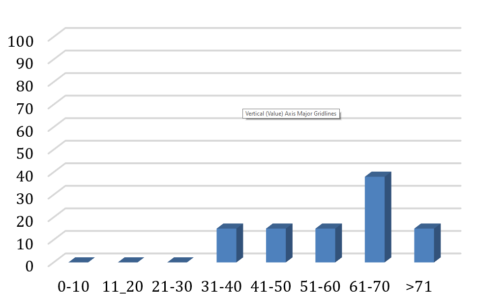
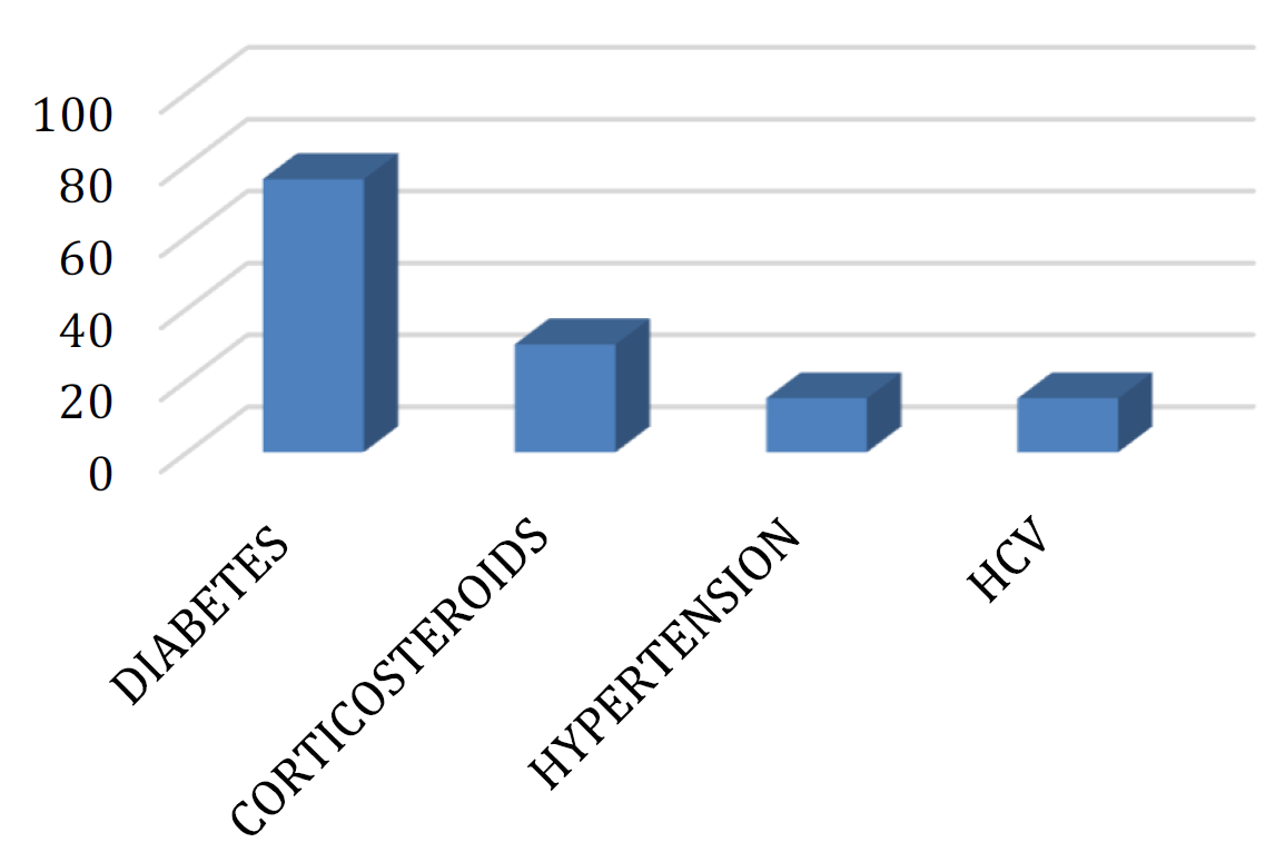
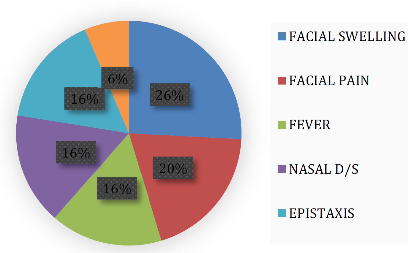
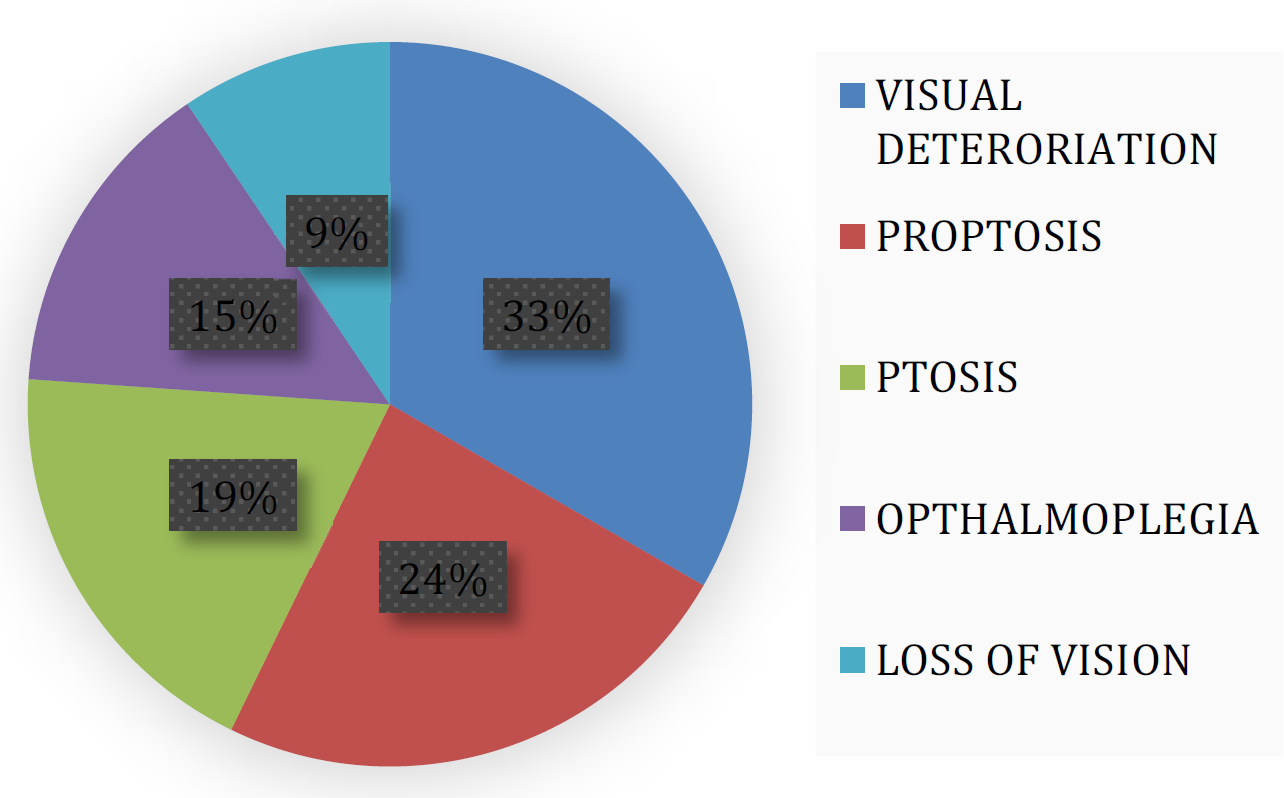
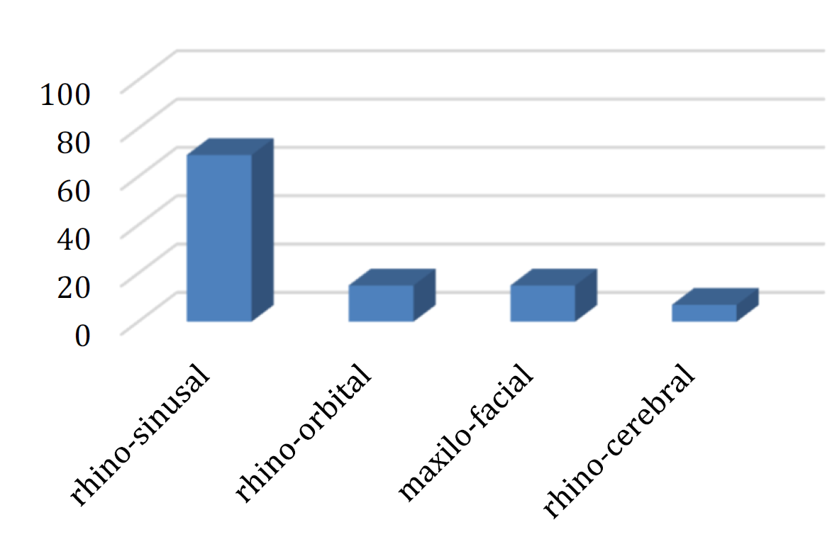
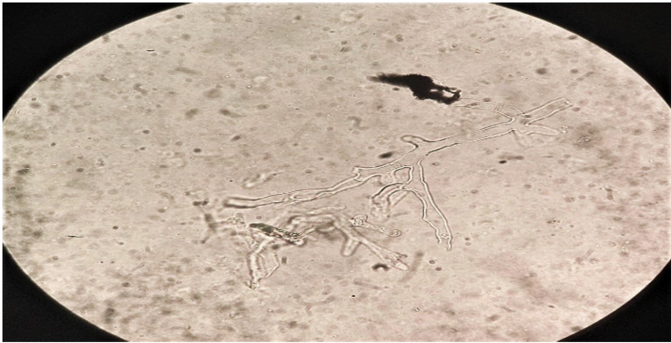
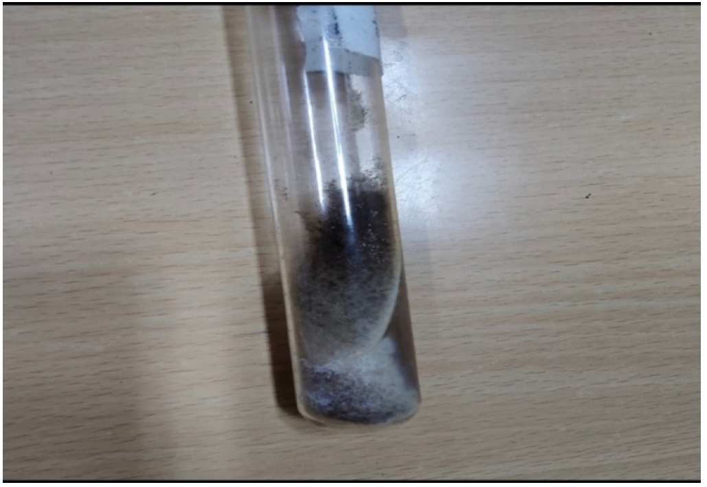
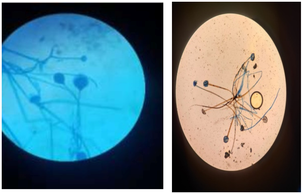
A total of 913 cases of covid-19 were admitted in the isolation unit during this period out of which 62(6.8%) were suspected of mucormycosis. Patients affected were mainly in the 5th (15%) and 6th (38%) decade of life Most common comorbid condition was diabetes10 (76%) followed by corticosteroids 4 (30%), hypertension 2 (15%) and hepatitis C virus 2 (15%) infection. Patients presented with fever, facial swelling, facial pain, nasal discharge, epistaxis, altered sensorium, visual deterioration, proptosis, ptosis, ophthalmoplegia and loss of vision. Involved sites in COVID-associated mucormycosis were rhino-sinusal 9 (69%), rhino-orbital 2 (15%), maxilo-facial 2 (15%) and rhino-cerebral 1 (7%). Initiation of antifungal therapy improved outcome in CAM. Overall mortality observed in COVID-associated mucormycosis was 32%. Broad, aseptate, thick-walled and ribbon-like hyaline hyphae with wide-angle branching was seen in KOH findings. Upon culture, in SDA, Rapidly growing mycelial colonies are floccose, dense and hairy in appearance. Initially it was white in color but later greyish in color. Of the 62 samples KOH and culture positive were 9(14%) and 11(18%). Among culture positive Rhizopus species wasobserved in10(77%)and Lichtheimia species in 1(7%) (Figure 1-10).
Figure 1: Age-Wise Distribution of CAM (n = 13)
Figure 2: Comorbidity Conditions in CAM (n=13)
Figure 3: Distribution of Rhino-Cerebral Presentation
Figure 4: Distribution of Rhino-Orbital Presentation
Figure 5: Showing Results of Site of Involvement in Mucormycosis (N =13)
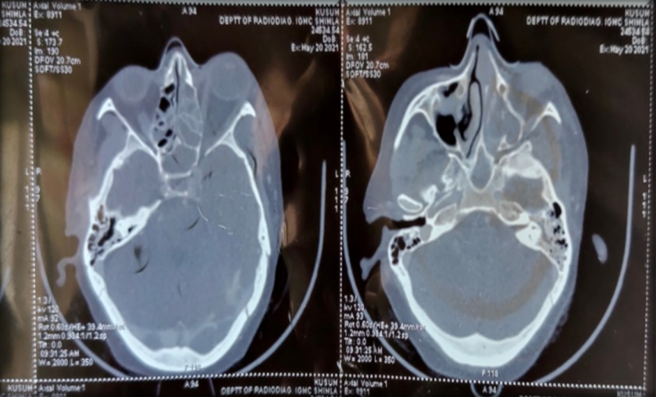
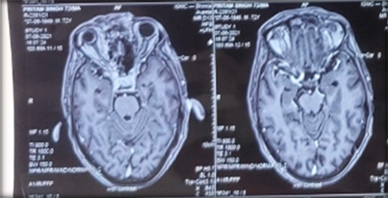
Figure 6: Head Computed Tomography – Showing Soft Tissue Density Content in Left Maxillary Sinus Extending into Intra-Orbital Region (Arrow)
Figure 7: Head Computed Tomography – Showing Cavernous Sinus Involvement along with Bilateral Optic Nerve. (Arrow)
Figure 8: KOH Mount: Hyphae Seen Under 40X
Figure 9: Rhizopus on SDA
Figure 10: LB Mount of Lichthemia as Seen under 40X
DISCUSSION
Patients affected were between the age group of 30 to 70 years. Most common comorbid condition was uncontrolled diabetes, followed by corticosteroids, hypertension and hepatitis C virus infection. Patients presented with fever, facial swelling, facial pain, nasal discharge, epistaxis, altered sensorium, visual deterioration, proptosis, ptosis, ophthalmoplegia and loss of vision. Involved sites were rhino-sinusal, rhino-orbital, maxilo-facial and rhino-cerebral. Rhizopus species was the most common isolate, followed by Lichtheimia species. Overall mortality observed in COVID-associated mucormycosis was 32%.
CONCLUSION
In COVID 19 infection with a comorbid condition like uncontrolled diabetes, there is a need to have high degree of suspicion for mucormycosis coinfection. COVID-associated mucormycosis is an emerging problem necessitating increased vigilance in COVID-19 patients, even those who have recovered.
REFERENCE
Singh, A.K. et al. “Mucormycosis in COVID-19: a systematic review of cases reported worldwide and in India.” Diabetes and Metabolic Syndrome: Clinical Research and Reviews, vol. 15, no. 4, 2021, pp. 102146.
Hussain, S. et al. “Global prevalence of COVID-19-associated mucormycosis (CAM): living systematic review and meta-analysis.” Journal of Fungi, vol. 7, no. 11, 2021, pp. 985.
Chander, J. Medical Mycology. 4th ed., Mehta Publishers, 2018.
Recommended Articles
Research Article
Knowledge of the Patient's Family about the Level of Emergency Based on Triage with the Satisfaction of the Patient's Family in the Emergency Department of the Sumedang Area General Hospital
Roganda Situmorang,
...
Raida Rihadatul Aisyi
Published: 30/03/2024
Download PDF
Research Article
The Influence of Murottal Al Qur'an Surat Ar Rahman Irama Hijaz Therapy on the Pain Scale of Post-Explorative Laparatomy Patients in the Icu Room of Bandung City Hospital
Andri Nurmansyah,
...
Fifi Siti Fauziah Yani
Published: 30/03/2024
Download PDF
Research Article
The role of IFN-g gene polymorphism (+874 T/A) as predisposing factor for pulmonary tuberculosis
Download PDF
Research Article
Caesarean Scar Pregnancy
Komal Sharma,
...
Jyotshna Sharma
Published: 02/02/2023
Download PDF
PO Box 101, Nakuru
Kenya.
Email: office@iarconsortium.org
Editorial Office:
J.L Bhavan, Near Radison Blu Hotel,
Jalukbari, Guwahati-India
Thakur, V., Sharma, S., Mehta, V., Sharma, V., sharma, G. & Verma, S. (2023). Covid-Associated Mucormycosis at Tertiary Care Institute. Scientific Research Journal of Clinical and Medical Sciences, 3(1), 1-3.
MLA
Thakur, Vibha, et al. "Covid-Associated Mucormycosis at Tertiary Care Institute." Scientific Research Journal of Clinical and Medical Sciences 3.1 (2023): 1-3.
Chicago
Thakur, Vibha, Swati Sharma, Vinod Mehta, Vineeta Sharma, Garima sharma and Santwana Verma. "Covid-Associated Mucormycosis at Tertiary Care Institute." Scientific Research Journal of Clinical and Medical Sciences 3, no. 1 (2023): 1-3.
Harvard
Thakur, V., Sharma, S., Mehta, V., Sharma, V., sharma, G. and Verma, S. (2023) 'Covid-Associated Mucormycosis at Tertiary Care Institute' Scientific Research Journal of Clinical and Medical Sciences 3(1), pp. 1-3.
Vancouver
Thakur V, Sharma S, Mehta V, Sharma V, sharma G, Verma S. Covid-Associated Mucormycosis at Tertiary Care Institute. Scientific Research Journal of Clinical and Medical Sciences. 2023 Jan;3(1):1-3.


 Download PDF
 Download XML