Background: The Ki-67 antigen and the D-type cyclins play a key role in the cell cycle regulation. Objective: The goal of this study was to evaluate an immunohistochemical expression status of Cyclin D1 and Ki-67 in basal cell carcinoma (BCC) of the skin and to find out whether there is a correlation between them. Material and Methods: The study included biopsy specimens from 29 BCCs, which were immunohistochemically stained for Cyclin D1 and Ki-67. The cases were graded on a semiquantitative scale based on the average proportion of immunoreactive cells: 0 (<1%), 1+ (1-10%), 2+ (11-50%) and 3+ (>50%). Results: All BCCs were positive for both proteins. As for Cyclin D1, the grades 1+, 2+ and 3+ were found in 4 cases (13.8%), 18 cases (62.1%) and 7 cases (24.1%). As for Ki-67, the grades 1+, 2+ and 3+ were found in 1 case (3.4%), 18 cases (62.1%) and 10 cases (34.5%). In adjacent epidermis, all cases exhibited a mild degree of Cyclin D1 and Ki-67 immunoreactivity. Comparing both markers, there were 22 BCCs (75.9%) showing the same semiquantitative grade of Ki-67 and Cyclin D1 expression and 7 BCCs (24.1%), in which Ki-67 grade was one degree higher than Cyclin D1 grade. Conclusion: All BCCs expressed Cyclin D1 and Ki-67 that were more pronounced in tumor tissue than in adjacent normal epidermis. This indicates, both markers play a role in the carcinogenesis of cutaneous BCC. The expression of Cyclin D1 appeared to have a positive correlation with Ki-67 expression, suggesting the production of Cyclin D1 protein is directly related to cell proliferation.
The cell cycle is a complex machinery of events during which a cell duplicates its genome, synthesizes the other constituents and eventually divides into two daughter cells. The eukaryotic cell cycle consists of the following phases: G1, S and G2 (collectively known as interphase) and mitotic phase [1,2]. Activation of each phase is dependent on the proper progression and completion of the previous one. The cells that have temporarily or reversibly stopped dividing are said to have entered a state of quiescence called G0 phase [1,2]. There are many intracellular and extracellular components that regulate or participate on the cell cycle regulation. Among them, the Ki-67 antigen and the D-type cyclins play a key role in this process [1,2]. Ki-67 antigen is a nonhistone nuclear protein that is expressed during active phases of cell cycle (G1, S, G2 and mitosis), being absent in quiescent phase [1]. Ki-67 levels are low in the G1 and S phases, than it markedly increases and reaches a peak in early mitosis [1]. In later phases of mitosis, a sharp decrease in the Ki-67 expression occurs [1]. Cyclin D1 (Bcl-1) is another nuclear protein that behaves as a proto-oncogene and whose activity is essential for the G1/S progression of the cell cycle [1,2]. During the G1 phase, it is synthesized rapidly and accumulates in the nucleus and is degraded as the cell enters the S phase [2]. Both the cell cycle regulators are excellent markers to determine the growth fraction and proliferation status of a given cell population using an immunohistochemistry and serve as prognostic biomarkers in many human malignancies [1,2].
Basal Cell Carcinoma (BCC) of the skin is the most common malignancy in humans with a rising incidence [3]. Etiology and pathogenesis of BCC is very complex and it is a good example of a disease caused by interaction between environmental, phenotypic and genetic factors [3]. In contrast to most other cancers, it is usually a slow-growing neoplasm with only minimal metastatic potential, but some subtypes grow aggressively causing an extensive tissue destruction [3]. Many studies done till present time have analyzed an immunohistochemical expression of Ki-67 [4-8] and cyclin D1 [9-12] in BCC of the skin. However, to the best of the author's knowledge, none of them has investigated both markers simultaneously. The objective of this study was to evaluate an immunohistochemical expression status of cyclin D1 and Ki-67 in cutaneous BCC and to find out whether there is a correlation between them.
Representative biopsy specimens from 29 cases of primary BCC of the skin were enrolled into this analysis. They were obtained from 22 patients (17 males, 5 females) in the age range of 46–92 years (mean age 76.2 y). The BCC samples included four histopathological subtypes: superficial (5 cases), nodular (11 cases), mixed nodular-infiltrative (10 cases) and infiltrative (3 cases). The superficial and nodular subtypes were considered the non-aggressive, while the nodular-infiltrative and infiltrative subtypes the aggressive tumor variants. The topographic locations of the lesions were as follows: head & neck (13 cases), trunk (12 cases) and upper/lower extremities (4 cases). Biopsy samples were routinely processed and all were simultaneously stained for Cyclin D1 (monoclonal rabbit anti-human antibody, clone EP12, DAKO, ready-to-use) and Ki-67 antigen (monoclonal mouse anti-human antibody, clone MIB1, DAKO, ready-to-use) according to manufacturer's instructions. The average relative fraction of positive cells with nuclear staining was determined as percentage and according to Liang, S.B. et al. [9], it was graded on a semiquantitative scale as follows: 0 (<1% of cells), 1+ (1-10% of cells), 2+ (11-50% of cells) and 3+ (>50% of cells). A small number of the cases investigated did not allow a statistical evaluation.
In our series, all BCCs were immunohistochemically positive for both, Cyclin D1 and Ki-67 antigen. Cyclin D1 showed a moderate to strong nuclear expression, which varied in the range of 5%–80 % of total tumor tissue (mean value 33.2 %). The grades 1+, 2+ and 3+ were found in 4 cases (13.8%), 18 cases (62.1%) and 7 cases (24.1%), respectively. In normal epidermis adjacent to the tumor, all cases exhibited a nuclear expression of Cyclin D1.
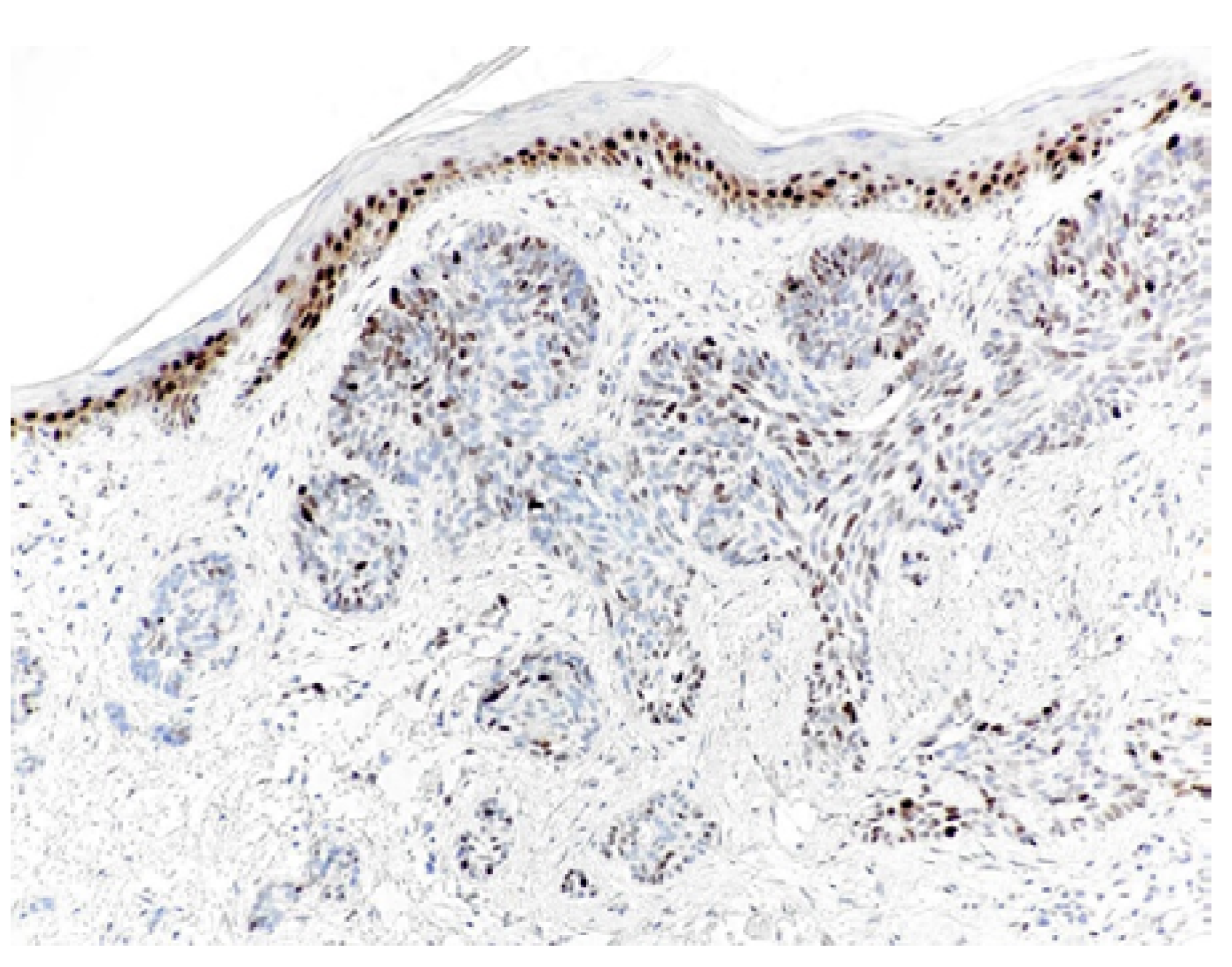
The Cyclin D1 positive keratinocytes were found in the basal and spinous layer and comprised up to 25 % of epidermal keratinocytes (Figure 1 and Figure 2).

Figure 1: Expression of Cyclin D1 (Grade 2+) in Nodular BCC. There is also a Continuous Nuclear Positivity in the Basal and Suprabasal Layers in Overlying Normal Epidermis (Magnification 40x)

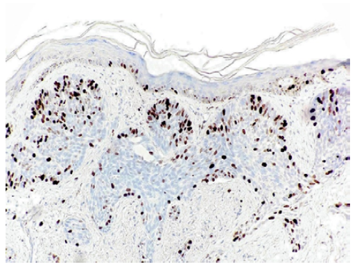
Figure 2: Expression of Ki-67 (Grade 2+) in Nodular BCC. (The Case is Corresponding to Figure 1). Overlying Epidermis Shows a Nuclear Immunostaining Only in a Few Basal Keratinocytes (Magnification 40x)
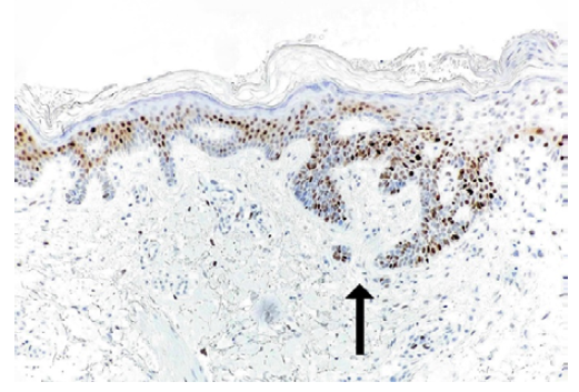
Ki-67 antigen showed a strong nuclear immunoreactivity, which ranged from 5% to 75% (mean value 44.5%). The grades 1+, 2+ and 3+ were found in 1 case (3.4%), 18 cases (62.1%) and 10 cases (34.5%), respectively. In adjacent normal epidermis, all cases exhibited a strong nuclear expression of Ki-67, but it was limited only to a few cell in the basal layer (about 5 % of epidermal keratinocytes) (Figure 3 and Figure 4).

Figure 3: Intense Expression of Cyclin D1 (Grade 3+) in Superficial BCC (Arrow). There is also a Striking Nuclear Positivity in Adjacent Normal Epidermis (Magnification 40x)
Figure 4: Almost Diffuse Expression of Ki-67 (grade 3+) in Superficial BCC (Arrow). (The Case is Corresponding to Figure 3). Adjacent Normal Epidermis Exhibits a Nuclear Immunostaining Only in Sporadic Keratinocytes (Magnification 40x)
A spatial distribution of both markers within the tumor nests was mostly irregular and areas with a high number of cells expressing Cyclin D1 or Ki-67 often occurred adjacent to areas with a lower number of the immunolabeled cells. Comparing both markers, there were 22 BCCs (75.9%) showing the same semiquantitative grade of Ki-67 and Cyclin D1 expression and 7 BCCs (24.1%), in which Ki-67 grade was one degree higher than Cyclin D1 grade. A detailed presentation of the immunohistochemical findings in our set of BCCs is in Table 1 and Table 2.
Table 1: A Status of Immunohistochemical Expression of Ki-67 and Cyclin D1 in a Set of Non-Aggressive BCC Variants
| BCC | Ki-67 | Cyclin D1 | |||
| Subtype | % | Grade | % | Grade | |
| 1. | superficial | 25 | 2+ | 35 | 2+ |
| 2. | superficial | 25 | 2+ | 20 | 2+ |
| 3. | superficial | 75 | 3+ | 70 | 3+ |
| 4. | superficial | 75 | 3+ | 80 | 3+ |
| 5. | superficial | 70 | 3+ | 15 | 2+ |
| 6. | nodular | 75 | 3+ | 60 | 3+ |
| 7. | nodular | 40 | 2+ | 35 | 2+ |
| 8. | nodular | 20 | 2+ | 20 | 2+ |
| 9. | nodular | 30 | 2+ | 30 | 2+ |
| 10. | nodular | 15 | 2+ | 25 | 2+ |
| 11. | nodular | 40 | 2+ | 25 | 2+ |
| 12. | nodular | 50 | 2+ | 25 | 2+ |
| 13. | nodular | 35 | 2+ | 20 | 2+ |
| 14. | nodular | 40 | 2+ | 40 | 2+ |
| 15. | nodular | 5 | 1+ | 5 | 1+ |
| 16. | nodular | 20 | 2+ | 20 | 2+ |
Table 2: A status of Immunohistochemical Expression of Ki-67 and Cyclin D1 in a Set of Aggressive BCC Variants
| BCC | Ki-67 | Cyclin D1 | ||
| Subtype | % | Grade | % | Grade |
| nodular-infiltrative | 40 | 2+ | 40 | 2+ |
| nodular-infiltrative | 60 | 3+ | 25 | 2+ |
| nodular-infiltrative | 40 | 2+ | 20 | 2+ |
| nodular-infiltrative | 75 | 3+ | 60 | 3+ |
| nodular-infiltrative | 70 | 3+ | 60 | 3+ |
| nodular-infiltrative | 40 | 2+ | 10 | 1+ |
| nodular-infiltrative | 25 | 2+ | 5 | 1+ |
| nodular-infiltrative | 35 | 2+ | 50 | 2+ |
| nodular-infiltrative | 15 | 2+ | 50 | 2+ |
| nodular-infiltrative | 40 | 2+ | 10 | 1+ |
| infiltrative | 70 | 3+ | 15 | 2+ |
| infiltrative | 70 | 3+ | 15 | 2+ |
| infiltrative | 70 | 3+ | 70 | 3+ |
Uncontrolled cell proliferation is the hallmark of malignant tumours. This paper describes an immunohistochemical expression status of two important cell cycle regulators, Cyclin D1 and Ki-67 antigen in a set of 29 cutaneous BCC. Despite the small number of the tumors investigated, some conclusions can be stated. First, all BCCs showed a nuclear positivity for both, Cyclin D1 and Ki-67 with a similar spatial distribution of immunolabeled cells. This finding was to be expected, since both proteins are produced in proliferating cells and coordinate the cell cycle via similar molecular pathways. Second, the immunoreactivity of Ki-67 antigen generally tended to have a slightly higher percentage and a stronger intensity compared to Cyclin D1. It may be at least partially explained by the fact, the G1 and G2 cell blocks, which may occur with DNA damage, may have different effects on the presence and intensity of the immunostaining of Ki-67 and Cyclin D1, because the maximal expression of these proteins occurs at different points in the cell cycle. While Ki-67 is maximally expressed at the G2/M interface [1], Cyclin D1 expression levels peak in the G1 phase [2]. Nevertheless, there was a tendency of a positive correlation between the semiquantitative grades of the Cyclin D1 and Ki-67 expression. Since no study has analyzed both markers simultaneously in cutaneous BCC, the author had no opportunity to compare the results with other observations. Therefore, this area may represent a promising subject for further research. Third, there was a constant basal and suprabasal immunoreactivity for both antigens in adjacent normal epidermis, but the Cyclin D1 was expressed in a larger proportion of keratinocytes than the Ki-67 antigen. This contradicts the results of some investigators. In the study of Staibano et al. [10] and Liang et al. [9], no immunostaining for the Cyclin D1 was found in morphologically normal epidermis adjacent to the excised BCCs. Koseoglu et al. [11], noticed the Cyclin D1 expression in normal epidermis in the vicinity of BCC in about half of the cases. Such results are quite surprising, because the epidermis consists of constantly recovering keratinocytes, that usually exhibit a certain degree of proliferation.
Many studies addressing the expression of Ki-67 and Cyclin D1 in cutaneous BCC have been published so far, but the data on their potential prognostic impact are controversial. In the research of Staibano, S. et al. [10] and Liang, S.B. et al. [9], the rates of Cyclin D1 positive BCCs were only 56.7% and 54.3%, respectively. This is in contrast with current paper, in which all lesions were positive for this marker. According to some authors [10,12], the different immunoreactivity for Cyclin D1 may depend on histologic subtypes or growth pattern of BCC. Staibano, S. et al. [10], investigated 60 BCCs divided into 30 non-aggressive and 30 aggressive forms. In the first group, Cyclin D1 was found only in four cases (13.3%). In contrast, high expression was seen in 11 cases of aggressive forms, moderate expression in 6 cases and low expression in the remaining 13 cases. Statistical analysis showed that Cyclin D1 overexpression was correlated with the occurrence of relapses and/or metastases and with a significant reduction of the disease-free interval. Similar findings were observed in the study of Sivrikoz, O.N. and Kandiloğlu, [12]. They found a statistically significant difference between histomorphologically non-aggressive (nodular subtype) and aggressive (micronodular and infiltrative subtype and basosquamous carcinoma) BCCs for this marker. Cyclin D1 was higher in the aggressive subgroup compared to the non-aggressive one. According to those results, the immunohistochemical analysis of Cyclin D1 in cutaneous BCC seems to constitute a promising tool for the identification of the subgroup of tumors with a more aggressive phenotype characterized by a worse prognosis than the tumors that do not express cyclin D1. On the other hand, the Turkish investigators [11], did not report any correlation between the grades of Cyclin D1 expression and histological BCC characteristics. Although the data in the present study are only indicative, there were not persuasive differences between these two tumor subgroups.
In what concerns to the Ki-67 antigen, the results have also been inconsistent. Although infiltrative (morpheic) BCCs generally demonstrate a higher growth fraction, we [4] and others [7,8], have not observed significant differences in the Ki-67 expression between aggresssive and non-aggressive BCC subtypes, implying that other factors might be more biologically significant. However, another recent papers [5,6], have showed a significantly higher Ki-67 index in the aggressive histopathologic subtypes of cutaneous BCC. The variation of the Ki-67 and Cyclin D1 immunoexpression in various studies can be attributed to the different processing technique and methodology, including the use of different antibody clones, as well as the tumor heterogeneity.
The present study demonstrates, all BCCs expressed Cyclin D1 and Ki-67 antigen, that were more pronounced in tumor tissue than in normal adjacent epidermis. This indicates, both markers play a role in the carcinogenesis of cutaneous BCC. The expression of Cyclin D1 appeared to have a positive association with Ki-67 expression, suggesting the production of Cyclin D1 protein is directly related to cell proliferation. To answer the question whether simultaneous immunohistochemical evaluation of both cell cycle regulators can more reliably reflect and predict biological behaviour and malignant potential of cutaneous BCC might require further studies.
Li, L.T. et al. “Ki67 is a promising molecular target in the diagnosis of cancer (Review).” Molecular Medicine Reports, vol. 11, no. 3, 2015, pp. 1566–1572.
John, R.R. et al. “Mini Review: Multifaceted role played by cyclin d1 in tumor behavior.” Indian Journal of Dental Research, vol. 28, no. 2, 2017, pp. 187–192.
Dika, E. et al. “Basal cell carcinoma: A comprehensive review.” International Journal of Molecular Sciences, vol. 21, no. 15, 2020, p. 5572.
Bartoš, V. et al. “Immunohistochemical evaluation of proliferative activity (Ki-67 Index) in different histological types of cutaneous basal cell carcinoma.” Biologia, vol. 67, no. 3, 2012, pp. 610–615.
Rao, C.R. et al. “EZH2, proliferation rate and aggressive tumor subtypes in cutaneous basal cell carcinoma.” JAMA Oncology, vol. 2, no. 7, 2016, pp. 962–963.
Florescu, D. et al. “Proliferative activity in basal cell carcinomas.” Current Health Sciences Journal, vol. 44, no. 1, 2018, pp. 55–60.
Esmaeili, R. et al. “Immunohistochemical expression of p53 and ki-67 in different histopathological variants of basal cell carcinoma.” Middle East Journal of Cancer, vol. 6, no. 1, 2015, pp. 29–34.
Kramer, E. et al. “Ki67 as a biologic marker of basal cell carcinoma: A retrospective study.” Israel Medical Association Journal, vol. 16, no. 4, 2014, pp. 229–232.
Liang, S.B. et al. “Overexpression of cyclin D1 in nonmelanocytic skin cancer.” Virchows Archiv, vol. 436, no. 4, 2000, pp. 370–376.
Staibano, S. et al. “DNA ploidy and cyclin D1 expression in basal cell carcinoma of the head and neck.” American Journal of Clinical Pathology, vol. 115, no. 6, 2001, pp. 805–813.
Koseoglu, R.D. et al. “Expressions of p53, cyclin D1 and histopathological features in basal cell carcinomas.” Journal of Cutaneous Pathology, vol. 36, no. 9, 2009, pp. 958–965.
Sivrikoz, O.N. and G. Kandiloğlu. “The effects of cyclin D1 and Bcl-2 Expression on aggressive behavior in basal cell and basosquamous carcinoma.” Iranian Journal of Pathology, vol. 10, no. 3, 2015, pp. 185–191.