Introduction: Fractures of the middle third of the face constitute a significant public health concern. The objective of this study is to elucidate the epidemiological characteristics associated with fractures occurring in the middle third of the facial anatomy. Materials and Methods: Retrospective, descriptive, single-center study covers the period from 2016 to 2022. The study focused on cases of depressed fractures involving the frontal, unguis, nasal bones, maxilla, mandible symphysis and parasymphysis fractures, all of which were admitted to our department. Excluded from the study were lateral third facial fractures not connected to the medial third, as well as cases with incomplete medical records. The investigated parameters encompassed the incidence, age distribution, gender distribution, occupational status, transportation mode, underlying causes, time lapse between injury and presentation, point of impact, imaging findings and fracture characteristics. Results: Out of a total of 358 patients, our study included 84 cases, resulting in a prevalence of 23.46%. The average age of the participants was 30 years. Men constituted the majority at 85.7%, yielding a sex ratio of 5.9. Among the cases, 38.1% were students and 32.1% arrived at the hospital via transportation. Notably, traffic accidents contributed to 54.8% of the reported instances. Prompt medical attention, within 24 hours, was sought by 23% of the patients. The area most frequently affected was the chin, observed in 34% of cases. Utilization of CT scans was prevalent, conducted in 76% of the cases. Predominantly, mandibular fractures accounted for 52.4% of the total occurrences. Conclusion: These fractures predominantly manifest in young males. The primary instigator is road traffic accidents. Accurate diagnosis entails the utilization of CT scans. Among these fractures, those involving the mandible take precedence in terms of frequency.
The medial third of the face is the part of the face that includes the frontal bone (frontal sinus), nasal bone, unguis (lacrimal bone), ascending branch of the maxilla, premaxilla, as well as the symphysis and parasymphysis of the mandible. This region is particularly vulnerable to trauma, resulting in fractures that pose a tangible public health concern due to their high occurrence rate. In a comprehensive study spanning three years, Razafindrabe [1] and Rocton [2], revealed that mandibular fractures stood out as the most prevalent, accounting for a substantial 77.27% and 65.1%, respectively.
Fractures affecting the middle third of the face predominantly affect young and active individuals. Motor vehicle accidents and physical assaults emerge as the primary causative factors [3]. Clinical manifestations vary depending on the specific area involved, often presenting as forehead swelling or depression, epistaxis, eyelid edema, pain in the maxillo-mandibular region, hemorrhagic wounds and consequential loss of both tissue and bone substance [4]. For diagnostic purposes, a range of techniques such as CT scans, Blondeau incidence, panoramic radiographs and Gosserez incidence are utilized. Notably, CT scans offer the unique advantage of comprehensive visualization across all facial levels simultaneously, facilitating clear and precise assessment of bone lesions. The overarching objective of this study is to delineate the comprehensive epidemiological profile of this specific fracture type.
This study constitutes a retrospective, descriptive, analytical, cross-sectional and single-center investigation, conducted within the Department of Dentistry and Maxillofacial Surgery at Owendo Teaching Hospital. The research spans a comprehensive period of six years, from March 2016 to March 2022. Encompassing cases that meet specific criteria, the study includes confirmed instances of middle third facial fractures, encompassing the depressed frontal bone, nasal bones, premaxilla, mandible (symphysis and parasymphysis) and unguis. Cases considered are those admitted to the department with complete and comprehensive medical records. Excluded from the analysis are incomplete files, patients diagnosed with malar fractures, fractures of the lateral third of the face unrelated to the middle third, as well as fractures occurring in the mandibular corpus and ramus. The parameters of interest for investigation encompass frequency, annual occurrence rate, age distribution, gender distribution, occupational status, mode of transportation and underlying etiology for establishing the epidemiological profile. Additionally, the study examines factors such as time elapsed before seeking medical consultation, the precise location of impact, imaging techniques employed and the nature of the fracture line to establish the clinical profile.
Data acquisition was executed through a structured data collection form, drawing information from the hospital admission records, consultation and hospitalization registers, as well as the operating theater intervention register. Subsequent data processing involved the utilization of Word and Excel 2010 software for comprehensive analysis.
For qualitative variables, numerical counts and corresponding percentages were utilized, providing an insight into relative occurrences. Quantitative variables, on the other hand, were characterized by their mean, minimum and maximum values, affording a more detailed view of the dataset's range and distribution.
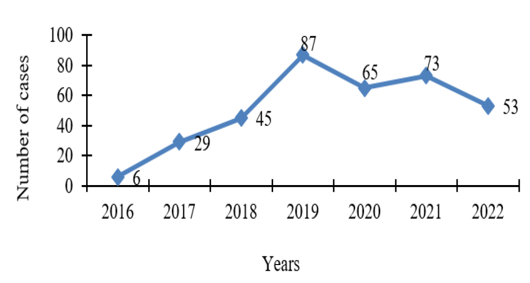
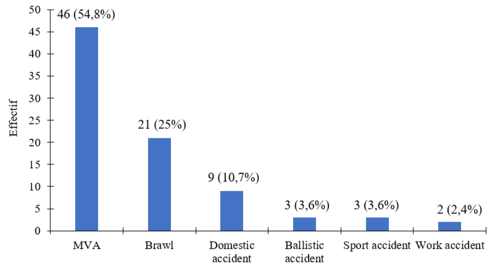
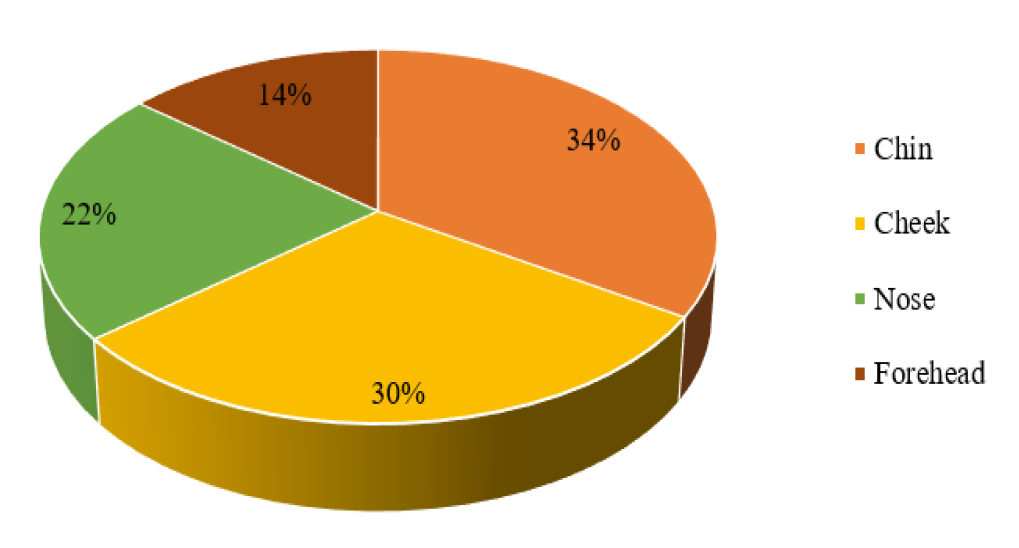
From a total pool of 358 patients admitted to the department, a sample of 84 cases was meticulously gathered, resulting in a prevalence rate of 23.46%. Notably, the year 2019 displayed the highest case count, closely pursued by 2021 with 87 and 73 cases, respectively (Figure 1). The collective mean age stood at 30.89 ± 12.76 years, spanning from the tender age of 2 to a mature 67 years. Gender distribution unveiled a conspicuous male dominance, comprising 85.7% (72 males), juxtaposed with 14.3% (12 females), thus establishing a sex ratio of 5.9. Within the cohort, students, pupils and the unemployed constituted 38.1% and 32.1% of cases, respectively (Table 1). Transportation modalities revealed a distribution of 32.1% by ambulance, 34.5% by taxi and 33.3% via private car. In the realm of causality, road traffic incidents emerged as the paramount etiology, contributing to 54.8% of cases, trailed by altercations (25%) and domestic mishaps (10.7%) (Figure 2). Temporal analysis demonstrated that a significant proportion, 70%, sought medical attention within 24 hours to one week. Comparatively, 23% sought help within the initial 24 hours, while 2% sought assistance between 8 and 14 days and 6% delayed care for more than 14 days. Focusing on anatomical sites, the chin registered the highest prevalence at 34% of cases, succeeded by the cheek at 30% and the nose at 22% (Figure 3).
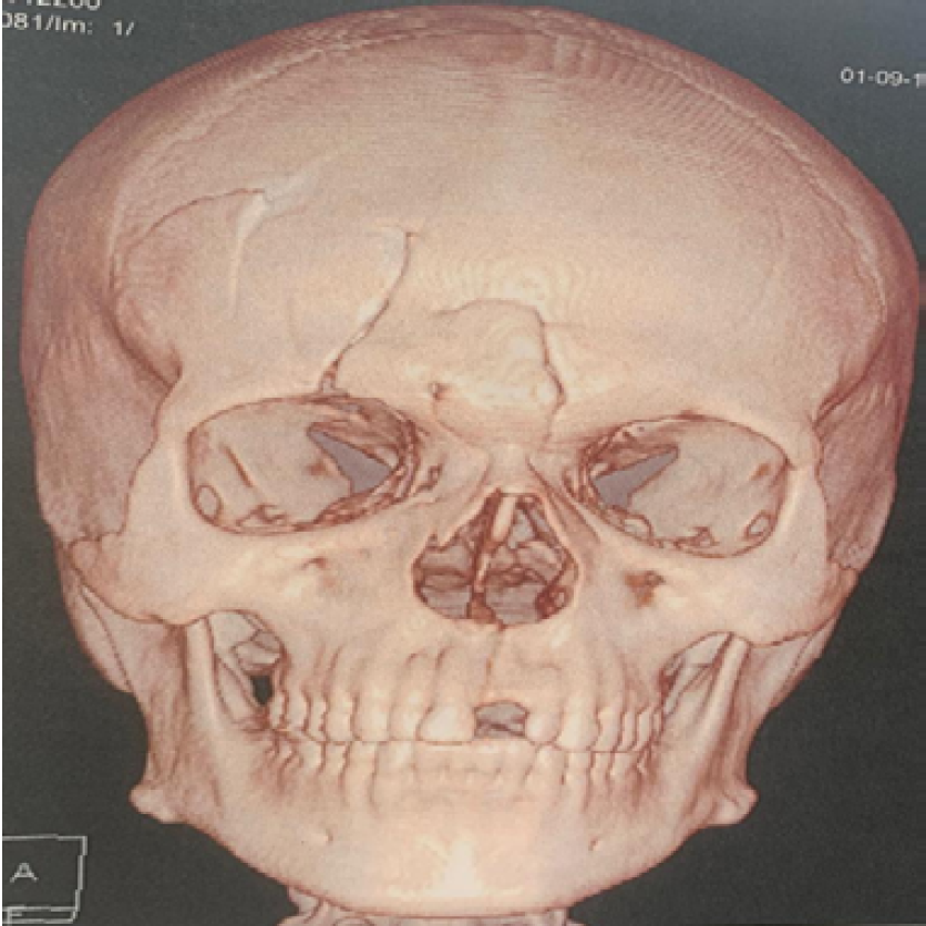
In terms of radiological assessment, the majority, 76.2%, underwent CT scans, while 23.8% opted for panoramic radiographs and 6% utilized Blondeau radiographs (Figure 4). Among the reported cases, fractures were localized in distinct regions, with maxillary, symphysis and parasymphysis fractures identified in 34.5%, 31% and 21.4% of cases, respectively (Table 2).

Figure 1: Yearly Distribution of Patients

Figure 2: Breakdown of Cases Based on the Causative Factors of Trauma

Figure 3: Distribution of Patients Based on Anatomical Site

Figure 4: Three-Dimensional CT Scan Illustrating a Frontal Bone Fracture CHUO Maxillofacial Service
Table 1: Arrangement of Patients Categorized by Occupation
| Categorized by occupation | n | % |
| Pupil and Student | 32 | 38,1 |
| Unemployed | 27 | 32,1 |
| Corporate representative | 13 | 15,5 |
| Law enforcement personnel | 5 | 6,0 |
| Administrator | 4 | 4,8 |
| Company Manager | 2 | 2,4 |
| Administrative manager | 1 | 1,2 |
| Total | 84 | 100 |
Table 2: Categorization of Patients by Fracture Types
| Fracture types | N | % |
| Fracture of the maxilla | 29 | 34,5 |
| Parasymphysis fracture | 26 | 31 |
| Symphysis fracture | 18 | 21,4 |
| Frontal bone fracture | 15 | 17,9 |
| Nasal bone fracture | 12 | 14,3 |
| Naso-maxillary fracture | 6 | 7,1 |
| LeFort II fracture | 5 | 6 |
| Lacrimal bone fracture | 4 | 4,8 |
| LeFort III fracture | 3 | 3,6 |
Middle third fractures constituted 20.54% of the total admissions to the department throughout the study duration. This prevalence can be attributed to the practice of referring certain patients to the departments of Ear, Nose and Throat (ENT) as well as Neurosurgery.
A clear male predominance emerged, encompassing 85.7% of the patient cohort, yielding a sex ratio of 5.9. This inclination can be ascribed to the fact that men tend to engage more frequently in daily activities that involve risks, such as sports, alcohol consumption and driving at high speeds. Notably, this predominance aligns with findings from other studies. For instance, Razanajato [5] in Antananarivo identified a male prevalence of 76.1%. Similarly, Bali [6] in India documented 81.09%, Salonen [7] in Finland found 80.15% and Lebeau [8] in Grenoble reported 76.14%, all of which underscored the dominance of male patients.
The average age of the patients was 30.89±12.76 years, ranging from 2 to 67 years. The age bracket most notably impacted was the 20 to 30-year range, accounting for 41.7% of cases. This average age distribution can be attributed to the demographic makeup of the population, which, as is common in many developing countries, is predominantly youthful. Consequently, this age group remains highly active and, regrettably, more exposed to confrontations and incidents of assault. Within the sample, 32.1% of cases were transported via ambulance, highlighting challenges in the initial response and communication during trauma events. This underscores the difficulties faced by the population in accessing emergency services within our country.
The prevailing cause behind these fractures was public road accidents, corroborating the findings of Kostakis in Greece (50.80% [9] and Bali in India (71.89% of cases [6]. Conversely, in other countries, road traffic incidents stand as the second most common contributor to facial trauma, as illuminated by Lebeau [8] whose research positioned road accidents at 23% after sports-related injuries. Similar trends were noted by Sasaki [10] in Japan (20.8%) and Carvalho [11] in Brazil (16.6%), where road traffic incidents ranked as the secondary cause. This disparity can be attributed to the varying conditions of road infrastructure and adherence to traffic regulations across different countries.
Within our study cohort, the duration from trauma to consultation spanned from 24 hours to one week. This range can be attributed to the circumstance that a significant portion of patients had initially been admitted to the emergency department or intensive care unit, given the nature of their admission (Motor Vehicle Accidents: MVA) and their clinical status. Notably, the chin emerged as the predominant site of fracture, constituting 34% of cases, closely trailed by the cheek at 30%. This tendency was particularly evident among young, unemployed adult patients.
CT scans of the facial region were conducted in 76.2% of the cases. In a study by Oliveira-Campos et al. in the USA in 2016, a similar practice was observed, where all patients also underwent facial CT scans [19]. In contrast, Moussa et al. study in Niger in 2020 reported a 70% utilization rate for facial CT scans [20]. Consequently, CT scans remain the preferred diagnostic modality, aligning with departmental practices in accordance with recommendations for facial traumatology. Dental panoramic radiographs were administered in 23.8% of cases. Conversely, Bali's study reported a higher frequency of 71.3% [6]. This suggests that dental panoramic radiography is particularly relevant and recommended for mandibular trauma. Regrettably, access to orthopantomogramme is limited within our country. The Blondeau incision was employed in 6% of cases. This relatively low frequency can be attributed to the widespread availability of CT scans, particularly in scenarios involving complex fractures.
The sequence of fracture types based on their frequency within our series is as follows: mandibular fractures constituted 52.4%, followed by maxillary fractures at 34.5%, depressed frontal bone fractures at 17.9%, nasal bone fractures at 14.3% and lacrimal bone fractures at 4.8%. These observations diverge from certain studies where nasal fractures ranked as the most prevalent maxillofacial fractures [8,12].
Mandibular involvement takes precedence, reflecting the predominant location of trauma across the mid and lower regions of the face. As the primary structural foundation of the face, the mandible consequently emerges as the primary site for facial fractures [13]. Maxillary fractures, the second most prevalent category, exhibit a higher incidence rate in comparison to literature-reported figures, which range from 6% to 23% [14,15].
The strategic positioning of the maxilla, acting as an additional protective barrier for the facial structures, substantiates our findings. The relatively modest incidence of frontal bone fractures in our series, differing from the 1-12% range typically noted in literature [8,14], can be rationalized by their management in neurosurgical departments. Craniofacial trauma often navigates through or is rerouted to these specialized units, operating at a distance from our department's focus. Such craniofacial fractures pose threats to the overlying neuro-encephalic structures, frequently involving osteo meningeal breaches that elevate the risk of meningitis for the patient. These injuries stem from exceptionally severe trauma, often co-occurring with multiple facial, visceral and orthopedic lesions in 57% to 77% of cases [16], thereby compounding prognostic challenges. El Khatib [17] noted that they were commonly linked with cranio-vertebral damage in 52.5% of cases, soft tissue injuries in 45% of cases and nasal fractures in 30% of cases. Although nasal fractures are relatively infrequent within our study, studies like Bedrume [18] indicate they constitute a significant proportion of craniofacial injuries, ranging from 45% to 74%. This disparity can be attributed to the frequent redirection of nasal fracture cases to Ear, Nose and Throat (ENT) departments for specialized care.
Fractures occurring in the middle third of the face exhibit a considerable prevalence among young males. Primarily stemming from road traffic accidents, these fractures are subsequently followed by incidents of altercations. Notably, the chin and cheek occupy the foremost positions as the most frequently impacted sites, closely trailed by the nose. As the preferred diagnostic approach, CT scans prove instrumental in confirming diagnoses. Mandibular and maxillary fractures dominate the fracture landscape, with frontal bone fractures completing the roster as a less frequent occurrence.
Razafindrabe, J.A.B. et al. “Epidemiology of mandible fractures treated at the Antananrivo hospital.” Revue Tropicale de Chirurgie, no. 1, 2007, pp. 33–35.
Rocton, S. et al. “Mandible fractures: epidemiology, therapeutic management and complications of a series of 563 cases.” Revue de Stomatologie et de Chirurgie Maxillo-Faciale, vol. 108, no. 1, 2007, pp. 3–12.
Barthelemy, Isabelle et al. UE 11 Maxillofacial Traumatology: Maxillofacial Surgery. 4th ed., Elsevier, 2017.
Oughegi, Omaima. Fractures of the Middle Floor of the Face (About 80 Cases): Experience of the Moulay Ismail Military Hospital of Meknes. 2022.
Njakasoa, Ramarozatovo and Razanajato Hoby. Epidemio-Clinical Aspect of Trauma of the Face and Neck. 2011.
Bali, R. et al. “A comprehensive study on maxillofacial trauma conducted in Yamunanagar, India.” Journal of Injury and Violence Research, 2013, pp. 108–116, https://doi.org/10.5249/jivr.v5i2.331.
Salonen, E.M. et al. “Violence-related facial trauma: analysis of multidetector computed tomography findings of 727 patients.” Dentomaxillofacial Radiology, vol. 39, no. 2, 2010, pp. 107–113, https://doi.org/10.1259/dmfr/67015359.
Lebeau, J. et al. “Facial trauma at Grenoble University Hospital.” Revue de Stomatologie et de Chirurgie Maxillo-Faciale, vol. 107, 2006, pp. 23–29.
Kostakis, G. et al. “Maxillofacial fractures and concomitant injuries: an epidemiologic analysis of 1,142.” Oral Surgery, Oral Medicine, Oral Pathology, Oral Radiology, vol. 114, no. 5, 2012, pp. 69–73.
Sasaki, R. et al. “Analysis of the pattern of maxillofacial fracture by five departments in Tokyo: a review of 674 cases.” International Journal of Oral Science, vol. 1, no. 1, 2009, pp. 1–7, https://doi.org/10.1016/S1348-8643(09)80008-6.
Cavalho, T.B. et al. “Six years of facial trauma care: an epidemiological analysis of 355 cases.” Brazilian Journal of Otorhinolaryngology, vol. 76, no. 5, 2010, pp. 565–574.
Ogundare, B.O., Bonnick, A. and Bayley, N. “Pattern of mandibular fractures in an urban major trauma center.” Journal of Oral and Maxillofacial Surgery, vol. 61, 2003, pp. 713–718.
Sojat, A.J. et al. “Epidemiology of mandible fractures treated at Toronto General Hospital: review of 246 cases.” Journal of the Canadian Dental Association, vol. 67, no. 11, 2001, pp. 640–644.
Brasileiro, B.F. and Passeri, L.A. “Epidemiological analysis of maxillofacial fractures in Brazil: a 5-year prospective study.” Oral Surgery, Oral Medicine, Oral Pathology, Oral Radiology and Endodontology, vol. 102, no. 2, 2006, pp. 28–34.
Zhang, J. et al. “Maxillofacial injury severity score: proposal of a new scoring system.” International Journal of Oral and Maxillofacial Surgery, vol. 35, 2006, pp. 109–114.
Madaliere, E. et al. “Aetiology and incidence of facial fractures sustained during sports: a prospective study of 140 patients.” International Journal of Oral and Maxillofacial Surgery, vol. 30, 2001, pp. 291–295.
El Khatib, K. et al. “The frontal sinus: a culprit or a victim? A review of 40 cases.” Journal of Cranio-Maxillofacial Surgery, vol. 32, 2004, pp. 314–317.
Bedrune, B. et al. “Fracture of the nose.” Encyclopédie Médico-Chirurgicale, Stomatologie, 2000, pp. 22.
Oliveira-Campos, G.H. et al. “Trends in Le Fort fractures at a South American trauma care center: characteristics and management.” Journal of Maxillofacial and Oral Surgery, 2016, pp. 32–37.
Moussa, M. et al. “Maxillo-facial trauma: Le Fort II fractures: experience from the Niamey national hospital.” Health Sciences and Disease, 2020, pp. 37–41.