This case report describes a unique instance of a spontaneous biloma in a 68-year-old male patient with choledocholithiasis, presenting with acute cholangitis and an upper gastrointestinal bleed. Initially misdiagnosed as a hydatid cyst through abdominal ultrasonography, the patient's condition progressed to require emergency surgery following the development of gastrointestinal bleeding. The subsequent surgical exploration and contrast-enhanced CT scan identified the biloma, leading to a necessary sleeve gastrectomy. This case underscores the diagnostic challenge of spontaneous bilomas, particularly when presenting with atypical symptoms and emphasizes the importance of considering them in differential diagnoses for patients with biliary tract disorders and cystic abdominal lesions. This report aims to raise awareness about the critical need for accurate diagnosis in such complex cases, where misidentification can lead to delayed or inappropriate treatment.
Keywords
Biloma
Spontaneous Bile Leak
ERCP
Choledocholithiasis
UGI Bleed
INTRODUCTION
Spontaneous bilomas, commonly referred to as nontraumatic bilomas, frequently originate from choledocholithiasis. This report details a distinctive case where a spontaneous biloma, initially misidentified as a hydatid cyst, resulted in a substantial upper gastrointestinal (UGI) hemorrhage. The patient, suffering from choledocholithiasis, presented with acute severe cholangitis.
Additionally, this report aims to highlight the diagnostic challenges and clinical implications associated with spontaneous bilomas, particularly in cases with overlapping symptoms and imaging findings akin to other abdominal pathologies. The case underscores the importance of considering spontaneous biloma in differential diagnoses, especially in patients with biliary tract disorders, to ensure timely and appropriate management. The complexity of this case illustrates the critical need for careful evaluation and a high index of suspicion in patients presenting with unusual abdominal symptoms and findings, thereby aiding in avoiding misdiagnosis and subsequent complications.
Case Details
A 68-year-old male patient presented with a one-month history of obstructive jaundice accompanied by high-grade fever, chills, rigors and reduced urine output for three days. There was no record of previous abdominal surgery, instrumentation, or trauma.
During the examination, the patient exhibited symptoms of fever and deep jaundice. He was in a hypotensive state with a blood pressure of 80/60 mm Hg and a rapid pulse rate of 132 per minute. The abdominal examination did not reveal any significant findings.
The laboratory results showed the following: Hemoglobin at 12.3 gm/dl, white blood cell count at 17200 per cubic millimeter, total Bilirubin at 14.1 mg/dl, conjugated bilirubin at 8.41 mg/dl, aspartate aminotransferase at 389 IU/L, alanine aminotransferase at 499 IU/L, alkaline phosphatase at 1512 IU/L, serum urea at 165 mg/dl, creatinine at 3.23 mg/dl and an INR of 1.8. Tests for viral and hydatid infections were negative.
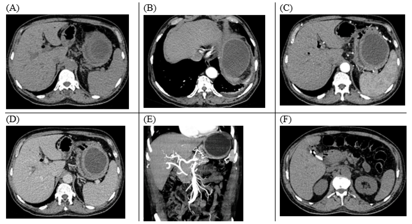
Abdominal ultrasonography revealed central intrahepatic dilation. A 13 mm stone was found in the common bile duct along with multiple gallbladder calculi. Additionally, a large hypoechoic lesion measuring 7x8 cm with internal septations was located adjacent to the left liver lobe, raising suspicions of a hydatid cyst (Figure 1).
The patient was evaluated for percutaneous biliary drainage after optimizing and correcting coagulopathy. However, no suitable peripheral duct was found for this procedure. Consequently, endoscopic retrograde cholangiopancreatography (ERCP) was performed. During this procedure, infected bile was aspirated and a 10 Fr x 5 cm double pigtail stent was placed. A bulge was observed in the posterior wall of the stomach through the side-viewing endoscope, though the gastric mucosa appeared normal.
Following the endoscopic drainage, the patient showed improvement and signs of resolving organ failure. However, he developed mild acute pancreatitis post-ERCP, which was managed conservatively. On the fourth day after ERCP, the patient experienced melaena, leading to a drop in hemoglobin from 12.3 to 7.9 gm/dl. A contrast-enhanced CT scan of the abdomen with angiography revealed a well-defined lesion in the left subphrenic area, displacing the stomach's fundus and greater curvature.
During upper gastrointestinal endoscopy, active bleeding was observed from the posterior gastric wall near the fundus and the stomach was filled with blood clots. The esophagus appeared normal with no varices. The patient's hemoglobin level further dropped to 3.2 gm/dl, necessitating emergency surgery.
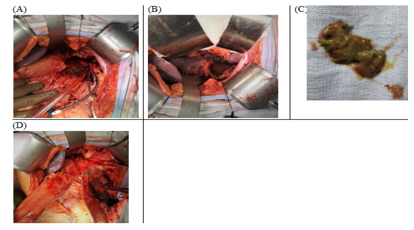
Upon abdominal exploration, signs of recent acute pancreatitis were noted. A cystic lesion was found in the left subphrenic area, adjacent to the left liver lobe, the posterior wall of the stomach near the fundus and the greater curvature. The hepatoduodenal ligament was rigid. The stomach's fundus and greater curvature, which were attached to the cyst, appeared inflamed and fragile. Upon performing gastrotomy, the stomach was found full of blood clots. The cyst was opened and bile-containing contents, resembling hydatid membranes, were removed.
A sleeve gastrectomy was performed to remove the damaged gastric wall and a feeding jejunostomy was added for early feeding. The patient recovered well and was discharged on the seventh postoperative day. Follow-up examinations showed good progress.
The histopathology report indicated transmural necrosis of the stomach wall. The cyst walls and contents showed a necro-inflammatory infiltrate mixed with biliophages, but no granuloma, hydatid membrane, or hooklets were observed (Figure 2).
Figure 1: CECT Abdomen: Axial Images of Abdomen, (A) NCCT, (B&C) Arterial Phase, (D) Venous Phase and (E) Sagittal Reconstruction Images Showing Well-Defined, Peripherally Enhancing, Hypodense Lesion in the left Subphrenic Region, Displacing Greater Curvature of the Stomach with Pneumobilia. (F) NCCT Axial Image Showing CBD Stent in Situ. Central Intrahepatic Bile Duct dilation with Pneumobilia is Evident if A, C, D and E.
Figure 2: (A) Unhealthy, Inflamed Gastric Wall, Ryle’s Tube Coming Out of Necrosed Part of the Stomach Near Greater Curvature and the Stomach was Full of BLOOd Clots. (B) De-Roofed Cystic Lesion. The lesion was Abutting the Left Lobe of the Liver, GE Junction and Stomach Fundus. (C) Bile Mixed Contents, Mimicking Hydatid Membrane Evacuated from Cystic Lesion. (D) Sleeve Gastrectomy done with the Help of Linear Stapler and Stapler Line Re-Inforced with Suture
DISCUSSION
In their 1997 study, Fujiwara et al. [1]. described a rare instance of spontaneous biloma in a 73-year-old woman with no apparent cause. They also reviewed 25 cases of spontaneous biloma documented in literature from 1979 to 1997. Bilomas are typically post-surgical phenomena, often occurring after procedures like cholecystectomy, various instrumental interventions such as percutaneous transhepatic cholangiography, liver biopsy, biliary drainage procedures, ERCP, or following trauma [2,3].
The exact mechanisms behind spontaneous biloma formation are not well understood. It is hypothesized that contributing factors could include increased intraductal pressure due to obstructions from stones, tumors, or spasms of the sphincter of Oddi. The clinical manifestation of bilomas varies widely, ranging from incidental imaging discoveries in asymptomatic patients to symptoms like abdominal fullness, pain, fever, jaundice and in rare instances, peritonitis without fever [4,5].
In the case we observed, the patient presented with a spontaneous biloma without any prior history of abdominal instrumentation. Initially, the lesion was assumed to be an incidental finding of a hydatid cyst on an ultrasonography. Typically, spontaneous bilomas are managed non-operatively, primarily through percutaneous drainage or endoscopic procedures. In this case, the misidentification of the biloma as a hydatid cyst precluded percutaneous drainage, which might have otherwise negated the need for emergency surgery. Additionally, a cholangiogram might have revealed a connection between the biloma and the biliary system, but this was missed due to limitation of ERCP in our Institute.
CONCLUSION
In conclusion, the occurrence of a biloma resulting in a significant upper gastrointestinal bleed, as seen in this case, is exceptionally rare. It highlights the necessity of including bilomas in the differential diagnosis when evaluating cystic lesions in the upper abdomen, especially among patients presenting with signs of biliary obstruction.
This case underscores the importance of accurate diagnosis and the potential implications of misidentifying bilomas, as in this instance, where the biloma was initially mistaken for a hydatid cyst, leading to critical delays in appropriate treatment.
REFERENCES
Fujiwara, H. et al. “Spontaneous rupture of an intrahepatic bile duct with biloma treated by percutaneous drainage and endoscopic sphincterotomy.” The American Journal of Gastroenterology, vol. 93, no. 11, 1998, pp. 2282–2284.
Vazquez, J.L. et al. “Evaluation and treatment of intraabdominal bilomas.” AJR American Journal of Roentgenology, vol. 144, no. 5, 1985, pp. 933–938.
Akhtar, M.A. et al. “Spontaneous idiopathic subcapsular biloma.” Journal of Hepato-Biliary-Pancreatic Surgery, vol. 14, no. 6, 2007, pp. 579–581.
Della Valle, V. et al. “Spontaneous biloma: a case report.” Journal of Ultrasound, vol. 18, no. 3, 2015, pp. 293–296.
Mueller, P.R. et al. “Detection and drainage of bilomas: special considerations.” AJR American Journal of Roentgenology, vol. 140, no. 4, 1983, pp. 715–720.
Recommended Articles
Research Article
Advantages of Two Clips Laparoscopic Cholecystectomy (TCLC) and Conventional Laparoscopic Cholecystectomy (CLC): A Comparative Study
Anshu Attri,
...
Kartik Syal
Published: 10/01/2022
Download PDF
Research Article
Bladder Brilliance: Unveiling the Tapestry of Knowledge on Bladder Cancer in District Kangra, Himachal Pradesh
Vandana Thakur,
...
Ankita Pandey
Published: 08/12/2023
Download PDF
Research Article
Shouldering the Pain: Public Knowledge Assessment of Rotator Cuff Injuries in Himachal Pradesh
Sidhant Korla,
...
Praveen Kumar Thakur
Published: 30/07/2024
Download PDF
Research Article
Awareness regarding Symptoms and Treatment Options for Benign hypertrophy of prostate among the male patients attending outpatient department of Civil hospital , Nurpur, Himachal Pradesh
Aarti Chaudhary,
Brish Bhanu Bhardwaj
Published: 26/05/2023
Download PDF
PO Box 101, Nakuru
Kenya.
Email: office@iarconsortium.org
Editorial Office:
J.L Bhavan, Near Radison Blu Hotel,
Jalukbari, Guwahati-India
Sharma, P. K., Negi, K., Dogra, H. & Ali, A. (2023). Spontaneous Biloma Mimicking Hydatid Cyst Leading to Upper Gastrointestinal Bleed: A Case Report. International Academic Research Journal of Surgery, 3(2), 1-3.
MLA
Sharma, Pardeep K., et al. "Spontaneous Biloma Mimicking Hydatid Cyst Leading to Upper Gastrointestinal Bleed: A Case Report." International Academic Research Journal of Surgery 3.2 (2023): 1-3.
Chicago
Sharma, Pardeep K., Kapil Negi, Harish Dogra and Afshan Ali. "Spontaneous Biloma Mimicking Hydatid Cyst Leading to Upper Gastrointestinal Bleed: A Case Report." International Academic Research Journal of Surgery 3, no. 2 (2023): 1-3.
Harvard
Sharma, P. K., Negi, K., Dogra, H. and Ali, A. (2023) 'Spontaneous Biloma Mimicking Hydatid Cyst Leading to Upper Gastrointestinal Bleed: A Case Report' International Academic Research Journal of Surgery 3(2), pp. 1-3.
Vancouver
Sharma PK, Negi K, Dogra H, Ali A. Spontaneous Biloma Mimicking Hydatid Cyst Leading to Upper Gastrointestinal Bleed: A Case Report. International Academic Research Journal of Surgery. 2023 Jul;3(2):1-3.


 Download PDF
 Download XML