Ethambutol use may lead to permanent vision loss by inducing a dose and duration dependent optic neuropathy. The present study is to compare toxic optic neuropathy among patients on anti-tubercular treatment according to age and sex. In the present study, the patients with ocular toxicity were significantly higher in age in comparison to the patients with no ocular toxicity (p = 0.014) and ocular toxicity was not significantly associated with sex (p = 0.356). As such all the patients and especially the elderly on anti-tubercular treatment must be followed up regularly to detect these adverse effects early.
Keywords
Toxic Optic Neuropathy
Anti Tubercular
Ocular Toxicity
INTRODUCTION
Ethambutol use may lead to permanent vision loss by inducing a dose- and duration-dependent optic neuropathy. This has been of concern to ophthalmologists and physicians both; however, ethambutol continues to be used because of its anti-mycobacterial action with relative systemic safety [1].
With the aim of eliminating TB by 2025, the Revised National Tuberculosis Control Programme (RNTCP) was revamped in 2016 with changes in the protocol for the management of TB cases. The three times a week regimen was changed to a daily regimen and ethambutol was made a part of both the intensive as well as the Continuation Phase (CP) of the treatment [2].
The incidence of ethambutol toxicity has been reported as varying from 0.5-4.3%n [3-5]. Toxicity with Ethambutol is quite rare and can occur after two months of therapy although the average time is around seven months. Prognosis is good following the cessation of the drug but recovery may take up to 12 months and a minority of patients may have residual visual impairments.
The present study is to compare toxic optic neuropathy among patients on anti-tubercular treatment according to age and sex.
Review of Literature
Ethambutol forms one of the first-line drugs for the treatment of tuberculosis. Despite being considered very safe and one of the least systemically toxic first-line drugs, it was shown to result in optic neuropathy within two years of its invention in 1961 [6]. Retro bulbar neuritis is the most common form of EON. While the exact mechanism of the ocular neurotoxic effect of ethambutol is unknown, chelating properties of ethambutol have been hypothesized to contribute to its neurotoxicity. It causes a calcium flux into the mitochondria and excitotoxicity [7].
Liebold [8] and Bobrowitz [9], reported dose related ocular toxicity of ethambutol, 18% in patients receiving >35 mg/kg/day, 5-6% with 25 mg/kg/day and <1% with 15 mg/kg/day of ethambutol for more than two months. Manifestation of ocular toxicity is usually delayed. Melamud et al. [10], reported toxicity does not develop until after treatment for at least 1.5 months. Variable mean interval three to five months between onset of therapy and toxic effects were reported. Manifestations of toxicity as late as 12 months after therapy initiation were reported.
MATERIALS AND METHODS
Study Design
A prospective single-center cohort study of newly diagnosed tuberculosis patients registered at the RNTCP-DOTS centre at Dr. RPGMC Kangra (Tanda) over one year and taking medicines from nearby DOTS centre.
Selection Criteria
Newly diagnosed TB patients coming to the institution’s RNTCP-DOTS centre, after their informed consent
All patients were above 18 years of age
Setting
The study was done in the Department of Ophthalmology, Dr. RPGMC Tanda, with the support of the Department of Pulmonary Medicine, Dr. RPGMC Tanda, after approval from the Institutional Ethical Committee (IEC).
Exclusion Criteria
Patients with other systemic or ocular diseases: affecting visual acuity, or which may contribute to colour vision defect.
Addiction: tobacco, alcohol, etc
Or, patients on medications causing colour vision defects
Or, the patients having colour vision defect /visual field defect, at the baseline examination
Methodology
The patients were examined on day-15 of ATT as baseline investigations, later there was a follow-up at 2-months and 6-months of treatment and in between if a patient complaint of decreased vision.
The patients were evaluated with special reference to:
Demographic information like name, age, sex (male/female), occupation, address
A detailed history of symptoms including the defective vision for distant and near, headache, double vision, transient visual obscuration, altered sensorium, nausea, vomiting
History of hypertension, diabetes and any other systemic illness
History of head injury, brain tumor
Past history of ocular infection, surgery, or trauma
Examination findings including general physical examination, pulse, blood pressure and thorough ophthalmic examination including:
Distant uncorrected visual acuity of all patients using Snellen’s chart of both eyes along with corrected visual acuity using a pinhole
Pupil size and reaction
Relative afferent pupillary defect by swinging flashlight examination
External eye examination for any swelling, erythema, protrusion, deviation of the eyeball
Extraocular movements both for ductions and versions in all cardinal positions.
Slit-lamp examination to see any opacity in media, anterior chamber depth, lens for evidence of cataract, pseudophakia, aphakia and anterior vitreous for pigment and cells
Colour vision by Ishihara chart
Visual fields evaluation using automated perimetry with Humphrey 30-2 program
Dilatation of pupils with one percent tropicamide eye drops
Fundus evaluation using direct ophthalmoscope (Heine beta 200s) and +90D aspheric volk lens and Haag Striet BM 900 slit lamp biomicroscope
Following features were noted in fundus examination: Size, shape and colour of the optic disc, cup size, cup-disc ratio, blurring of disc margins, hemorrhages over the disc, tortuosity of a vein, venous pulsation
Fundus photograph- It was taken using fundus camera VISUCAM:
Optical coherence tomography-OCT of the optic nerve head was done using iVue 100
SD-OCT version 2016.3. The subjects were asked to gaze at the fixation light within the machine and foveolar fixation is confirmed by observing the retina through the infrared monitoring camera
The good quality scan was done. During the examination, the operator ensures exact motion-free centration. Measurements of peripapillary RNFL thickness from 3 scans were averaged to provide a mean measurement of peripapillary RNFL thickness average in the different quadrants
RESULTS
In newly diagnosed TB patients, HRZE given for 2-months and HRE is given for next 4-months. Injection streptomycin to be added in IP phase in previously treated drug sensitive patients (Table 1).
Table 1: Drug Dosage for Adult Tb Under RNTCP
Parameters
Number of tablets (FDCs) (mg)
Inj. Streptomycin
Intensive phase
Continuation phase
HRZE
HRE
Weight category
75/150/400/275
75/150//275
Gm
25-39 Kg
2
2
0.5
40-54 Kg
3
3
0.75
55-69 Kg
4
4
1
>69 Kg
5
5
1
Comparison of toxicity with age
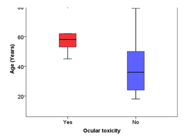
In the present study, the patients with ocular toxicity were significantly higher in age in comparison to the patients with no ocular toxicity (p = 0.014) (Table 2, Figure 1).
Table 2: Comparison of Toxicity with Age
Variables
Ocular toxicity (n = 5)
No ocular toxicity (n = 77)
p-value
Age (Years)
58.0 (49.0, 71.0)
36.0 (24.0, 50.0)
0.014
Data expressed as median (Q1, Q3)
Figure 1: Box-Plot Showing Comparison of Age (Years) Between the Patients with Ocular Toxicity. Lower and Upper Boundary of Each Box Represent First and Third Quartile Respectively while Middle Black Line Represents Median Value
Comparison of Toxicity with Sex
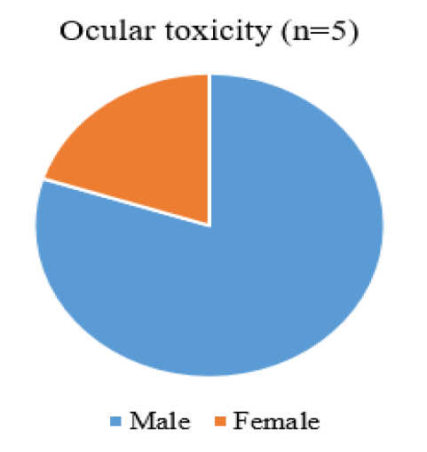
In the present study, ocular toxicity was not significantly associated with sex (p = 0.356) (Table 3, Figure 2).
Figure 2: Gender distribution of patients with ocular toxicity (n = 5)
Table 3: Comparison of Toxicity with Sex
Variables
Ocular toxicity (n = 5)
No ocular toxicity (n = 77)
p-value
Male
4
37
0.356
Female
1
40
DISCUSSION
ATT-induced ocular toxicity in India has been studied earlier; however, with shifting from thrice-weekly regimen to daily regimen and increased duration of ethambutol treatment from two months to six months in newly diagnosed TB patients, there could be a higher incidence of ATT-induced ocular toxicity. Hence, the present study evaluated the incidence of ocular complications in patients treated with ATT under RNTCP in newly diagnosed TB patients taking treatment at DOTS centre, Dr. RPGMC Tanda and nearby DOTS centres. In the present study, the patients received ethambutol from 15.27 mg/kg to 20.625mg/kg body weight.
In our study, the incidence of ocular toxicity was found to be 6.09% (10/164). This included changes in visual acuity, visual field defects, colour vision abnormality, or fundus changes. Our results are in concordance with Mathur et al. [11], who reported incidence of ocular toxicity as 6.3% (3/47). In the present study, the patients with ocular toxicity were significantly higher in age in comparison to the patients with no ocular toxicity (p = 0.014). In the present study, ocular toxicity was not significantly associated with sex (p = 0.356). Narang et al. [12], in their study of 640 cases treated by Ethambutol of 25 mg/kg/day of the drug, along with companion drugs came across only four cases of ocular toxicity (0.62%), Solu et al. [13], in their study reported that out of 94 study participants on standard HRZE regimen, 14 eyes of seven patients (7.44%) had a drop in visual acuity from baseline (p-value = 0.03).
CONCLUSION
This study was an attempt to assess the incidence of ocular toxicity in patients on anti-tubercular treatment. In our study, we found that the overall incidence of ocular toxicity in the patients on anti-tubercular treatment was 6.09%. Although ocular toxicity was more common in patients above the age of 60 years. All the patients and especially the elderly on anti-tubercular treatment must be followed up regularly to detect these adverse effects early. However, studies with larger sample size and long-term follow-up are required to generalize the findings.
Financial Disclosure
The study did not receive any financial assistance. The subjects included in the study were not provided any financial support and no financial burden was placed on the study subjects.
REFERENCES
Saxena, R. et al. “Response to comment on: Ethambutol toxicity: Expert panel consensus for the primary prevention, diagnosis and management of ethambutol-induced optic neuropathy.” Indian Journal of Ophthalmology, vol. 70, no. 3, 2022, pp. 1070–1071. https://doi.org/10.4103/ijo.IJO_88_22.
“Revised national tuberculosis control programme.” National Health Portal of India, www.nhp.gov.in/revised-national-tuberculosis-control-programme_pg.
Polak, B.C. et al. “Blue yellow colour vision changes as early symptoms of ethambutol toxicity.” Ophthalmologica, vol. 191, 1985, pp. 223–226.
Trusiewicz, D. “Farnsworth 100 hue test in diagnosis of ethambutol-induced damage to optic nerve.” Ophthalmologica, vol. 171, 1975, pp. 425–431.
Barron, G.J. et al. “Ocular toxicity from glutametheol.” American Journal of Ophthalmology, vol. 77, no. 2, 1974, pp. 257–260.
Carr, R.E. and P. Henkind. “Ocular manifestations of ethambutol: Toxic amblyopia after administration of an experimental antituberculous drug.” Archives of Ophthalmology, vol. 67, 1962, pp. 566–571. https://doi.org/10.1001/archopht.1962.00960020566009.
Kerrison, J.B. “Optic neuropathies caused by toxins and adverse drug reactions.” Ophthalmology Clinics of North America, vol. 17, 2004, pp. 481–488.
Leibold, J.E. “The ocular toxicity of ethambutol and its relation to dose.” Annals of the New York Academy of Sciences, vol. 135, 1966, pp. 904–909.
Bobrowitz, I.D. “Ethambutol in the retreatment of pulmonary tuberculosis.” Annals of the New York Academy of Sciences, vol. 135, 1966, pp. 796–822.
Melamud, A. et al. “Ocular ethambutol toxicity.” Mayo Clinic Proceedings, vol. 78, no. 11, 2003, pp. 1409–1411.
Mathur, K.C. and J.S. Sankhla. “Ophthalmic manifestations of the toxicity of ethambutol.” Indian Journal of Ophthalmology, vol. 24, 1976, pp. 6–9.
Narang, R.K. and B.M. Varma. “Ocular toxicity of ethambutol (a clinical study).” Indian Journal of Ophthalmology, vol. 27, 1979, pp. 37–40.
Solu, T.M. and K. Panchal. “Ocular side effects of anti-tubercular drugs in patients receiving anti-TB treatment at a tertiary care center.” IP International Journal of Ocular Oncology and Oculoplasty, vol. 6, no. 3, 2020, pp. 187–191.
Recommended Articles
Research Article
Advantages of Two Clips Laparoscopic Cholecystectomy (TCLC) and Conventional Laparoscopic Cholecystectomy (CLC): A Comparative Study
Anshu Attri,
...
Kartik Syal
Published: 10/01/2022
Download PDF
Research Article
Bladder Brilliance: Unveiling the Tapestry of Knowledge on Bladder Cancer in District Kangra, Himachal Pradesh
Vandana Thakur,
...
Ankita Pandey
Published: 08/12/2023
Download PDF
Research Article
Shouldering the Pain: Public Knowledge Assessment of Rotator Cuff Injuries in Himachal Pradesh
Sidhant Korla,
...
Praveen Kumar Thakur
Published: 30/07/2024
Download PDF
Research Article
Awareness regarding Symptoms and Treatment Options for Benign hypertrophy of prostate among the male patients attending outpatient department of Civil hospital , Nurpur, Himachal Pradesh
Aarti Chaudhary,
Brish Bhanu Bhardwaj
Published: 26/05/2023
Download PDF
PO Box 101, Nakuru
Kenya.
Email: office@iarconsortium.org
Editorial Office:
J.L Bhavan, Near Radison Blu Hotel,
Jalukbari, Guwahati-India
Kumar, S., Bhatnagar, R., Chauhan, N. & Parmar, V. (2023). Comparison of Ocular Toxicity According to Age and Sex among Patients Taking Anti Tubercular Treatment. International Academic Research Journal of Surgery, 3(2), 1-4.
MLA
Kumar, Sunil, et al. "Comparison of Ocular Toxicity According to Age and Sex among Patients Taking Anti Tubercular Treatment." International Academic Research Journal of Surgery 3.2 (2023): 1-4.
Chicago
Kumar, Sunil, Richa Bhatnagar, Nikita Chauhan and Vipul Parmar. "Comparison of Ocular Toxicity According to Age and Sex among Patients Taking Anti Tubercular Treatment." International Academic Research Journal of Surgery 3, no. 2 (2023): 1-4.
Harvard
Kumar, S., Bhatnagar, R., Chauhan, N. and Parmar, V. (2023) 'Comparison of Ocular Toxicity According to Age and Sex among Patients Taking Anti Tubercular Treatment' International Academic Research Journal of Surgery 3(2), pp. 1-4.
Vancouver
Kumar S, Bhatnagar R, Chauhan N, Parmar V. Comparison of Ocular Toxicity According to Age and Sex among Patients Taking Anti Tubercular Treatment. International Academic Research Journal of Surgery. 2023 Jul;3(2):1-4.



 Download PDF
 Download XML