Many pathogenic genes found in Pantoea spp. contribute to the development of numerous human illnesses. Therefore, in the present study, a total of 60 samples from canned and packaged pet food products were examined. Out of these, 10 Pantoea spp. isolates were confirmed using 16S rRNA gene Polymerase Chain Reaction (PCR) technology. Subsequently, all the Pantoea spp. isolates were analyzed for the occurrence of specific virulence genes, namely cbl and cytR, using the PCR technique. The results indicated that the cbl gene was prevalent in 90.0% of the isolates, with nine out of ten isolates showing its presence. On the other hand, the cytR gene was found in 78.8% of the Pantoea spp. isolates, specifically in seven out of ten samples.
Keywords
Pantoea spp.
Cbl and Cytr
PCR
INTRODUCTION
The genus Pantoea comprises multiple species that are commonly linked to plants, either living on their surface (epiphytes) or causing diseases (pathogens). Additionally, some of these species have the potential to cause illnesses in humans. Among them, Pantoea agglomerans is the most frequently encountered species in human-related cases. It can be found widely in nature, inhabiting various environments such as plants, water, soil, humans and animals. It is commonly associated with plants, acting as an epiphyte or an endophyte and certain strains have been identified as pathogenic agents causing tumors [1, 2].
Since high-throughput sequencing became available, PCR amplification has been widely used to generate Operational Taxonomic Units (OTUs) from 16S sequences based on their similarity. These OTUs are typically clustered and their representative sequences are compared with reference databases to determine their likely taxonomy. Despite being convenient and effective, this approach requires certain assumptions. For example, it has historically been assumed that sequences with over 95% identity belong to the same genus, while sequences with over 97% identity belong to the same species [3].
Low-throughput techniques have been utilized to differentiate strains, also known as subspecies, by analyzing 16S sequences and identifying polymorphisms within the gene. By examining Single-Nucleotide Polymorphisms (SNPs), researchers have been able to trace clinically relevant strains or, if these SNPs are consistently associated with other parts of the bacterial haplotype, predict certain phenotypic traits [4].
Virulence genes are genetic elements within the bacterial genome that encode virulence factors, which are molecules or proteins that contribute to the pathogenicity of the bacterium. These genes play crucial roles in colonization, adhesion, invasion, immune evasion, toxin production and other mechanisms that enable the bacteria to establish infections and cause disease in the host [5]. Studies have extensively investigated the virulence genes of Enterobacteriaceae (including pantoea spp.) to gain insights into their pathogenic potential and the mechanisms by which they cause infections [6].
The study aimed to isolate bacteria in pet food products and detect them by PCR using the 16S rRNA gene, along with investigating some virulence genes of bacteria.
Collection of Samples
In Iraq, a total of 60 samples were gathered from various pet food products manufactured by different companies, including Jungle, Hellaw, Pado, Vitus, LoLo, Paw, Pedigree and Dr. Clauders. For each sample, a subset of 1 gram was taken and homogenized in a sterile blender bag with 225 ml of 1% buffered peptone water for 60 seconds. To determine the number of Colony-Forming Units (CFU) per gram of pet food, the homogenates and their decimal dilutions in 0.9% NaCl were streaked onto plate count agar. After incubation at 37 °C for 18-24 hours, the colonies were counted. In order to identify the isolated bacteria, visual examinations were conducted to detect growth, pigmentation and colonial morphology. Subsequently, the isolated bacteria were purified through repeated subculture on XLD agar plates and incubated at 37°C for 24 hours. For further identification, biochemical tests were performed using the Vitek 2 system on the pure bacterial cultures, which were also stored at 4 °C. To specifically identify Pantoea spp., a genetic test using 16S rRNA gene PCR was conducted. The bacteria were inoculated in a nutrient broth and then placed in a test tube for this genetic analysis (Table 1).
Table 1: Primers: Used in This Study
Primer Name
Seq.
Annealing Temp. (ºC)
Pantoea spp.
16s-Rrna-F
5- TCGACTTGGAGGTTGTTCCC -3
60
16s-Rrna-R
5- AGGGCCATGATGACTTGACG -3
CytR-F
5- GTGCACATCGACAACCTGAC-3
59
CytR-R
5- GTCATCAGCTGGTTCATGGC-3
Cbl-F
5- CTCAGCGAGCCAACCAGTAA-3
59
Cbl-R
5- CTGGCATTTTCACGGCAACA-3
DNA Primers
Design PCR primers that are specific for the target gene or sequence of interest in Pantoea spp. There are several genes that can be targeted for PCR detection of Pantoea, including the 16S rRNA gene.
DNA-Amplifying PCR Thermocycler Programs
Polymerization enzyme chain reaction, commonly known as PCR, was employed with a PCR thermocycler, following the procedure as Shown in Table 2.
Table 2: Temperature Conditions Used in the PCR
Steps
°C
m: s
Cycle
Initial Denaturation
95
05:00
1
Denaturation
95
00:20
40
Annealing
60
00:20
Extension
72
00:20
Final Extension
72
10:00
1
RESULTS AND DISCUSSION
Counting Pantoea spp. and Isolation
According to the results of the current investigation, the pet food in particular was subjected to a total bacterial count determination to determine the bacterial burden of Pantoea spp. The results showed a range of log Colony-Forming Units (CFU) per milliliter (mL) of 9.2 x 10⁶ to 2.7 x 10⁷ total bacterial counts. Enterobacteriaceae presence and yeast/mold contamination in dry pet food products, as per EU Regulation No. 142/2011, pet food samples, excluding canned pet food, that has an Enterobacteriaceae count exceeding 3 × 10² colony-forming units per gram (CFU/g) are deemed unsatisfactory in terms of microbial hygiene [7].
Microorganisms can cause food spoilage and pose a risk to consumer health. However, there are currently no stringent regulations in place regarding the maximum limits of bacterial and fungal contamination in pet food [8,9]. Kukier et al., [10], stated that, in terms of livestock feed, the Total Aerobic Microbial Count (TAMC) should not exceed 10^6 Colony-Forming Units per gram (CFU/g). This emphasizes the need for guidelines and standards to ensure the microbiological quality and safety of pet food.
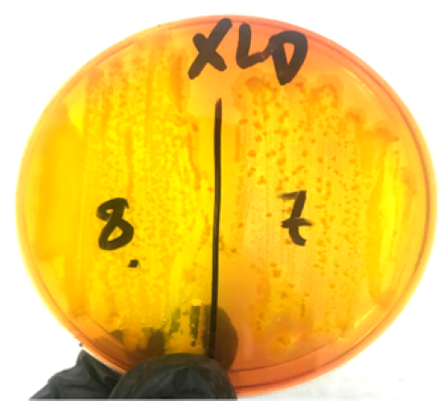
Figure 1, presents the morphology, cultural characteristics and staining characteristics of isolated Pantoea spp. The isolates were cultured on Xylose-Lysine Deoxycholate (XLD) Agar, the colonies appeared yellow.
Figure 1: Pantoea Yellow Colonies XLD Agar Acidify the Medium, Turning It Yellow
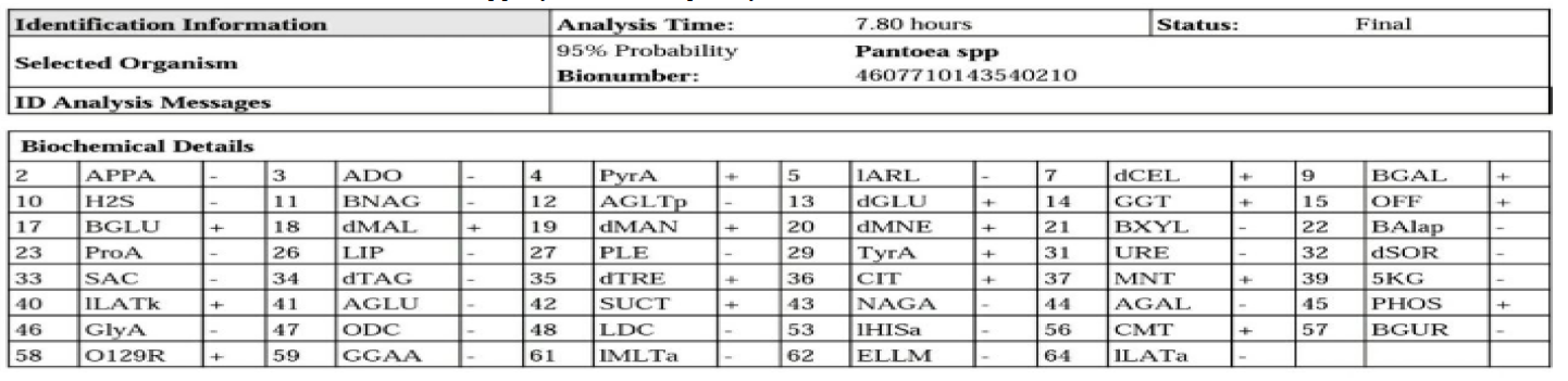
Using the Vitek 2 technique, which correctly recognized all isolates as Pantoea with a 95% accuracy rate, the diagnosis of Pantoea was finally established. Table 3 contains the full findings of this investigation. The Pantoea species can be determined by the Vitek 2 system using a mix of biochemical processes, growth patterns and metabolic activities. To establish the most likely identification, the algorithm evaluates how the bacterial isolate responds to various substrates and develops a profile that is compared to a large database.
Table 3: Identification Information of Pantoea spp. By Vitek 2 Compact System
Diagnosis of Pantoea spp. Using PCR Technology
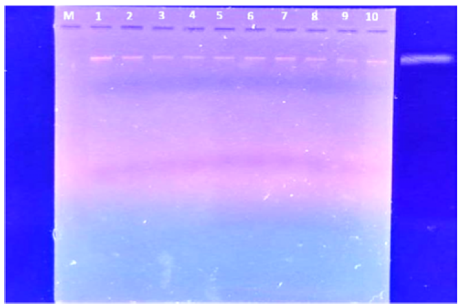
In this work, the isolates of Pantoea spp. were identified using the 16S rRNA gene and the polymerase chain reaction (Monoplex PCR) method. The findings in Figure 2 demonstrate that the 16S rRNA gene, the diagnostic gene for these bacteria, is present in all isolates and has a molecular weight of (1500 bp). This demonstrates that the isolates come from Pantoea species. During this study, the PCR method has a 100% sensitivity rate for isolating isolates.
Figure 2: Total Genomic DNA Extracted from Isolated Cells Using 1% Agarose Gel Electrophoresis (Purified DNA of Pantoea spp.)
According to the study by Cheng et al., [11], 18 isolates were tested for Pantoea spp. using the 16S rRNA gene, confirming that 9 isolates belonged to this bacterial family. However, a study of Delétoile et al., [12], Pantoea spp. isolates determined that 95.6% of them had the 16S rRNA gene, making them pathogenic to humans and animals.
Figure 2 shows that the extracted DNA samples underwent PCR amplification targeting the 16S rDNA gene. Upon comparison to the molecular DNA ladder, all 10 bacterial isolates displayed a single band approximately 1500 bp in size. To identify Gram-negative strains, representatives of various bacteria underwent analysis using the 16S rRNA sequence. The genomic DNAs from these bacterial strains were prepared following previously established protocols [13].
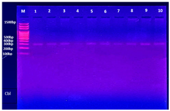
Figure 3: Agarose Gel Electrophoresis of the PCR Assay Shows Results of the Cbl Gene in Pantoea spp., Whereas M: Marker Ladder 1500-100bp and Drilling Number (1, 2, 3, 5, 6, 7, 8, 9, 10), Isolates the Germ of Pantoea spp. Positive for the Gene with a Length of 300 bp.
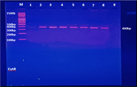
Table 4 presents data on the prevalence of virulence genes in Pantoea spp. isolates. The table includes isolate numbers, the specific Pantoea spp. strains tested, the percentage of isolates containing the gene and the corresponding virulence gene. The results show that the Cbl gene was present in 90.0% of tested isolates (nine out of ten), while the cytR gene was detected in 78.8% of the isolates (seven out of nine). Isolate number 4 did not show the presence of the cbl gene and isolates 1 and 9 did not exhibit the presence of the cytR gene. These findings provide valuable insights into the distribution of these virulence genes among Pantoea spp. strains, which can contribute to our understanding of their potential pathogenicity and virulence mechanisms.
Table 4: The Distribution of Virulence Genes Among Pantoea spp. Isolates
No.
Isolate
Pantoea spp. Containing the gene (%)
Gene
1-
(1,2,3, 5, 6, 7,8,9, 10)
9 (90.0%)
cbl
(4)
Not-detected 1 (10.0%)
2-
(2,3,4 5, 6, 7,8)
7 (78.8%)
cytR
(1,9)
Not- detected 2 (22.2%)
The Cbl gene, also known as CysB, is a gene that encodes a protein called Cystathionine beta-lyase. This gene is found in various organisms, including bacteria, plants and animals [14]. The CysB gene codes for the CBL enzyme, which catalyzes the breakdown of cystathionine, an intermediate in the transsulfuration pathway, into cysteine, α-ketobutyrate and ammonia. This reaction is essential for maintaining the balance of sulfur-containing amino acids in the body [15] (Figure 4).
Figure 4: Agarose Gel Electrophoresis of the PCR Assay Shows Results of the CytR Gene in Pantoea spp., Whereas M: Marker Ladder 1500-100bp and Drilling Number (2, 3, 4, 5, 6, 7, 8), Isolates the Germ of Pantoea spp. Positive for the Gene with a Length of 400 bp
CytR Gene
Figure 4 illustrates the outcomes of the gel electrophoresis examination of the PCR products. The ethidium bromide-stained agarose gel clearly displays well-defined bands corresponding to the amplified 400 bp fragment. In this study, most of the Pantoea isolates that were examined exhibited the presence of the Cytr gene. In a previous investigation carried out by Jia et al. [19], utilizing the TAIL-PCR method, a mutated gene was identified, which showed similarity to cytR. This gene codes for a cytidine repressor protein known as CytR, which acts as a bacterial transcription factor involved in a complex regulatory process. The mutation in cytR led to negative regulation of indole, resulting in an increased expression of the downstream gene deoC. The deoC gene encodes a critical enzyme called deoxyribose-phosphate aldolase, which plays a significant role in pentose metabolism. Several studies have investigated the role of CysB in Entero-bacteriaceae. For example, research on Escherichia coli has shown that CysB regulates the expression of the cysJIH operon, which encodes enzymes involved in the biosynthesis of cysteine. It also controls the expression of the cysK and cysP genes, which are responsible for cysteine transport [16-18].
The study also revealed that DeoC is one of the regulatory proteins in Pantoea agglomerans that helps to counteract starvation. Interestingly, under starvation conditions, the expression of deoC was induced while there was a concurrent decrease in cytR expression. This finding suggests the presence of a regulatory interplay between cytR, deoC and the response to nutrient availability in P. agglomerans [19].
Pedersen et al. [20], discovered that the regulatory region of the Escherichia coli nupG gene contains one CytR-binding site and four CRP-binding sites. The unique aspect of their study was the identification of a novel structure for the CytR-binding site, unlike previously known targets. Interestingly, when the CytR repressor bound to its operator, it caused a repositioning of a cAMP-CRP complex in the −35 region of the promoter. This suggests that the formation of repression and activation complexes at the nupG promoter involves specific subsets of CRP-binding sites. These findings demonstrate the bacterium's employment of both positive and negative regulatory mechanisms to intricately regulate the expression of CytR- and cAMP-CRP-regulated genes.
REFERENCES
Grimont, P.A.D. and F. Grimont. “Genus Pantoea.”Bergey’s Manual of Systematic Bacteriology, vol. 2, 2005, pp. 713–720.
Weinthal, D.M. et al. “Distribution and replication of the pathogenicity plasmid pPATH in diverse populations of the gall-forming bacterium Pantoea agglomerans.” Applied and Environmental Microbiology, vol. 73, no. 23, 2007, pp. 7552–7561.
Schloss, P.D. and J. Handelsman. “Introducing DOTUR, a computer program for defining operational taxonomic units and estimating species richness.” Applied and Environmental Microbiology, vol. 71, no. 3, 2005, pp. 1501–1506.
Fitz-Gibbon, S. et al. “Propionibacterium acnes strain populations in the human skin microbiome associated with acne.” Journal of Investigative Dermatology, vol. 133, no. 9, 2013, pp. 2152–2160.
Albaaj, A.A.A. and S.K. Al-Ramahy. “Molecular study of bacteria Pantoea spp. in Diwaniyah City, Iraq.” Journal of Physics: Conference Series, vol. 1294, no. 6, 2019, article 062060.
Kępińska-Pacelik, J. and W. Biel. “Microbiological hazards in dry dog chews and feeds.” Animals, vol. 11, no. 3, 2021, p. 631.
Kazimierska, K. et al. “Evaluation of nutritional value and microbiological safety in commercial dog food.” Veterinary Research Communications, vol. 45, nos. 2–3, 2021, pp. 111–128.
Kukier, E. et al. “Microbiological quality of compound feed used in Poland.” Bulletin of the Veterinary Institute in Pulawy, vol. 56, no. 3, 2012, pp. 349–354.
Cheng, A. et al. “Bacteremia caused by Pantoea agglomerans at a medical center in Taiwan, 2000–2010.” Journal of Microbiology, Immunology and Infection, vol. 46, no. 3, 2013, pp. 187–194.
Delétoile, A. et al. “Phylogeny and Identification of Pantoea Species and Typing of Pantoea agglomerans Strains by Multilocus Gene Sequencing.” Journal of Clinical Microbiology, vol. 47, no. 2, 2009, pp. 300–310.
Ni, Y. et al. “16S rDNA and 16S–23S internal transcribed spacer sequence analyses reveal inter- and intraspecific Acidithiobacillus phylogeny.” Microbiology, vol. 154, no. 8, 2008, pp. 2397–2407.
Ejim, L.J. et al. “Cystathionine β-lyase is important for virulence of Salmonella enterica Serovar Typhimurium.” Infection and Immunity, vol. 72, no. 6, 2004, pp. 3310–3314.
Stipanuk, M.H. and I. Ueki. “Dealing with methionine/homocysteine sulfur: Cysteine metabolism to taurine and inorganic sulfur.” Journal of Inherited Metabolic Disease, vol. 34, 2011, pp. 17–32.
van der Ploeg, J.R. et al. “Sulfonate-sulfur metabolism and its regulation in Escherichia coli.” Archives of Microbiology, vol. 176, 2001, pp. 1–8.
van der Ploeg, J.R. et al. “The Escherichia coli ssuEADCB gene cluster is required for the utilization of sulfur from aliphatic sulfonates and is regulated by the transcriptional activator Cbl.” Journal of Biological Chemistry, vol. 274, no. 41, 1999, pp. 29358–29365.
Iwanicka-Nowicka, R. and M.M. Hryniewicz. “A new gene, cbl, encoding a member of the LysR Family of transcriptional regulators belongs to the Escherichia coli cys Regulon.” Gene, vol. 166, no. 1, 1995, pp. 11–17.
Jia, M. et al. “The Cytidine Repressor Participates in the Regulatory Pathway of Indole in Pantoea agglomerans.”Research in Microbiology, vol. 168, no. 7, 2017, pp. 636–643.
Pedersen, H. et al. “Gene-regulatory modules in escherichia coli: nucleoprotein complexes formed by camp–crp and CyTr at the NuPg promoter.” Molecular Microbiology, vol. 17, no. 5, 1995, pp. 843–853.
Recommended Articles
Research Article
Advantages of Two Clips Laparoscopic Cholecystectomy (TCLC) and Conventional Laparoscopic Cholecystectomy (CLC): A Comparative Study
Anshu Attri,
...
Kartik Syal
Published: 10/01/2022
Download PDF
Research Article
Bladder Brilliance: Unveiling the Tapestry of Knowledge on Bladder Cancer in District Kangra, Himachal Pradesh
Vandana Thakur,
...
Ankita Pandey
Published: 08/12/2023
Download PDF
Research Article
Shouldering the Pain: Public Knowledge Assessment of Rotator Cuff Injuries in Himachal Pradesh
Sidhant Korla,
...
Praveen Kumar Thakur
Published: 30/07/2024
Download PDF
Research Article
Awareness regarding Symptoms and Treatment Options for Benign hypertrophy of prostate among the male patients attending outpatient department of Civil hospital , Nurpur, Himachal Pradesh
Aarti Chaudhary,
Brish Bhanu Bhardwaj
Published: 26/05/2023
Download PDF
PO Box 101, Nakuru
Kenya.
Email: office@iarconsortium.org
Editorial Office:
J.L Bhavan, Near Radison Blu Hotel,
Jalukbari, Guwahati-India
Aswad, D. A. & Hadi, S. H. (2023). cbl and cytR virulence genes of pantoea spp. bacteria from pet food products. International Academic Research Journal of Surgery, 3(2), 1-4.
MLA
Aswad, Dhulfiqar A. and Sabaa H. Hadi. "cbl and cytR virulence genes of pantoea spp. bacteria from pet food products." International Academic Research Journal of Surgery 3.2 (2023): 1-4.
Chicago
Aswad, Dhulfiqar A. and Sabaa H. Hadi. "cbl and cytR virulence genes of pantoea spp. bacteria from pet food products." International Academic Research Journal of Surgery 3, no. 2 (2023): 1-4.
Harvard
Aswad, D. A. and Hadi, S. H. (2023) 'cbl and cytR virulence genes of pantoea spp. bacteria from pet food products' International Academic Research Journal of Surgery 3(2), pp. 1-4.
Vancouver
Aswad DA, Hadi SH. cbl and cytR virulence genes of pantoea spp. bacteria from pet food products. International Academic Research Journal of Surgery. 2023 Jul;3(2):1-4.



 Download PDF
 Download XML