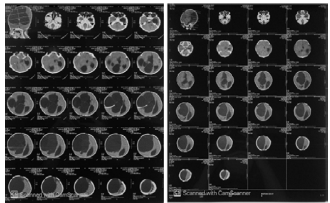
Hydrocephalus is a condition characterized by an excess of Cerebrospinal Fluid (CSF) in the ventricles of the brain. Cerebrospinal fluid is produced by the choroid plexus of the ventricles of the brain and is a sterile fluid. Cerebrospinal normally flows through the ventricular system, starting from the lateral ventricles to the third and then fourth ventricles through ducts to the circulation around the brain, then this fluid is absorbed [1]. Whereas, loculated hydrocephalus is when a compartment is filled with a discrete fluid formed in or in conjunction with the system of the ventricles of the brain. Loculated hydrocephalus was first introduced as a diagnosis entity by Cushing. Loculated hydrocephalus commonly presents in patients suffering from diseases such as bacterial or fungal meningitis, intraventricular bleeding, post-shunt infection, or other inflammatory processes, as a complication from the diseases [2]. A type of loculated hydrocephalus is uniloculated hydrocephalus. Uniloculated hydrocephalus is a cystic dilation that happens on one segment of the ventricular system [2].
Most patients with these various conditions may need repeat surgery to revise the traditional shunt. Other following procedures such as stereotactic aspiration, Endoscopic Third Ventriculostomy (ETV), septostomy, endoscopic fenestration of cystic septa and walls, aqueductoplasty, or a combination of several surgical principles. Thus, there is yet to be any perfect treatment for loculated hydrocephalus [2].
The full incidence of hydrocephalus is unknown, while in Indonesia it reaches 10 per mil1 per year. The incidence of acquired hydrocephalus is also unknown, possibly due to the many types of diseases that can cause hydrocephalus [2].
The etiology of congenital hydrocephalus is unknown. Acquired hydrocephalus mostly caused by tumor obstruction, trauma, intracranial hemorrhage and infection. Some of the first modern reports of multi-located hydrocephalus were noted in patients with a history of neonatal meningitis. This finding is thought to be due to the tendency of neonates to develop ventriculitis resulting in intraventricular gliosis [1].
Several clinical manifestations that can be found in children are delayed mental capacity, headache, neck pain indicating a tonsillar herniation, morning vomiting, blurred vision due to papilledema or advanced papil atrophy, diplopia due to the paralysis of nervus cranialis VI, delayed sexual growth and maturity, walking difficulty due to spasticity. The signs are papilledema, cracked pot on head percussion, gait dysfunction, macrocephaly [1].
Lumbar puncture can be used to measure intracranial pressure, but only after radiological examination reveals an obstruction. This examination is helpful in establishing the diagnosis.
Loculated hydrocephalus can be treated pharmacologically by giving 25 mg/kg/day of acetazolamide in 3 doses or 1 mg/kg/day of furosemide in 3 doses. Surgery procedure is also a better option for the management of loculated hydrocephalus. The procedures are choroid plexotomy, opening the aquaductus stenosis, tumor excision, endoscopy phenestration [4].
Most of the hydrocephalus patients undergo shunt installation. The principle of shunt installation is to maintain the connection between the CSF and the drainage cavity (peritoneum, right atrium, pleura). Several alternatives to shunt installation include Ventriculo-Peritoneal (VP) shunt, Ventriculo-Atrial (VA) shunt, lumbo-Peritoneal shunt and Ventriculo-Pleural shunt [5].
Currently, the treatment options for loculated hydrocephalus are fenestrated microsurgery of the septate compartment with open or endoscopic craniotomy, shunt surgery which includes the placing of multiple catheters in a multilobulated compartment and combinations of these modalities [6].
A shunt installation is highly required, even if the patient had received a successful fenestration by craniotomy or endoscopy. The main goal of open or endoscopic microsurgery is to unify multiple compartments into a single CSF compartment. This would allow the drainage to go through a single shunt catheter system. The best treatment option for MLH is yet to be defined up to this day. It is important to relieve intracranial hypertension in MLH patients. Another significance of MLH treatment is also to minimize the needs for surgery and complications related to multiple shunt catheter, thus the morbidity can be decreased.
Ventriculo-Peritoneal shunt is a surgical procedure performed to relieve intracranial pressure caused by too much cerebrospinal fluid (hydrocephalus). Fluid flows from the ventricles in the brain to the peritoneal cavity. These complications include infection, block, subdural hematoma, ascites, CSSoma, gastrointestinal tract obstruction, hollow organ perforation, malfunction, or migration of the shunt.



 Download PDF
 Download XML