Fetus in Fetu (FIF) usually occurs in infants, presenting as an abdominal mass. It is a condition where a foetoid tumor is found withing the body of an infant. To minimize the negative effects of FIF, physicians need to acknowledge its diagnosis and management immediately. We report a case of a four-month-old boy presenting with an abdominal distension. From serology, it is known that the level of Alpha Fetoprotein (AFP) was >400.00 IU/mL and from abdominal ultrasonography we found a right retroperitoneal mass that pushes the right kidney inferiorly and extends to the left side. The patient underwent a surgery and the mass was resected. The gross pathology specimen measured 20 x 20 x 15 cm3. It contains partly formed appendicular bone, cranium and vertebrae. The patient's postoperative diagnosis was FIF.
Fetus in Fetu (FIF) was first introduced by Meckels on 1800. Up to this day, this clinical condition is still considered rare. Fetus in fetu was then defined by Willis on 1953 as a tumor mass which contains axial vertebrae and often other organs or limbs. The incidence of this condition is one in every 500.000 livebirths [1]. FIF is a pedunculated and capsulated vertebrae tumor. The majority of the cases are anencephalic, however, almost in all cases, vertebrae and limbs were found in 91% and 82,5% cases, respectively [2].
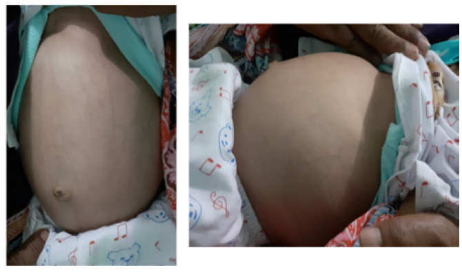
A four-month-old boy presenting with a chief complaint of a mass on the right abdomen. The mass was first noticed by his parents when he was just two-month-old. The patient was delivered via normal vaginal delivery in the gestational age of 40 weeks. Physical examination showed an abdominal distention. The laboratory examination revealed hyponatremia, hypoalbuminemia and hypocalcemia. The patient’s alpha fetoprotein level was >400.000 IU/mL.
Abdominal ultrasonography revealed the right retroperitoneal mass that pushes the right kidney inferiorly and extends to the left side. Other than that, a bilateral spermatic cord hydrocele and ascites were found. No abnormalities were seen in other intra-abdominal organs, such as liver, spleen, gallbladder, pancreas, kidneys and urinary bladder. The CT scan examination showed a solid lesion with lipid components and retroperitoneal calcification that pushes the right kidney posteriorly and aorta abdominal to the contralateral side, liver, spleen, intestines to the superolateral side, pancreas and bladder adhered to the hilus of the right kidney, liver capsule and intestine wall, supporting the appearance of retroperitoneal teratoma. A spina bifida was also found in vertebra sacralis 1-4 (Figure 1).

Figure 1. Physical Examination Revealed an Abdominal Distention
The patient’s initial diagnosis was intrabdominal mass with retroperitoneal teratoma as the differential diagnosis. The patient was also undernourished. The management as done by pediatrician and surgeon. The patient underwent a surgery for a tumor incision. The surgery procedure was done by making a supraumbilical incision, on the peritoneum cavity, ±500 cc of cylus liquid was found.
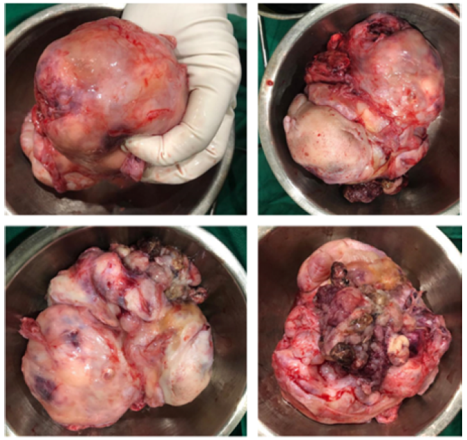
The tumor was located in the posterior duodenum, with the size of 20x20x15 cm3. The macroscopic appearance of the tumor revealed an appendicular bone, cranium and vertebrae.
Furthermore, the patient’s postoperative diagnosis was fetus in fetu. The pathology anatomy examination showed an intraabdominal solid tumor containing mesoderm components (cartilage tissue), fat tissue and muscle, entoderm (the epithelial gland of gastrointestinal tissue) with no signs of malignancy (Figure 2).

Figure 2. Gross Pathology Specimen of Fetus in Fetu
Fetus in fetu is an anomaly condition consists of monozygotic malformation, diamniotic twin found in the twin’s body. FIF is most commonly found in abdominal cavity (retroperitoneum), mediastinum posterior, sacrococcygeal region and neck [3]. The majority of FIF is located retroperitoneally along the ventral midline, meanwhile the location that is rarely reported are brain ventricle, liver, pelvis, scrotum and mediastinum [4]. The commonly visible organs in FIF can be vertebra, limbs, central nervous system, gastrointestinal tract, blood vessel and genitourinary tract. Several differential diagnoses of FIF are teratoma, meconium pseudocyst and neuroblastoma. Patients usually present with signs and symptoms related to the effects of the mass, such as abdominal distention, constipation, eating difficulty, pressure effect on kidney system and dyspnea. Alpha Fetoprotein (AFP) serum level and human chorionic Gonadotropin (HCG) level might be normal or increased. To establish the diagnosis, the patients usually receive plain radiograph, ultrasonography, CT scan and MRI. FIF is usually managed with surgical incision. Postoperative observation is done with USG and AFP serum level to assume further recurrence and malignant transformation [5].
The difference between FIF and adult teratoma is from the existence of axial bones and well-differentiated tissue [6]. According to Willis definition, the condition of FIF consists of separated vertebral column and full development of the original organ. The separated vertebral column shows the finished process of primary phase after gastrulation, involving the formation of neural tube and the symmetrical development around its axis. Furthermore, Spencer explained the characteristics of FIF, among others are enclosed in different sac, fully or partially enclosed by the skin, attached to the host with only several big blood vessels and may be placed directly adjacent to one of the attachment sites of the conjoint twins or connected to the neural tube or digestive system [5].
Meanwhile, teratoma is a condition where there is an accumulation of pluripotent cells that lacks organogenesis or vertebral segmentation. AFP levels may be increased in older children because of their greater likelihood of malignancy. Unlike teratomas, FIF rarely transforms into malignancy [5].
Differentiating FIF lesions from teratomas is very important, because teratomas, mainly the retroperitoneal kind, can have a malignancy rate up to 10%. Pathologically, mature teratomas are tumors that involve the pluripotent cells that have an unorganized arrangement. In mature teratomas, all three germ lines are represented by the pluripotent cells. In FIF, tissue growth is more regular and must show axial bones with metameric segmentation. If the limb bud array is visible, this may support the diagnosis. The mass karyotype must be identical to that of the host [6].
FIF is benign. However, it requires a surgical incision to establish the diagnosis pathologically and to rule out a malignant teratoma [6]. During the surgery procedure of FIF, it is highly important to perform a complete incision along the capsule to prevent malignant recurrence. A relief from the obstruction and a prevention of further compression can also be achieved by surgical incision. FIF is usually highly vascular, thus the procedure of FIF removal remains challenging. The procedure can be more difficult when the size of the FIF is large due to its high probability of injuring the surrounding structure. To assist in identifying the vascularization and feasibility of the surgery, CT angiography can be given to the patients [1].

FIF is a rare clinical condition that usually presents as an abdominal mass in infants. Although there are many theories regarding the development of this lesion, treatment is still carried out by means of a surgical incision to rule out malignancy.
Yaacob, R. et al. “The entrapped twin: A case of fetus-in-fetu.” BMJ Case Reports, 2017, article no. bcr2017220801, https://doi.org/10.1136/bcr-2017-220801.
Dutta, H.K. et al. “Fetus in fetu in a neonate: report of a case.” Surgery Today, vol. 43, 2013, pp. 547–549.
Arlikar, J.D. et al. “Fetus in fetu: Two case reports and review of literature.” Pediatric Surgery International, vol. 25, no. 3, 2009, pp. 289–292, https://doi.org/10.1007/s00383-009-2328-8.
Ji, Y. et al. “Fetus in fetu: Two case reports and literature review.” BMC Pediatrics, vol. 14, no. 1, 2014, article no. 88, https://doi.org/10.1186/1471-2431-14-88.
Kumar, B. et al. “Is definition of ‘fetus in fetu’ needs revision: A case report and review of literature.” Journal of Pediatric Surgery Case Reports, vol. 12, 2016, pp. 24–26.
Landmann, A. et al. “Fetus-in-fetu presenting as a cryptorchid testis and abdominal mass: A report of a case and review of the literature.” Journal of Pediatric Surgery Case Reports, vol. 13, 2016, pp. 38–40, https://doi.org/10.1016/j.epsc.2016.07.010.