The present study was done to evaluate the correlation between Standardised Uptake Value maximum (SUVmax) and Prostate Imaging Reporting and Data System (PIRADS) in Prostate Cancer Patients. Material and Methods: Forty-four patients with suspected PCa on clinical assessment and/or raised serum PSA levels (>4ng/mL) were prospectively recruited. Patient who underwent prostatic biopsy prior to 68Ga-PSMA PET/CT and mpMRI, deranged renal function tests or coagulogram and refused consent were excluded. Out of total 44, 33 patients also underwent mpMRI. Out of the total 44 patients, 33 underwent biopsy. Only 22 out of these 33 patients underwent mpMRI. The images were interpreted and mpMRI reporting was done according to PIRADS v.2. SUVmaxof the lesions were measured for quantization. Results: Mean age of patients was 66.61±9.3 years and Malignancy was detected in 16/33 (48.5%) patients who underwent biopsy. Out of these 22 patients, 9 were interpreted as PIRADS II, 3 patients were PIRADS III and 10 were given PIRADS score of IV/V. No patient was given a PIRADS score of 1. 11/22 (50%) patients who underwent mpMRI were diagnosed with malignancy. When PIRADS III-V were considered as positive lesions on mpMRI, the sensitivity, specificity, PPV, NPV and accuracy of mpMRI was 100%, 81.82%, 84.62%, 100% and 90.91% respectively to detect the adenocarcinoma PCa. The median SUVmax for cases with confirmed malignancy (n = 16) was found out to be 17.6 (IQR 18.9). Median SUVmax for cases with no evidence of malignancy was 6.3 (IQR 2.80). In 22 patients who underwent mpMRI, PSMA PET/CT and biopsy, significant correlation was found between PIRADS score and SUVmaxwith correlation coefficient rho of .555 and P value of .007. Conclusion: Present study concluded that significant correlation was noted between PIRADS score and SUVmax in prostate cancer patients.
Keywords
Correlation SUVmax
PIRADS
Prostate Cancer Patients
INTRODUCTION
68Ga PSMA PET/CT is a relatively new hybrid functional imaging modality which has been extensively used in PCa in the last decade. The basis of 68Ga PSMA PET/CT is targeting of PSMA receptors which are over expressed (100-1000x) in PCa as compared to normal prostate cells [1-4]. Initial studies on bio distribution of 68Ga PSMA PET/CT reported tracer uptake (PSMA expression) in normal prostate as measured by SUVmax. The reported SUVmax values in a normal prostate ranged from 2.5-8.8. 68Ga PSMA PET/CT has shown encouraging results in staging, restaging, response assessment and evaluation of biochemical recurrence in PCa [5-7].
Multiparametric MRI (mpMRI) is used to detect and evaluate the clinical significance of prostatic lesions, to assess loco-regional extension (T stage) and metastasis to lymph node or bone marrow in pelvic region. The usage of Diffusion-Weight Imaging (DWI) with Dynamic Contrast Enhanced Imaging (DCE) in mpMRI has shown to improve the sensitivity of MRI to 90%, specificity over 70% with negative predictive value of 95% for more than 3+3 Gleason score tumors. Although mpMRI has a structured reporting system called Prostate Imaging Reporting and Data System (PIRADS), its wide clinical application is limited by inter observer variability and lack of reproducibility. Therefore, on account of the moderate specificity, the only way to make diagnosis in case of cancer remains biopsy of the prostate. Due to limitations of MRI, there is need of other imaging modalities which can be in this clinical scenario [8-12].
Aims and Objectives
To evaluate the correlation between Standardised Uptake Value maximum (SUVmax) and Prostate Imaging Reporting and Data System (PIRADS) in Prostate Cancer Patients.
MATERIALS AND METHODS
Study Design
Prospective pilot study.
Place of Study
This study has been conducted at the Department of Nuclear Medicine, PGIMER Chandigarh in collaboration with the Department Urology, Radiodiagnosis and Histopathology, PGIMER, Chandigarh.
Period of Study
The study was done during the period between January, 2018 and June, 2019.
Inclusion Criteria
Patients having suspicion of carcinoma prostate on clinical assessment and/or raised serum PSA levels(>4ng/mL)
Patient not suffering from bleeding diathesis or renal failure
Patient consent to participate in the study
Exclusion Criteria
Patient with prior diagnosed PCa or who underwent prostatic biopsy priorto68Ga-PSMA PET/CT and mpMRI
Patients with deranged renal function tests orcoagulogram
Patient refused to participate in the study
Study Population
A total of 44patients suspected for PCa on the basis of clinical assessment and/or raised PSA were recruited for the study.
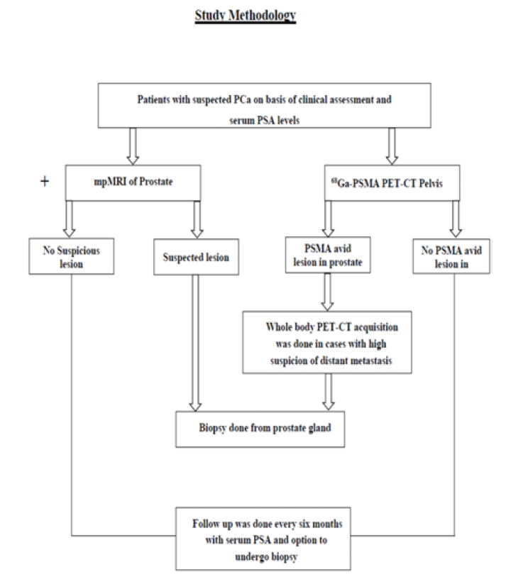
Patients suspected for prostate adenocarcinoma and/or serum PSA levels >4ng/mL and willing to participate in the study were recruited. Study was approved by Institutional Ethics Committee, PGIMER Chandigarh. Written informed consent was obtained. After clinical assessment, mpMRI and 68Ga-PSMA PET/CT were done. Biopsy was done in cases with high clinical suspicion of PCa (Figure 1).
Statistical Analysis
Qualitative variables are described using number and percentages. Normally distributed continuous quantitative data is described using mean and standard deviation. Skewed quantitative data is described using median and interquartile range. The normality of data will be checked by measures of Kolmogorov-Smirnov tests of normality. The sensitivity, specificity, NPV and PPV of 68Ga-PSMA PET/CT and mpMRI was estimated using a 2x2 table.
Figure 1: Study Methodology
To see correlation between different variables, Pearson or Spearman correlation coefficient was calculated, depending on data being analyzed. The statistical analysis was conducted using the IBM SPSS Statistics (version 23.0).
RESULTS
In this prospective study, a total of 44 patients suspected to have PCa were recruited on the basis of clinical assessment and serum PSA levels. All patients were subjected to 68Ga-PSMA PET/CT acquisition and SUVmax of the lesions were measured for quantitation. Out of total 44, 33 patients also underwent mpMRI. Out of the total 44 patients, 33 underwent biopsy. The remaining 11 patients did not undergo biopsy after revised clinical assessment based on negative 68Ga-PSMA PET/CT and mpMRI and were kept on follow up. Therefore, 33 patients underwent 68Ga-PSMA PET/CT and biopsy.
Only 22 out of these 33 patients underwent mpMRI. The images were interpreted and were categorised based on PIRADS V2. Out of these 22 patients, 9 were interpreted as PIRADS II, 3 patients were PIRADS III and 10 were given PIRADS score of IV/V. No patient was given a PIRADS score of 1.
In this study, malignancy was detected in 16/33 (48.5%) patients on biopsy. In 17/33 (51.5%), no evidence of malignancy was noted.
11/22 (50%) patients who underwent mpMRI were diagnosed with malignancy. When PIRADS III-V were considered as positive lesions on mpMRI and compared with gold standard, the analysis revealed sensitivity, specificity, PPV, NPV and accuracy of mpMRI to be 100%, 81.82%, 84.62%, 100% and 90.91% respectively to detect the adenocarcinoma PCa. Table 1 showed the mpMRI results.
The median SUVmax for cases with confirmed malignancy (n = 16) was found out to be 17.6 (IQR 18.9). Median SUVmax for cases with no evidence of malignancy was 6.3 (IQR 2.80).
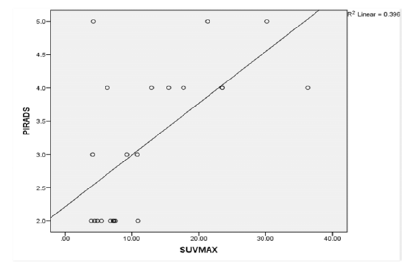
In 22 patients who underwent mpMRI, PSMA PET/CT and biopsy, significant correlation was found between PIRADS score and SUVmaxwith correlation coefficient rho of 0.555 and p-value of 0.007 (Figure 2).
Table 1: Showing mpMRI (Multiparametric Magnetic Resonance Imaging) Results Compared with Biopsy
Figure 2: Scatter Plot Between SUVmax (Standardised Uptake Value Maximum, X Axis) and PIRADS (Prostate Imaging Reporting And Data System, Y Axis)
DISCUSSION
In the present study, we found significant correlation between PIRADS score and SUVmax (r 0.555, p-value 0.007). Till date, no prospective study has been done to assess the correlation between PIRADS and SUVmax in suspicious PCa. This denotes higher PSMA expression in lesions with high PIRADS score. Similarly, high concordance was found between mpMRI and 68Ga PSMA PET/CT. Findings were concordant in 19/22 patients who underwent both investigations. Only 3/22 of the patients were having discordant findings, with mpMRI false positive in two of them and true positive in one of them.
CONCLUSION
Present study concluded that significant correlation was noted between PIRADS score and SUVmax in prostate cancer patients.
REFERENCES
Von Eyben, F.E. et al. “68Ga-labeled prostate-specific membrane antigen ligand positron emission tomography/computed tomography for prostate cancer: A systematic review and meta-analysis.” European Urology Focus, vol. 4, no. 5, 2018, pp. 686–693.
Zhang, J. et al. “Diagnostic performance of 68Ga-PSMA PET/CT in the detection of prostate cancer prior to initial biopsy: Comparison with cancer-predicting nomograms.” European Journal of Nuclear Medicine and Molecular Imaging, vol. 46, no. 4, 2019, pp. 908–920.
Bray, F. et al. “Global cancer statistics 2018: GLOBOCAN estimates of incidence and mortality worldwide for 36 cancers in 185 countries.” CA: A Cancer Journal for Clinicians, vol. 68, no. 6, 2018, pp. 394–424.
Alizadeh, M. and S. Alizadeh. “Survey of clinical and pathological characteristics and outcomes of patients with prostate cancer.” Global Journal of Health Science, vol. 6, no. 7, 2014, pp. 49–57.
Bancroft, E.K. et al. “Targeted prostate cancer screening in BRCA1 and BRCA2 mutation carriers: Results from the initial screening round of the IMPACT study.” European Urology, vol. 66, no. 3, 2014, pp. 489–499.
Uprimny, C. et al. “68Ga-PSMA-11 PET/CT in primary staging of prostate cancer: PSA and Gleason score predict the intensity of tracer accumulation in the primary tumour.” European Journal of Nuclear Medicine and Molecular Imaging, vol. 44, no. 6, 2017, pp. 941–949.
Demirci, E. et al. “Normal distribution pattern and physiological variants of 68Ga-PSMA-11 PET/CT imaging.” Nuclear Medicine Communications, vol. 37, no. 11, 2016, pp. 1169–1179.
Dola, E.F. et al. “Assessing the validity of Prostate Imaging Reporting and Data System version 2 (PI-RADS v2) scoring system in diagnosis of peripheral zone prostate cancer.” European Journal of Radiology, vol. 21, no. 4, 2017, pp. 19–26.
Tan, C.H. et al. “Dynamic contrast-enhanced MRI for the detection of prostate cancer: Meta-analysis.” American Journal of Roentgenology, vol. 204, no. 4, 2015, pp. W439–W448.
Berger, I. et al. “68Ga-PSMA PET/CT vs. mpMRI for locoregional prostate cancer staging: Correlation with final histopathology.” Prostate Cancer and Prostatic Diseases, vol. 21, no. 2, 2018, pp. 204–211.
Heidenreich, A. et al. “EAU guidelines on prostate cancer. Part 1: Screening, diagnosis and local treatment with curative intent—Update 2013.” European Urology, vol. 65, no. 1, 2014, pp. 124–137.
Carter, H.B. “American urological association (AUA) guideline on prostate cancer detection: Process and rationale.” BJU International, vol. 112, no. 5, 2013, pp. 543–547.
Recommended Articles
Research Article
Advantages of Two Clips Laparoscopic Cholecystectomy (TCLC) and Conventional Laparoscopic Cholecystectomy (CLC): A Comparative Study
Anshu Attri,
...
Kartik Syal
Published: 10/01/2022
Download PDF
Research Article
Bladder Brilliance: Unveiling the Tapestry of Knowledge on Bladder Cancer in District Kangra, Himachal Pradesh
Vandana Thakur,
...
Ankita Pandey
Published: 08/12/2023
Download PDF
Research Article
Shouldering the Pain: Public Knowledge Assessment of Rotator Cuff Injuries in Himachal Pradesh
Sidhant Korla,
...
Praveen Kumar Thakur
Published: 30/07/2024
Download PDF
Research Article
Awareness regarding Symptoms and Treatment Options for Benign hypertrophy of prostate among the male patients attending outpatient department of Civil hospital , Nurpur, Himachal Pradesh
Aarti Chaudhary,
Brish Bhanu Bhardwaj
Published: 26/05/2023
Download PDF
PO Box 101, Nakuru
Kenya.
Email: office@iarconsortium.org
Editorial Office:
J.L Bhavan, Near Radison Blu Hotel,
Jalukbari, Guwahati-India
Sharma, V., Chadha, R. & Guleria, K. S. D. S. (2022). Correlation between Standardised Uptake Value Maximum (SUVmax) and Prostate Imaging Reporting and Data System (PIRADS) in Prostate Cancer Patients. International Academic Research Journal of Surgery, 2(2), 1-3.
MLA
Sharma, Viper, Rishabh Chadha and Kunwar S. D. S. Guleria. "Correlation between Standardised Uptake Value Maximum (SUVmax) and Prostate Imaging Reporting and Data System (PIRADS) in Prostate Cancer Patients." International Academic Research Journal of Surgery 2.2 (2022): 1-3.
Chicago
Sharma, Viper, Rishabh Chadha and Kunwar S. D. S. Guleria. "Correlation between Standardised Uptake Value Maximum (SUVmax) and Prostate Imaging Reporting and Data System (PIRADS) in Prostate Cancer Patients." International Academic Research Journal of Surgery 2, no. 2 (2022): 1-3.
Harvard
Sharma, V., Chadha, R. and Guleria, K. S. D. S. (2022) 'Correlation between Standardised Uptake Value Maximum (SUVmax) and Prostate Imaging Reporting and Data System (PIRADS) in Prostate Cancer Patients' International Academic Research Journal of Surgery 2(2), pp. 1-3.
Vancouver
Sharma V, Chadha R, Guleria KSDS. Correlation between Standardised Uptake Value Maximum (SUVmax) and Prostate Imaging Reporting and Data System (PIRADS) in Prostate Cancer Patients. International Academic Research Journal of Surgery. 2022 Jul;2(2):1-3.


 Download PDF
 Download XML