Background: There were many cases of thyroid cancer proven from histopathology test after lobectomy from single thyroid nodules, which were not being predicted in advanced. The challenge for the clinician is thus to predict the malignancy preoperative to prevent unnecessary second completion thyroidectomy. Objective: This study aimed to determine the cancer probability risk of a single thyroid nodule by using preoperative parameters. Methods: This cohort study was conducted in Sanglah General Hospital, from 2016 to 2021. All data was obtained by Cancer Registry and electronic medical records. There were nine variables that being analyzed in this study, such as age, gender, side, size, consistency, vascularization, node, and cytology. The data was pooled and analyzed in SPSS and R studio. Smart phone application was developed using Android studio. Data were analyzed by logistic regression multivariate analysis. Results: From 2016-2021, there were 198 subjects with single thyroid nodules underwent surgeries in Sanglah General Hospital, Denpasar, Bali, Indonesia. As many as 98 subjects were histopathology proven cancer (follicular, papillary) after surgery.From multivariate analysis, the only left significant variables were vascularization, calcification, age, consistency, and cytology. Further, nomogram was developed to plot the probability of cancer from significant variables.By using only cytology parameter, the cancer predictive value was only less than 10%. Even age and consistency parameter by physical examination gave a higher value of 30% predictive value. The ultrasound parameters, combining the presence of calcification and vascularization, had a good predictive value of 60%. Conclusion: The combination of the ultrasound characteristics, age and physical examination able to accurately predict thyroid cancer.
Keywords
Vascularization
Predict Thyroid Cancer
Android Studio
INTRODUCTION
Thyroid nodule was one of the common diseases of the endocrine system. In 2010, the prevalence of TNs was 18.6%, based on the epidemiological survey of ten cities in China [1]. In recent studies, the cases were found to be emergingly increased due to the lifestyle modification. In current situation, cytology from fine-needle aspiration and thyroid ultrasonography are still becoming the main examination for thyroid nodule, deciding the role of surgery, either lobectomy or total thyroidectomy [2]. However, the rate calculation of thyroid cancer probability in Indonesian subjects has not yet been carried out.
There are a number of well-established predictors of malignancy in thyroid nodules including physical, radiological, and cytology examination. Not all parameters being tested in previous study could be applied in developing country such as Indonesia. Thus, this study aimed to determine the cancer probability risk of a single thyroid nodule by using parameters before surgery. This study was carried out to reduce the need of second surgery for completion thyroidectomy if malignancy is found in advanced.
MATERIALS AND METHODS
Study Design
This cohort prospective study was conducted in Sanglah General Hospital, from 2016 to 2021. All data was obtained by Cancer Registry and electronic medical records. All subjects were consented and signed the informed consent.
Study Population
In this study, we included all the subjects with single thyroid nodule underwent surgery in Surgical Oncology Department. The subject with multiple nodules examined physically or radiologically and with presence of other malignancy was excluded from this study. The eligible
subjects were categorized to case (cancer) and control (benign) group by the final histopatholgy results after surgery. There were nine variables that being analyzed in this study, such as age, gender, side, size, consistency, vascularization, node, and cytology.
Study Flow
All eligible subjects were followed from the outpatient clinic, operating theatre, and until the histopathology results obtained. In outpatient clinic, authors did history taking about age and gender. Physical examination was done for nodule side (location), largest diameter size, and its consistency. Then, we collected the status of vascularization and presence of neck lymph nodes from thyroid ultrasound. The ultrasound was carried out by two radiologists, which if the interpretation was different, both radiologists will discuss until a final imaging judgement was done. In all patients, surgery was carried out by our Surgical Oncology team. Surgical specimen was sent to Pathology laboratory for histopathology examination. The radiologist, pathologist, and data collector were blinded to this study.
Data Analysis
All data were recorded in SPSS. Categorical data were presented as number and proportion. Numerical data were presented as mean and standard deviation. For age and size variables, Youden Index in ROC analysis was calculated to determine the cut off points. Side dan consistency was determined by physical examination and categorized as hard or firm. Thyroid ultrasound was used to determine the presence of nodule size, calcification, vascularization, and node. Chi square test was carried out for analyzing the difference risk of cancer in each variable. Then, all the significance variabels were further analyzed in regression logistic model. Nomogram was developed by R studio using rms package.
RESULTS
From 2016-2021, there were 198 subjects with single thyroid nodules underwent surgeries in Sanglah General Hospital, Denpasar, Bali, Indonesia. As many as 98 subjects were histopathology proven cancer (follicular, papillary) after surgery. About 14.3% was follicular cancer, 75.5% was papillary cancer, 2% was medullary cancer, and 8% was mixed papillary-follicular cancer.
Among those, only 22 subjects were detected to be cancer by cytology examination and all the subjects underwent total thyroidectomy. The remaining subjects (77.5%) underwent two times surgeries, lobectomy, then completion surgery.
This study analyzed eight parameters, as described in methods section. Most subject with cancer had age older than 42 years old, right side of thyroid nodule, nodule size less than 4 cm, hard consistency, presence of calcification, high vascularity, presence of node, and malignant cytology (either cancer or follicular neoplasm). From these parameters, nodule side and size were not significant after Chi square analysis. The age, consistency, calcification, vascularization, and cytology were continued to logistic regression analysis (Table 1).
This logistic regression model was developed using SPSS. Due to unsignificant result of node parameter after the first logistic regression, it was being eliminated and the new model by only significant variable, age, consistency, calcification, vascularzation, and cytology was developed. This model was considered appropriate to be tested (Hosmer Lemeshow test’ p = 0.459) with accuracy of model 88.8%. The overall effect size of this model was 74.5% (Nagelkerke R square test). The most significant variable was vascularization, followed by calcification, age, consistency, and cytology (Table 2).
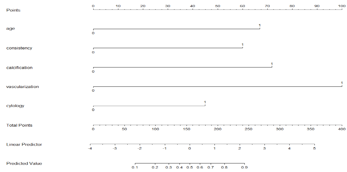
Further, nomogram was developed to plot the probability of cancer from significant variables (Figure 1). By using only cytology parameter, the cancer predictive value was only less than 10%. Even age and consistency parameter by physical examination gave a higher value of 30% predictive value. The ultrasound parameters, combining the presence of calcification and vascularization, had a good predictive value of 60%. Overall, the cancer prediction score had to be combined by using all parameters to receive a better predictionof cancer and reducing the need of second surgery.
Table 1. Decription of Parameters in Thyroid Cancer and Benign Nodule
Parameters
Thyroid cancer
Thyroid benign nodule
Significance
Odds ratio
Age
<42 years old
61
35
<0.001
3.02
>42 years old
37
64
-
(1.69-5.39)
Gender
Male
8
13
0.356
0.588
Female
90
86
-
(0.232-1.489)
Side
Right
61
63
0.883
0.94
Left
37
36
-
(0.53-1.68)
Nodule size
>4 cm
43
45
0.886
0.94
<4 cm
55
54
(0.54-1.65)
Consistency
Hard
65
8
<0.001
22.4
Firm
33
91
-
(9.71-51.67)
Calcification
Yes
82
17
<0.001
24.72
No
16
82
-
(11.7-52.24)
Vascularization
Yes
74
8
<0.001
35.07
No
24
91
-
(14.89-82.63)
Node
Yes
65
21
<0.001
7.66
No
32
78
-
(4.04-14.54)
Cytology
Malignant
81
33
<0.001
9.53
Benign
17
66
-
(4.89-18.61)
Table 2: The model of Logistic Regression of Thyroid Cancer Parameters
Parameter
B
SE
p
Exp(B)
Age
1.669
0.545
0.002
5.309
Consistency
1.495
0.617
0.015
4.461
Calcification
1.791
0.542
0.001
5.996
Vascularization
2.492
0.544
0.001
12.082
Cytology
1.121
0.531
0.035
3.068
Constant
-3.865
0.625
0.001
0.021
Figure 1: The Development of Nomogram for Thyroid Cancer Probability
DISCUSSION
Thyroid cancer is the most common endocrine malignancy in Indonesia. Indonesian Cancer Registration reported that thyroid cancer still ranked the ninth out of tenth most common malignancies [2]. The challenge for the clinician is to determine and predict which thyroid nodules are malignant, thus preventing unnecessary intervention for benign nodules.
In preoperative condition, the prediction of malignancy in thyroid nodules could be determined by physical and laboratory examinations. The characteristics of nodule, includinga firm, hard, rapidly enlarging nodule, have traditionally been associated with an increased malignancy risk [3]. The noninvasive examination, ultrasonography, was still the main imaging study for thyroid nodules. The imaging provided the diameter size of thyroid nodule, its vascularity and margin, presence of calcification, and assessment of lymphadenopathy [4]. Then, fine needle aspiration biopsy served as minimal invasive examination for cytology analysis of nodule. However, the terminology in interpreting cytology result varied between institutions making it was not generalized [5]. Few thyroids cancer prediction model has been developed but most studies focused in including molecular factor that was not available in Indonesia. In the modern era, clinicians are now incorporating artificial intelligence to aid the process in the decision making of management. The development of nomogram should provide an approximate rate of an outcome prediction by a formulated model. In this study, we have produced an accurate nomogram for the prediction of malignancy for a single thyroid nodule.
CONCLUSION
The combination of the ultrasound characteristics, age and physical examination able to accurately predict thyroid cancer.
Acknowledgment
The authors had nothing to acknowledge
Conflict of Interest
This study has no conflict of interest to be declared.
REFERENCE
Jiang, H. et al. “The prevalence of thyroid nodules and an analysis of related lifestyle factors in beijing communities.” International Journal of Environmental Research and Public Health, vol. 13, no. 4, 2016, pp. 442. https://doi.org/10.3390/ijerph13040442.
Wahidin, M. et al. “Population-based cancer registration in Indonesia.” Asian Pacific Journal of Cancer Prevention, vol. 13, no. 4, 2012, pp. 1709–1710.
Zheng, L. et al. “An epidemiological study of risk factors of thyroid nodule and goiter in Chinese women.” International Journal of Environmental Research and Public Health, vol. 12, no. 9, 2015, pp. 11608–11620.
Papini, E. et al. “Risk of malignancy in nonpalpable thyroid nodules: Predictive value of ultrasound and color-doppler features.” Journal of Clinical Endocrinology & Metabolism, vol. 87, no. 5, 2002, pp. 1941–1946.
Baloch, Z.W.et al. “Diagnostic terminology and morphologic criteria for cytologic diagnosis of thyroid lesions: A synopsis of the national cancer institute thyroid fine-needle aspiration state of the science conference.” Diagnostic Cytopathology, vol. 36, no. 6, 2008, pp. 425–437
Recommended Articles
Research Article
Advantages of Two Clips Laparoscopic Cholecystectomy (TCLC) and Conventional Laparoscopic Cholecystectomy (CLC): A Comparative Study
Anshu Attri,
...
Kartik Syal
Published: 10/01/2022
Download PDF
Research Article
Bladder Brilliance: Unveiling the Tapestry of Knowledge on Bladder Cancer in District Kangra, Himachal Pradesh
Vandana Thakur,
...
Ankita Pandey
Published: 08/12/2023
Download PDF
Research Article
Shouldering the Pain: Public Knowledge Assessment of Rotator Cuff Injuries in Himachal Pradesh
Sidhant Korla,
...
Praveen Kumar Thakur
Published: 30/07/2024
Download PDF
Research Article
Awareness regarding Symptoms and Treatment Options for Benign hypertrophy of prostate among the male patients attending outpatient department of Civil hospital , Nurpur, Himachal Pradesh
Aarti Chaudhary,
Brish Bhanu Bhardwaj
Published: 26/05/2023
Download PDF
PO Box 101, Nakuru
Kenya.
Email: office@iarconsortium.org
Editorial Office:
J.L Bhavan, Near Radison Blu Hotel,
Jalukbari, Guwahati-India
Sudarsa, I. W., Aryanti, C., Tusta Adiputra, P. A. & Budhi Setiawan, I. G. (2022). The development of nomogram for predicting thyroid cancer in subject with single thyroid nodule in Bali, Indonesia. International Academic Research Journal of Surgery, 2(1), 1-4.
MLA
Sudarsa, I. Wayan, et al. "The development of nomogram for predicting thyroid cancer in subject with single thyroid nodule in Bali, Indonesia." International Academic Research Journal of Surgery 2.1 (2022): 1-4.
Chicago
Sudarsa, I. Wayan, Citra Aryanti, Putu Anda Tusta Adiputra and I. Gede Budhi Setiawan. "The development of nomogram for predicting thyroid cancer in subject with single thyroid nodule in Bali, Indonesia." International Academic Research Journal of Surgery 2, no. 1 (2022): 1-4.
Harvard
Sudarsa, I. W., Aryanti, C., Tusta Adiputra, P. A. and Budhi Setiawan, I. G. (2022) 'The development of nomogram for predicting thyroid cancer in subject with single thyroid nodule in Bali, Indonesia' International Academic Research Journal of Surgery 2(1), pp. 1-4.
Vancouver
Sudarsa IW, Aryanti C, Tusta Adiputra PA, Budhi Setiawan IG. The development of nomogram for predicting thyroid cancer in subject with single thyroid nodule in Bali, Indonesia. International Academic Research Journal of Surgery. 2022 Jan;2(1):1-4.


 Download PDF
 Download XML