Single-incision laparoscopic cholecystectomy (SILC) has been developed to further minimize the invasiveness of laparoscopic cholecystectomy by reducing the number of incisions. Knowledge of preoperative clinical factors to predict difficult SILC may be used for the preoperative counselling of the patients regarding the successful outcome of the surgery as well as to predict the risk of conversion preoperatively for selected patients, prepare the patient psychologically and minimise the procedure related cost. Objective: The present study was conducted to know about radiological predictors for difficult SILC. Methods: The study was a hospital based prospective observational study where 50 patients undergoing elective laparoscopic cholecystectomy due to gall stone diseases were included in the study. Multiple logistic regression analysis was done to predict difficult single incision laparoscopic cholecystectomy. Results: Out of 50 patients 15 patients underwent difficult SILC, while 35 patients had undergone easy single incision laparoscopic cholecystectomy independent risk factors in multivariate logistic regression analysis for difficult SILC were: contracted or distended gall bladder on ultrasound, longer umbilicus to Calot’s triangle distance (UCD) on MRI. Conclusion: RadiologicalFactors associated with difficult SILC contracted or distended gall bladder on ultrasound and longer umbilicus to Calot’s triangle distance on MRI.
Keywords
Single Incision Laparoscopic Cholecystectomy
Gall Stone Diseases
Risk Factors
Magnetic Resonance Imaging
INTRODUCTION
Laparoscopic cholecystectomy has now become the procedure of choice for symptomatic gall stone disease [1]. Recently, a single-incision laparoscopic cholecystectomy (SILC), also called as trans umbilical laparoscopic cholecystectomy or laparoendoscopic single site (LESS) cholecystectomy, has been developed to further minimise the invasiveness of laparoscopic cholecystectomy by reducing the number of incisions [2,3]. Various randomised controlled trials have shown that SILC can provide better cosmetic results, shorter duration of hospital stay and early recovery as compared with conventional laparoscopic cholecystectomy [4].
Considering that the main benefit of single incision laparoscopic cholecystectomy (SILC) appears to be improved cosmesis, it is important to complete the procedure via a single incision. Because the number of incisions is the primary concern for patients who hope to undergo SILC, the ability to identify an individual patient’s risk for needing additional ports is important. If conversion is necessary for whatever reason, the benefit of the minimal access concept is lost. Therefore, every effort should be made to increase the probability of successful completion of the laparoscopic procedure to be attempted through single incision [5].
Some factors which can be accessed preoperatively to reliably predict the feasibility of successful single incision laparoscopic cholecystectomy or the requirement for additional ports have been observed in previous studies [6-9].
Thus, for surgeons it would be helpful to establish criteria that would assess the risk of conversion preoperatively and risk of postoperative complications. Factors, which can be assessed preoperatively can reliably predict feasibility of the successful single incision laparoscopic cholecystectomy and the requirement of extra ports. Knowledge of these factors, may be used for the preoperative counselling of the patients regarding the successful outcome of the surgery as well as to predict the risk of conversion preoperatively for selected patients, prepare the patient psychologically, minimize the procedure related cost, help overcome financial constraints, which is a significant problem in developing countries and possibility of the conversion so that needful arrangements can be made by the patients [10,11].
Therefore, present study was planned to know about radiological predictors for difficult single incision laparoscopic cholecystectomy.
MATERIALS AND METHODS
Subjects and Methods
Study design, settings and participants: It was a hospital based prospective observational study conducted over a period of one year from June 2020 to June 2021 in general surgery department of a tertiary care teaching hospital in Himachal Pradesh, India. All the patients undergoing elective laparoscopic cholecystectomy due to gall stone diseases were included in the study. Patients with common bile duct calculus, suspicion of carcinoma gall bladder, features of obstructive jaundice, cholelithiasis during pregnancy absolute contraindications to laparoscopic cholecystectomy were excluded from the study. So, total 50 patients undergoing single incision laparoscopic cholecystectomy were enrolled for study.
Operational Definition
Single incision laparoscopic cholecystectomy was considered as difficult if any of the following 4 criteria was fulfilled during the surgery:
Time taken from skin incision to skin closure more than 60 minutes
Time taken for adhesiolysis at Calot’s triangle more than 20 minutes
Time taken for adhesiolysis of gall bladder at gall bladder fossa more than 20 minutes
Addition of ports (either one, two or three)
Estimation of UCD from Preoperative MRI
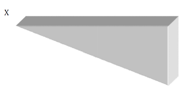
In order to calculate umbilicus to calot’s triangle distance from MRI we used Pythagoras’ theorem in a right-angled triangle.
Pythagoras' theorem states that in a right angled triangle the square of the hypotenuse (the side opposite the right angle) is equal to the sum of the squares of the other twosides.
(Hypotenuse)2 = (Horizontal)2 + (Vertical)2
Vertical distance (X) was measured by number of sections between an image that contains the neck of the gallbladder and one that contains the umbilicus × thickness of each section + space between the two consecutive sections × number of spaces between an image that contains the neck of the gallbladder and one that contains the umbilicus.
Thickness of each section of MRI was 1.2mm
Space between two consecutive sections in MRI was 6 mm
Figure 1: UCD MR (Umbilicus to Calot’s Triangle Distance)
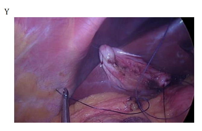
Figure 2: Suture Taken Through the Fundus of GB
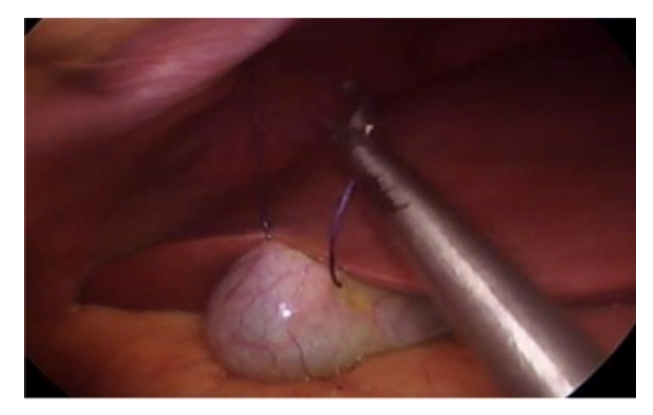
Figure 3: Suture Taken Through Hartmann’s Pouch
Horizontal distance (Y) was measured between the umbilicus and the same coordinate point as the neck of the gallbladder on an image that contains the umbilicus
Hypotenuse is the umbilicus to calot’s triangle distance (UCD- MR)
(UCD-MR)2 = (X)2 + (Y)2
Surgical Technique
Patient was placed supine on the table with the legs split apart. Both arms were placed at an angle less than 90 degree to the torso. Umbilicus was everted and an infra umbilical curved (smiling) incision 2.0 to 2.5 cm in length was given. This was deepened through fat and flaps were undermined to expose fascia. A Veress needle was inserted through this incision and 12 mm Hg pneumoperitoneum with CO2 was induced and maintained. We used two ports one 5 mm port for camera and another 10 mm working port through which laparoscopic needle holder, Maryland forceps and extractor were introduced at the various steps of SILC procedure. The camera port was at the left margin (patient’s) of 10 mm port.
Placement of Traction Sutures
This was the key step of our SILC technique. GB fundus was recognized by moving omentum away from right upper quadrant with the help of grasper and dissector (Figure 2). Needle was introduced through intercostal space above the costal margin which was introduced into peritoneal cavity with the help of laparoscopic needle holder. This needle was then taken through fundus of GB and then back to the same intercostal space. This traction suture helped in elevating GB fundus. Another traction suture was introduced at epigastrium just below xiphisternum. This suture was then passed through the Hartmann’s pouch, and was brought out at subcostal parietal wall at anterior axillary line (Figure 3). This helped in lifting the Hartmann’s area and helped in better dissection.
was held by grasper at neck and extracted through umbilical 10 mm port. Careful closure of fascial incision was done to prevent port site hernia. The edges of fascial incision were grasped with Kelly’s haemostat. Rectus sheath was closed with Vicryl no.1 suture. The fascia and the skin were infiltrated with local anaesthesia and the skin was closed using monocryl 3-0 subcuticular stitches.
Statistical Analysis
Data were analyzed and statistically evaluated using SPSS software, version 25 (Chicago II, USA). Quantitative data was expressed in mean, standard deviation and difference between two comparable groups were tested by student’s t-test (unpaired) while qualitative data were expressed in percentage. Statistical differences between the proportions were tested by chi square test or Fisher’s exact test. Multiple logiSstic regression analysis was done to predict difficult single incision laparoscopic cholecystectomy for all factors who were found significant in univariate analysis and odds ratio along with 95% CI was calculated. P’ value less than 0.05 was considered statistically significant.
Ethical Issues
All participants were explained about the purpose of the study. Confidentiality was assured to them along with informed written consent. The study was approved by the Institutional Ethical Committee.
RESULTS
Out of 50 patients 15 patients underwent difficult single incision laparoscopic cholecystectomy, while 35 patients had undergone easy single incision laparoscopic cholecystectomy. Out of 15 patients undergoing difficult SILC, SILC was successfully completed through a single incision in 11 patients (73.33%), whereas additional ports were required in 4 patients (26.66%).
Table 1 represent association of different radiological factors with difficult SILC. Among different radiological factors contracted or distended gallbladder on ultrasound and longer UCD were found to be significantly associated with difficult SILC (Table 2,3).
Table 1 : Radiological Factors to Predict Difficult SILC
Gall Bladder status on Sonography
Easy (n = 35)
Difficult (n = 15)
Total (n = 50)
p value
No.
Percentage
No.
Percentage
No.
Percentage
Neither contracted nor distended
20
57.1
3
20.0
23
46.0
<0.01
Contracted
12
34.3
5
33.3
17
34.0
Distended
3
8.5
7
46.7
10
20.0
Table 2: Radiological Factors Contracted or Distended Gallbladder on Ultrasound
Multiple Stones in Gall Bladder on USG
Easy (n = 35)
Difficult (n = 15)
Total (n = 50)
p value
No.
Percentage
No.
Percentage
No.
Percentage
No
6
17.1
2
13.3
8
16.0
1.0
Yes
29
82.9
13
86.7
42
84.0
Table 3: Longer UCD were Found to be Significantly Associated with Difficult SILC
Wall Echo Sign on USG
Easy (n = 35)
Difficult (n = 15)
Total (n = 50)
p value
No.
Percentage
No.
Percentage
No.
Percentage
No
31
88.6
12
80.0
43
86.0
0.41
Yes
4
11.4
3
20.0
7
14.0
Impacted stone on USG
Easy (n = 35)
Difficult (n = 15)
Total (n = 50)
p value
No.
%
No.
%
No.
%
No
29
82.9
9
60.0
38
76.0
0.08
Yes
6
17.1
6
40.0
12
24.0
Parameters
Total (n = 50)
Easy (n = 35)
Difficult (n = 15)
p value
Mean±SD
Mean±SD
Mean±SD
Gall bladder wall thickness (mm)
2.49±0.74
2.44±0.69
2.59±0.87
0.73
Umbilicus to calot’s triangle distance
Total (n=50)
Easy (n=35)
Difficult (n=15)
p value
Mean±SD
Mean±SD
Mean±SD
On MRI (in mm)
210.22±15.50
200.77±5.70
232.27±4.26
<0.001
DISCUSSION
In the present study, SILC was completed with a single incision in 46 patients (92%), whereas at least one additional port was required in 4 patients (8%).
Various authors have reported significant correlation of contracted and distended gall bladder with difficult SILC. Contracted gall bladder is a potential risk for conversion and patient having thickened gall bladder have significantly increased duration of surgery, difficult access, difficult dissection of calot’s triangle, gall bladder fossa dissection and extraction of gall bladder. Thus, we also noted significant association of contracted and distended gall bladder on USG with the difficult single incision laparoscopic cholecystectomy (p value < 0.01).
Arumugam et al. [12] found that gall bladder packed with multiple stones was associated with increased risk of difficult laparoscopic cholecystectomy. He explained that multiple stones are likely to make difficult grasping of gall bladder, narrows the Calot’s triangle with multiple adhesions over Calot’s triangle and also poses difficulty in extraction of the gall bladder. However, in our study we did not find any significant association between multiple stones in gallbladder and difficult SILC.
Wall echo sign is sign of chronic inflammation of gall bladder. Arora B.K. et al.[13] observed on laparoscopic view, recorded point that wall echo shadow complex can be a predictor of difficult cholecystectomy. We do not have the same results for WES as a predictor for difficult SILC.
Impacted calculus at the neck of gall bladder poses difficulty in grasping the neck of gall bladder to allow adequate retraction to perform dissection at the Calot’s triangle and during the extraction of gall bladder from the port. Due to impacted stone, the gall bladder is distended with mucus forming the mucocele of gall bladder which is even more difficult to hold. Many authors[14,15] have reported impacted calculus to be associated with difficult laparoscopic cholecystectomy. However, our study showed impacted stone is statistically not significant factor in predicting the difficulty of the procedure (p value 0.08).
Thickened gall bladder wall due chronic cholecystitis is difficult to handle and manipulate. Increased gall bladder wall thickness is associated with difficult dissection of the gall bladder at Calot’s triangle as well as from its fossa. The thickened gall bladder implies features of chronic cholecystitis and sometimes it becomes difficult for the surgeon to perform single incision laparoscopic cholecystectomy. Various studies[16,17] have reported increased gall bladder wall thickness more than 4mm as a preoperative ultrasonographic predictor of difficult laparoscopic cholecystectomy. In our study higher gall bladder wall thickness is statistically not significant factor in predicting the difficulty of the procedure (p value 0.73). In single incision laparoscopic surgery, the operator’s hands and the scope get closer to each other in the extracorporeal space, resulting in a narrower workspace and decreased surgical maneuverability when the distance from a port to a target organ is long. In 2018 Masafumi Ohira[18]examined the relationship between the distance from the port to the target organ and surgical maneuverability or procedural difficulty in SILC. They investigated the correlation between the umbilicus to Calot’s triangle distance (UCD), which may represent the port-to-target distance in SILC, and operative time as a proxy for procedural difficulty. Their findings revealed a strong positive correlation between the UCD and operative time.Similar results are obtained in our study with significant P value of <0.001. Therefore, longer port-to-target distance resulted in increased procedural difficulty in SILC.
CONCLUSION
Conclusion and Recommendations
It can be concluded from the study that USG evidence of contracted/distended gall bladder and longer port to target distance were found statistically significant in predicting difficult single incision laparoscopic cholecystectomy. Hence, an experienced team should be arranged to tackle the difficulties before planning surgery and patient should be counselled accordingly before surgery to avoid legal consequences.
Acknowledgment
The authors are grateful to all the participants for their support and contribution.
REFERENCE
Sachdeva, S. et al. “Lifestyle and gallstone disease: scope for primary prevention.” Indian Journal of Community Medicine, vol. 36, no. 4, October 2011, pp. 263–267.
Al-Mulhim, A.A. “Current trends in laparoscopic cholecystectomy.” Journal of Family and Community Medicine, vol. 4, no. 2, July 1997, pp. 33–40.
Navarra, G. et al. “One-wound laparoscopic cholecystectomy.” British Journal of Surgery, vol. 84, no. 5, 1997, pp. 695.
Piskun, G. and S. Rajpal. “Transumbilical laparoscopic cholecystectomy utilizes no incisions outside the umbilicus.” Journal of Laparoendoscopic & Advanced Surgical Techniques A, vol. 9, 1991, pp. 361–364.
Tyagi, S. et al. “Single-incision laparoscopic cholecystectomy: Less scar, less pain.” Journal of Minimal Access Surgery, vol. 13, no. 2, 2017, pp. 118–123.
Soltes, M. and J. Radoňak. “A risk score to predict the difficulty of elective laparoscopic cholecystectomy.” Wideochirurgia i Inne Techniki Małoinwazyjne, vol. 9, no. 4, December 2014, pp. 608–612.
Sato, N. et al. “Preoperative factors predicting the need for additional ports during single-incision laparoscopic cholecystectomy.” Asian Journal of Endoscopic Surgery, vol. 9, no. 3, August 2016, pp. 192–197.
Khambaty, F. et al. “Laparoscopic versus single-incision cholecystectomy.” World Journal of Surgery, vol. 35, 2011, pp. 967–972.
Ikumoto, T. et al. “Feasibility of single-incision laparoscopic cholecystectomy for acute cholecystitis.” World Journal of Gastrointestinal Endoscopy, vol. 7, no. 9, 2015, pp. 1327–1333.
Corr, P. et al. “Preoperative ultrasound to predict technical difficulties and complications of laparoscopic cholecystectomy.” American Journal of Surgery, vol. 168, no. 1, 1994, pp. 54–56.
Sandhu, G. et al. “Preoperative prediction of difficult laparoscopic cholecystectomy: A scoring method.” Indian Journal of Applied Research, vol. 6, no. 6, 2016, pp. 481–487.
Arumugam, R. and K. Pandurengan. “Preoperative prediction of difficult laparoscopic cholecystectomy.” International Surgery Journal, vol. 2, no. 4, 2015, pp. 584–589.
Arora, B.K. et al. “Laparoscopic cholecystectomy in wall echo complex gall stone disease: A study.” International Surgery Journal, vol. 4, 2017, pp. 1309–1312.
Randhawa, J.S. and A.K. Pujahari. “Preoperative prediction of difficult lap chole: A scoring method.” Indian Journal of Surgery, vol. 71, no. 4, 2009, pp. 198–201.
Agrawal, N. et al. “Preoperative prediction of difficult laparoscopic cholecystectomy: A scoring method.” Nigerian Journal of Surgery, vol. 21, no. 2, 2015, pp. 130–133.
Strasberg, S.M. et al. “An analysis of the problem of biliary injury during laparoscopic cholecystectomy.” American Journal of Surgery, vol. 180, 1995, pp. 101–123.
Hutchinson, C.H. et al. “Laparoscopic cholecystectomy: Do preoperative factors predict the need to convert to open?” Surgical Endoscopy, vol. 8, 1994, pp. 875–878.
Ohira, M. et al. “Correlation between port-to-target distance and procedural difficulty in single-incision laparoscopic cholecystectomy: An observational study.” Asian Journal of Endoscopic Surgery, vol. 26, September 2018.
Recommended Articles
Research Article
Advantages of Two Clips Laparoscopic Cholecystectomy (TCLC) and Conventional Laparoscopic Cholecystectomy (CLC): A Comparative Study
Anshu Attri,
...
Kartik Syal
Published: 10/01/2022
Download PDF
Research Article
Bladder Brilliance: Unveiling the Tapestry of Knowledge on Bladder Cancer in District Kangra, Himachal Pradesh
Vandana Thakur,
...
Ankita Pandey
Published: 08/12/2023
Download PDF
Research Article
Shouldering the Pain: Public Knowledge Assessment of Rotator Cuff Injuries in Himachal Pradesh
Sidhant Korla,
...
Praveen Kumar Thakur
Published: 30/07/2024
Download PDF
Research Article
Awareness regarding Symptoms and Treatment Options for Benign hypertrophy of prostate among the male patients attending outpatient department of Civil hospital , Nurpur, Himachal Pradesh
Aarti Chaudhary,
Brish Bhanu Bhardwaj
Published: 26/05/2023
Download PDF
PO Box 101, Nakuru
Kenya.
Email: office@iarconsortium.org
Editorial Office:
J.L Bhavan, Near Radison Blu Hotel,
Jalukbari, Guwahati-India
Chaudhary, A., Chauhan, R. & Jhobta, R. (2022). Radiological Predictors for Difficult Single Incision Laparoscopic Cholecystectomy. International Academic Research Journal of Surgery, 2(1), 1-5.
MLA
Chaudhary, Amita, Rohit Chauhan and R.S. Jhobta. "Radiological Predictors for Difficult Single Incision Laparoscopic Cholecystectomy." International Academic Research Journal of Surgery 2.1 (2022): 1-5.
Chicago
Chaudhary, Amita, Rohit Chauhan and R.S. Jhobta. "Radiological Predictors for Difficult Single Incision Laparoscopic Cholecystectomy." International Academic Research Journal of Surgery 2, no. 1 (2022): 1-5.
Harvard
Chaudhary, A., Chauhan, R. and Jhobta, R. (2022) 'Radiological Predictors for Difficult Single Incision Laparoscopic Cholecystectomy' International Academic Research Journal of Surgery 2(1), pp. 1-5.
Vancouver
Chaudhary A, Chauhan R, Jhobta R. Radiological Predictors for Difficult Single Incision Laparoscopic Cholecystectomy. International Academic Research Journal of Surgery. 2022 Jan;2(1):1-5.


 Download PDF
 Download XML