Understanding coagulation disorders is essential, especially for obstetricians and intensive care providers managing parturients, as these disorders can have significant consequences for the mother and child during the peripartum period. Von Willebrand-Jürgens syndrome or von Willebrand disease is the most common congenital bleeding disorder. It is subject to autosomal inheritance with variable penetrance and expressivity. The cause of the disease lies in quantitative (type 1, type 3) or qualitative (type 2) abnormalities of vonWillebrand factor (vWF). vWF has two important functions in coagulation. On the one hand, it circulates in plasma as a carrier protein for factor VIII and thus has a protective function; on the other hand, it is responsible for platelet adhesion to the subendothelium of injured vessels and platelet aggregation. The risk of bleeding varies according to the severity of the injury. The therapeutic management strategy depends on the type of disease. In this paper, we report the observation of two patients with von Willebrand disease who had vaginal deliveries.
Keywords
Peripartum
Vonwildebrand Disease
INTRODUCTION
Von Willebrand-Jürgens syndrome or von Willebrand disease is the most common congenital bleeding disorder. It is subject to autosomal inheritance with variable penetrance and expressivity. The cause of the disease lies in quantitative (type 1, type 3) or qualitative (type 2) abnormalities of vonWillebrand factor (vWF) [1].
The risk of bleeding varies according to the severity of the injury. The therapeutic management strategy depends on the type of disease.
In this paper, we report the observation of two patients with von Willebrand disease who had vaginal deliveries.
Case 1
Mrs. H.H.A, 31 years old, born of a non-consanguineous marriage and being followed since childhood for a type 2 von Willebrand disease without major hemorrhagic manifestations, and never required specific treatment for her disease.
As for her obstetrical history, she was at her second pregnancy with a live 5-year-old child whose delivery had been completed without major hemorrhagic incidents, with prophylactic treatment with Tranexamic acid and Desmopressin. The patient was admitted in labor to the delivery room of the Souissi Maternity Hospital for a planned vaginal delivery after a full-term pregnancy of 40 weeks gestation. Her obstetrical examination revealed an evolving singleton pregnancy with normal fetal heart activity and rate. The general examination revealed a patient in good general condition, with a blood pressure of 120 mmHg arterial systolic pressure and 68 mmHg arterial diastolic pressure, a heart rate of 80 bpm, normal colored conjunctiva, without signs of external bleeding.
The delivery took place 25 minutes after admission and was uneventful, except for a few small cervical lesions that were sutured during an under-valve examination.
Medical management consisted of the administration of Tranexamic acid at a rate of 15mg/kg per 8 hours with close monitoring of bleeding and hemodynamic parameters during the first 2 hours in the delivery room and then in the intensive care unit where Desmopressin was introduced at a rate of 2 nasal sprays every 12h. Early breastfeeding was initiated.
A biological workup was requested immediately postpartum, including a CBC and a specific hemostasis workup revealed a hemoglobin of 10.2 g/dl, platelets of 243,000/µl, the prothrombin rate was 100% and the activated partial thromboplastin time was 1.3 times the control, and the fibrinogen level was 4.6 g/l. Factor VIII activity was 11%, von Willebrand factor activity was collapsed to 10.5% and vWF antigen activity was 89.4%.
This appearance was consistent with type 2 von Willebrand disease. The search for multimers was not performed, as it was not possible to differentiate between subtypes 2A and 2M.
The patient stayed in the intensive care unit for 3 days, during which time she was closely monitored. The postpartum period was simple and no unusual bleeding was reported. Thus, we did not use specific treatments (FFP or factor VIII-VWF concentrates).
Case 2
We report the observation of a 23-year-old parturient, born of a consanguineous marriage (third degree consanguinity), primiparous at 41 weeks of gestation + 1 day admitted urgently for delivery, known to be a carrier of Von Willebrand disease type 1 diagnosed at the age of 10 years following an ENT bleeding after tonsillectomy.
The clinical symptomatology was typical, with mucosal bleeding such as epistaxis and gingivorrhagia, ecchymosis and menorrhagia from adolescence onwards, which required a blood transfusion on one occasion and was responsible for a microcytic hypochromic anemia, which was put on iron therapy.
The somatic examination did not reveal any hemorrhagic syndrome, but found a slight skin and mucous membrane pallor, mildly discolored conjunctivae, and free, mobilizable and painless joints.
The haemogram revealed a microcytic hypochromic anaemia (Hb = 8.8 g/dl; MCV = 78 m3; MCHC = 29%), with a normal platelet and white blood cell count, The obstetrical ultrasound was without anomaly and the delivery was planned by vaginal delivery, Biological workup: Factor VIIIc (Chronometry): 9%, VWF (Immunoturbidemetry): 21% VWF(Aggregometry) / VWF ristocetin cofactor activity: <10%, Two hours before delivery a desmopressin 0.3 ug/kg test was performed with a control at 15 min, 45 min and 60 min after the end of this infusion. No hemorrhagic or cardiovascular complications were noted, and no impact on diuresis.
In view of the efficacy of the therapeutic test, the same dose of desmopressin was injected 60 min before the procedure with control of the levels of FVIII, vWF: Ag and vWF: Rco on the same day (D0) and on the day of discharge (D3).
DISCUSSION
Von Willebrand factor is a multimeric protein synthesized by endothelial cells and megakaryocytes with a half-life of 12 hours and has two major functions: platelet adhesion to the injured vascular wall and factor VII transport and protection. There are three types of vWD [2]: type 1 is the most frequent form representing 60 to 80% of cases. It is characterized by a partial quantitative deficiency of vWF; type 2 corresponds to a quantitatively normal or subnormal synthesis of vWF which is qualitatively abnormal.
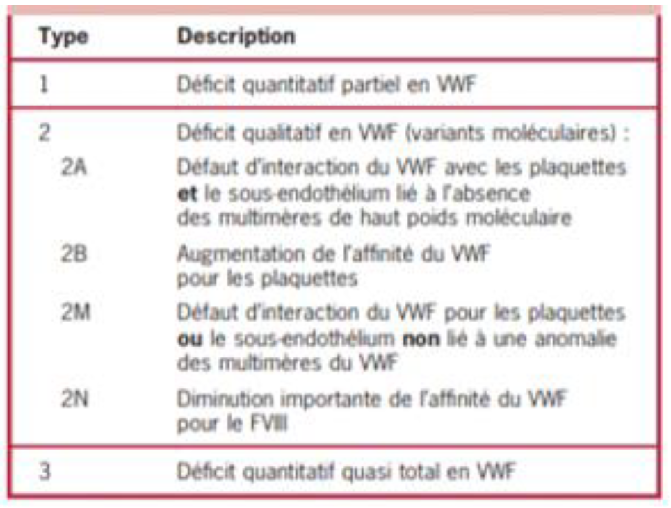
Four variants are described (Table 1) and type 3 is the most severe form, and the least frequent (less than 5%), characterized by extremely low or undetectable levels of vWF and its activity.
During pregnancy, vWF and factor VIII levels increase from 16 weeks' gestation to three to four times the pre-pregnancy value. This physiological rise in coagulation factors may be sufficient for good hemostasis during pregnancy and delivery in pregnant women with congenital bleeding disorders. However, these factors drop rapidly after delivery. Thus, bleeding disorders are rare during pregnancy and occur mostly during delivery and postpartum [3].
Patients with this disorder are at greater risk of bleeding, particularly during surgical procedures, hence the necessity of multidisciplinary perioperative management requiring collaboration between the anesthesiologist, the hematologist, the biologist and the surgeon [4].
Management begins with a meticulous history-taking to determine the circumstances of the diagnosis, the phenotypic status of the disease, and the existence of a history of bleeding and its management. The biological haemostasis work-up, including initially the PT, APTT and platelet count with, if necessary, bleeding time, VWF and factor VIII assays, allows the severity of the disease to be quantified [9].
There are two agents used to prevent peripartum bleeding:
Desmopressin and plasma VWF and FVIII concentrate. The choice depends on the type of disease, the response to desmopressin and the nature of the procedure [5]
Desmopressin (an antiduiretic hormone analog) has the property of mobilizing FVIII and VWF from their intracellular storage sites to plasma. The recommended dose is 0.3μg/kg, administered over 30 minutes intravenously. This results in a 3- to 5-fold rise in baseline VWF and factor VIII levels within 30 minutes of injection. Levels usually remain elevated for 6 to 8 hours. Administration can be repeated every 12 to 24 hours [6]
Table 1: Classification of Willebrand Disease 100
Nasal administration of high-dose desmopressin acetate (300 μg if weight ≥50 kg and 150 μg if weight <50 kg) is often effective for minor bleeding, but the IV route is the preferred route for prophylaxis of surgical hemorrhage and for treatment of major bleeding; Nasal absorption is variable and all patients with VWD who are sensitive to desmopressin should be tested after nasal instillation for FVIII and VWF response before use. When used for epistaxis, nasal administration is delivered into the non-bleeding nostril [7]. Desmopressin can also be administered subcutaneously (sc). The effective dosage is identical to the IV dose, but the SC preparation is not available. The response to desmopressin decreases with repeated dosing, probably due to depletion of the VWF storage compartment [7].
Contraindications to the use of desmopressin are age less than 2 years and patients with vascular conditions [8].
Replacement therapy, based on VWF alone or combined with factor VIII, should be reserved for patients who do not respond to desmopressin or who have contraindications to it, and for highly hemorrhagic procedures (because of the difficulty of procurement and high cost). These concentrates are administered intravenously at a dosage of 35-50 IU/kg, every 12 hours or 24 hours depending on the risk of bleeding [5]. In low-risk bleeding situations, desmopressin is indicated in responding patients.
Adjuvant antifibrinolytic treatment with tranexamic acid can be added in all types and in all types of surgery. The dosage is 10mg/kg, three times a day. Administered immediately in our patient.
In type 1 VwD with FVIII: VWF C less than 30 U/dL, administration of desmopressin usually after umbilical clamping and for 3-4 days thereafter is necessary especially when episiotomy is required. The same approach, with fewer infusions can be applied to those with VWF> 30 and <50 U/dL. Recent experience suggests the possibility of starting the treatment immediately before delivery, without obvious side effects for the mother and the newborn. The use of desmopressin in the first trimester of pregnancy to cover chorionic villus sampling or amniocentesis appears to be feasible and safe, without risk of miscarriage [12].
In type 3 VWD, VWF and FVIII do not increase during pregnancy and therefore VWF/FVIII concentrates may be necessary during pregnancy to control intermittent vaginal bleeding and at delivery or for cesarean section. The latter should be reserved only for routine obstetrical indications. Daily doses of 50 IU/kg VWF are required to maintain FVIII: C level > 50 U/dL for 3-4 days. Usual thromboprophylaxis treatment with LMWH should be implemented in patients at high risk of venous thrombosis during replacement therapy in case of cesarean section. If performed, VWF: RCo and peak FVIII: C should be > 50 U / dL [12].
In type 2A, 2B and 2M patients generally VWF: RCo does not reach normal levels and therefore replacement therapy is still advised. However, in type 2B the abnormal VWF increase may cause or worsen thrombocytopenia and a platelet transfusion might also be required at parturition.
FVIII and VWF fall to baseline levels soon after delivery, and therefore oral anti-fibrinolytics (e.g., tranexamic acid 1 gr every 8 hours for 3-4 days) can be used during this period to prevent bleeding due to heavy lochia [12].
The choice of anesthetic technique (general anesthesia/local or neuraxial anesthesia) depends mainly on the preoperative preparation and therefore on the preoperative VWF level. A recent systematic review of the literature of 507 techniques of locoregional anesthesia (spinal anesthesia, combined spinal epidural, epidural analgesia and paravertebral block) for patients with hemophilia and Von Willebrand disease, reports only one complication in which the bleeding diathesis was undiagnosed before needle insertion [10]. This was a case of spinal hematoma after lumbar puncture in a hemophiliac patient leading to permanent paraplegia [10].
Currently there are no recommendations on the practice of locoregional anesthesia techniques, in particular neuraxial anesthesia, as well as on the minimum level of VWF required.
Post-operative analgesia involves the use of different levels of analgesics. Non-steroidal anti-inflammatory drugs should be avoided as they increase the risk of bleeding.
Thrombo-prophylaxis should be implemented according to recommendations at the same time as supplementation with Willebrand factor concentrate. If prolonged anticoagulation is necessary, Willebrand factor supplementation is also prolonged. In our case analgesia was provided by paracetamol and thrombo-prophylaxis by mechanical measures.
CONCLUSION
Von Willebrand-Jürgens syndrome or von Willebrand disease is the most common congenital bleeding disorder. vWF has two important functions in coagulation. On the one hand, it circulates in plasma as a carrier protein for factor VIII and thus has a protective function; on the other hand, it is responsible for platelet adhesion to the subendothelium of injured vessels and platelet aggregation. In this paper, we’ve presented 2 cases of of bleeding varies according to the severity of the injury, and talked about the therapeutic management strategy.
REFERENCE
Ruggeri, Z.M. and J.Ware. “The structure and function of von willebrand factor.” Thrombosis and Haemostasis, vol. 67, no. 6, June 1992, pp. 594–599.
Nichols, W.L. et al. “Von willebrand disease (VWD): Evidence-based diagnosis and management guidelines, the national heart, lung, and blood institute (NHLBI) expert panel report (USA).” Haemophilia, vol. 14, no. 2, 2008, pp. 171–232.
Castaman, G. “Changes of von willebrand factor during pregnancy in women with and without von willebrand disease.” Mediterranean Journal of Hematology and Infectious Diseases, vol. 5, no. 1, 2013, e2013052.
Terraube, V. et al. “Role of von willebrand factor in tumor metastasis.” Thrombosis Research, vol. 120, suppl. 2, 2007, pp. S64–S70.
Schwinn, D.A. et al. “Prise en charge périopératoire de l’hémophilie et de la maladie de willebrand.” Conférences d’Actualisation, 2002, pp. 147–156.
Castaman, G. et al. “Response to desmopressin is influenced by the genotype and phenotype in type 1 von willebrand disease (VWD): Results from the european study MCMDM-1VWD.” Blood, vol. 111, 2008, pp. 3531–3539.
Leissinger, C. et al. “Desmopressin (DDAVP) in the management of patients with congenital bleeding disorders.” Haemophilia, vol. 20, no. 2, March 2014, pp. 158–167.
Fossard, J.P. and P.Vivin. “Utilisation préopératoire de la desmopressine chez une femme traitée par ticlopidine.” Annales Françaises d’Anesthésie et de Réanimation, vol. 9, no. 5, 1990, pp. 468. Righini, M. and F.Becker. “Maladie de willebrand: De la biologie au traitement.” Revue Française des Laboratoires, no. 349, 2003.
Choi, S. and R.Brull. “Neuraxial techniques in obstetric and non-obstetric patients with common bleeding diatheses.” Anesthesia & Analgesia, vol. 109, no. 2, 2009, pp. 648–660.
Sadler, J.E. et al. “Update on the pathophysiology and classification of von willebrand disease: A report of the subcommittee on von willebrand factor.” Journal of Thrombosis and Haemostasis, vol. 4, 2006, pp. 2103–2114.
Baouahi, H. et al. “Le Rôle du Médecin Anesthésiste-Réanimateur dans la Prise en Charge de la Femme Enceinte Porteuse de la Maladie de Von Willebrand.” Pan African Medical Journal, vol. 22, 2015, pp. 335.
Recommended Articles
Research Article
Advantages of Two Clips Laparoscopic Cholecystectomy (TCLC) and Conventional Laparoscopic Cholecystectomy (CLC): A Comparative Study
Anshu Attri,
...
Kartik Syal
Published: 10/01/2022
Download PDF
Research Article
Bladder Brilliance: Unveiling the Tapestry of Knowledge on Bladder Cancer in District Kangra, Himachal Pradesh
Vandana Thakur,
...
Ankita Pandey
Published: 08/12/2023
Download PDF
Research Article
Shouldering the Pain: Public Knowledge Assessment of Rotator Cuff Injuries in Himachal Pradesh
Sidhant Korla,
...
Praveen Kumar Thakur
Published: 30/07/2024
Download PDF
Research Article
Awareness regarding Symptoms and Treatment Options for Benign hypertrophy of prostate among the male patients attending outpatient department of Civil hospital , Nurpur, Himachal Pradesh
Aarti Chaudhary,
Brish Bhanu Bhardwaj
Published: 26/05/2023
Download PDF
PO Box 101, Nakuru
Kenya.
Email: office@iarconsortium.org
Editorial Office:
J.L Bhavan, Near Radison Blu Hotel,
Jalukbari, Guwahati-India
Abdelaziz, E. H., Hajar, E. H., Mohammed, A., Youness, N., abidi, A., Salma, J. & tazi, A. S. (2022). Peripartum Management of Vonwildebrand Disease: A Case Report. International Academic Research Journal of Surgery, 2(1), 1-4.
MLA
Abdelaziz, El H., et al. "Peripartum Management of Vonwildebrand Disease: A Case Report." International Academic Research Journal of Surgery 2.1 (2022): 1-4.
Chicago
Abdelaziz, El H., El H. Hajar, Afrikh Mohammed, N'joumi Youness, Asmae abidi, Jebli Salma and Anas S. tazi. "Peripartum Management of Vonwildebrand Disease: A Case Report." International Academic Research Journal of Surgery 2, no. 1 (2022): 1-4.
Harvard
Abdelaziz, E. H., Hajar, E. H., Mohammed, A., Youness, N., abidi, A., Salma, J. and tazi, A. S. (2022) 'Peripartum Management of Vonwildebrand Disease: A Case Report' International Academic Research Journal of Surgery 2(1), pp. 1-4.
Vancouver
Abdelaziz EH, Hajar EH, Mohammed A, Youness N, abidi A, Salma J, tazi AS. Peripartum Management of Vonwildebrand Disease: A Case Report. International Academic Research Journal of Surgery. 2022 Jan;2(1):1-4.


 Download PDF
 Download XML