Background: Synovial sarcoma (SS), a rare carcinoma of soft tissues (including ligaments and muscles) around the big joints of the body, poses significant diagnostic and management challenges in low- and middle-income countries especially if it presented when metastasis had occurred. When there is metastasis to the bones, it mimics multiple myeloma on radiological skeletal survey. The objective of this report is to draw attention to this rare carcinoma which, usually, presents as a small painless nodule within the first three decades of life. We report this rare case of SS in a middle-aged Nigerian lady who presented initially at National Orthopaedic Hospital (NOHE) with chronic severe right hip pain in which the initial radiological skeletal survey strongly suggested multiple myeloma. Definitive diagnosis and management of this case of synovial sarcoma was very challenging to the patient and managing unit.
Conclusion: Early diagnosis of SS is critical at which point surgical excision or radiotherapy may be the preferred treatment option to achieve a favorable outcome. When metastasis is extensive, chemotherapy becomes the therapy of choice and it does not come cheap or easily available.
Keywords
Bone Metastasis
Multiple Myeloma
Nigeria
Synovial Sarcoma
INTRODUCTION
Synovial sarcoma (also known as synovial cell sarcoma) is a rare soft tissue cancer that can occur in any part of the body of children and young adults [1,2]. It is cancer of soft tissues (including that of muscles and ligaments) near joints of the hips, knee, wrist or ankle. It can, also, form in soft tissues in the lungs, abdomen or in any part of the body but the most common locations are the legs, arms and throat [3]. A rare case of SS has been reported in the scalp [4], mediastinum [5] and in the hand [6]. Despite its name, SS is not related to the synovium and it is not associated with the synovial joint. Synovial sarcoma got its name from the similarities between the cells of SS and primitive synoviocytes [7]. Three histologic subtypes of SS are known and they include biphasic tumours which contain both epithelial and spindle cell components, the monophasic tumour which has only the spindle cell component and the poorly differentiated SS [8]. As regards aetiology, there is a specific chromosomal translocation [9] between chromosome X and chromosome 18. Being the 3rd commonest soft tissue tumour in young adults, SS afflicts female and male sexes equally, no family history has been documented and it occurs sporadically in people [10]. Synovial sarcoma usually occurs within the first three decades of life and, typically, it presents as a small nodule [11]which is deep seated, painless, often ignored and overlooked. Most commonly, SS is situated in the lower extremities around the knee but it can occur in the regions of the hand and feet. Initially, it is asymptomatic but later, it can increase rapidly in size over several months or years and cause numbness and or pain if it presses on nearby nerves [11]. Another common symptom is limitation of range of motion if adjacent to a joint. Symptoms of SS may be mistaken for arthritis or bursitis [11]. The tumour spreads along fascial planes and may have spread wider than thought on initial evaluation [11].
A third of patients with SS will be diagnosed when they are less than 30 years [12]. Because SS is a slow growing tumour, it's diagnosis can be delayed or take years to be made [12]. Diagnosis of SS [12] is made with imaging studies such as radiography, ultrasound, computed tomography scan or Magnetic Resonance Imaging (MRI). Magnetic Resonance Imaging has the advantage of showing involvement of blood vessels and nerves by SS. Excision biopsy followed by histological examination and immunohistochemistry is utilized by pathologists to make a diagnosis of SS while cytogenetic testing is an important diagnostic tool to detect the specific chromosomal translocation between chromosome X and 18.
Surgery is the recommended treatment modality for SS when it has not spread beyond the site of origin [13]. A tumour-free margin of 1-3cm is recommended during the surgery. If metastasis has taken place to other tissues, adjuvant chemotherapy for metastatic SS may be Doxorubicin and Cyclophosphamide based therapy [13] or Doxorubicin and bolus ifosfamide. Radiation therapy [14] using external beam radiation is another therapy option but its timing is controversial ie whether it should be before or after surgical excision of tumour. Radiation by a local implant (brachytherapy) or Intensity Modulated Radiation Therapy (IMRT) are alternative means of delivering radiation therapy in SS [11,14]. Like most surgeries, complications of surgical excision of SS include wound infection, wound breakdown, injury to nerves and blood vessels, haematoma, local recurrence and distant metastasis [13].
In general, SS has a survival rate of 50-60% at 5 years and 40-50% at 10 years [10,15]. It is characterized by a tendency to metastasize to other tissues [7] – disease outcome depends on tumour size, anatomic site and histologic grade, patient age at diagnosis, negative surgical margins and adjuvant radiotherapy [16-18]. Anatomic site of SS is correlated with survival thus: distal extremity tumours (involving the hands, feet and ankles) have a better prognosis than proximal extremity (arms, forearms, thighs and legs) or truncal tumours (involving the head, neck, thorax, abdomen and pelvis). Campbell et al reported the following good prognostic factors of SS – biphasic histologic pattern, location in hand or foot, size less than 5cm, female sex, age < 50 years and absence of metastasis on initial presentation [19].
There is a dearth of published reports from Nigeria on SS. With this rare case presenting to us at NOHE, we, therefore, set out to draw the attention of the medical and scientific community to this rare soft tissue sarcoma which, possibly, may be under diagnosed or underreported in Nigeria. In this, we report a rare case of SS in a 57-year-old Nigerian lady managed at NOHE to highlight the challenges of diagnosis and management of this soft tissue sarcoma in a resource poor setting. Approval was obtained from the Health Research Ethics Committee of ABSUTH, Aba and written consent was obtained from the patient to use her clinical details.
Case Report
Mrs CN, a known hypertensive and diabetic on medications, presented to a tertiary health facility in Abia state about 2017 for a painless subcutaneous swelling measuring about 4x6.5cm on the anteriolateral aspect of her right knee which had started insidiously as a small progressively increasing nodule years prior to presentation. Excision biopsy was done and post operative healing was good. Histological examination of the specimen was not done. About a year later, she presented to the Orthopaedic unit of the same tertiary hospital in Abia state on account of chronic severe right hip pain for which a routine radiograph showed multiple lytic (punched out) lesions on the neck of the right femur.
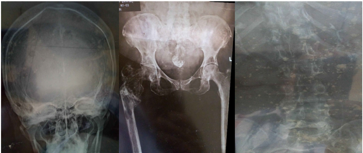
She promptly presented at NOHE where a radiological skeletal survey was ordered and it showed multiple punched out lesions in the pelvis, spine, skull and right femur just above the knee (Figure 1). A tentative diagnosis of multiple myeloma was entertained even with no myeloma defining illnesses. In the light of the above, she was referred to the haematological oncologists at University of Nigeria teaching Hospital (UNTH), Enugu. Liver function test, kidney function test, full blood count and erythrocyte sedimentation rate (FBC and ESR), normal urinalysis and negative Bence Jones protein in urine, serum electrophoresis, bone marrow aspiration and biopsy were done and results were not compatible with multiple myeloma (MM) (Table 1). Abdominal, chest and brain CT scans were requested but it did not show any primary source of the bone lesions in the viscera. With MM and any primary visceral source of metastatic lesions excluded, Mrs CN was referred back to NOHE as a case of primary bone metastatic disease. At NOHE, under general anaesthesia, she was subjected to an open biopsy on the neck of the right femur at which time, curetting from the right femur neck region was obtained. Curetting samples were divided into three – 2 samples were sent to 2 different pathologists and one sample sent for immunohistochemistry. A diagnosis of SS was finally made at which point surgical excision and radiotherapy were not favourable treatment options because of the extensive metastasis of the SS. The standard chemotherapeutic agents (Doxorubicin and ifosfamide) were not available at NOHE by mid-2020 during the covid-19 pandemic period when there were global lockdown. On the contrary, the patient opted out of chemotherapy by choice and has been on supportive therapy and follow-up routine clinic visits. Pain has been the most worrisome of her complaints.
Table 1: Relevant Laboratory Results of Mrs CN on Presentation at NOHE
Patient's Parameters at Presentation
Results
Reference Values
HbA1C
9.3%
<7%
Na
137mmol/l
135-148
K
4.5mmol/l
3.5-5.5
Cl
94mmol/l
96-107
HCO3
26mmol/l
24-32
Urea
27mg/dl
15-45
Creatinine
0.8mg/dl
0.6-1.5
FBC + ESR
Hb =13.2mg/dl, TWBC= 8.5x10⁹/l
Neutrophil=58%
Lymphocyte= 37%
Basophil 3%
Monophil= 2%
ESR = 23mm/1st hour
-
Urine Bence Jones protein
Negative
-
Urine analysis
Glucosuria +
Protein - Negative
Ketone - Negative
Nitrite Negative
Pus cells 0-2/hpf
-
Pelvis, spine and skull radiograph
Lytic punched out lesions as shown in Figure 1 below mimicking MM lesions.
-
Liver function tests including the enzymes
Normal
-
Serum electrophoresis
Normal
-
Bone marrow aspiration and later, bone marrow biopsy
Normal
-
Keywords: HbA1C = Glycated Haemoglobin, Na = Serum Sodium, K = Serum Potassium, Cl = Serum Chloride, HCO3 = Serum Bicarbonate, FBC = Full Blood Count, TWBC = Total White Blood Cell Count, ESR = Erythrocyte Sedimentation Rate, Hb = Haemoglobin Concentration
Figure 1: Lytic (punched out) Bone Lesions on the Skull Bone, Lumbar Spine and the Right Femur Neck and Pelvis of Mrs C.N
DISCUSSION
Synovial sarcoma has its highest frequency in the lower extremities of children and young adults even though it can afflict any tissues of the body. The patient in this report was, however, middle aged when her diagnosis was confirmed. It is possible that the initial tumour on the index patient's right knee may have started as a painless (asymptomatic) nodule decades before metastasis of the SS occurred to the right hip and other bones of the body. The implication of this is that painless nodules on any part of the body especially around joints of the lower extremities should not be treated with kid gloves. An excision biopsy should be done promptly and histological examination insisted on.
Synovial sarcoma with metastasis predominantly in the bones is a strong differential diagnosis of MM. The index patient had to be referred to the haematological Oncology unit of another centre based on the radiological skeletal survey findings. A lesson from this experience is to be open minded in trying to clinch the diagnosis of a patient. As regards MM, this experience with the index patient underscores the importance of seeking the myeloma-defining illnesses whenever the radiological skeletal survey points in that direction. In the setting where cost of laboratory investigations and imaging studies are paid out of pocket, clinching a diagnosis of metastatic SS in the index patient was quite challenging. Reason why the excision biopsy specimen on our patient's right knee was not subjected to histological examination is not clear but it could be that the patient considered the painless, slowly growing nodule to be innocuous. Cost, ignorance and superstition could, also, be contributory. It is probable, therefore, that SS may be under-diagnosed or under-reported in Nigeria and the other low-income countries especially if the patient cannot afford investigations including immunohistochemistry profile and cytogenetic screening. Considering the age, sex and initial radiological skeletal survey of the index patient, one is in order to consider MM as the diagnosis. One unique feature of this case is that up to the time of reporting the case, metastasis was primarily to the bones. The viscera were spared.
The index patient presented when metastasis had occurred to several bones of the body such that her treatment modality had to be adjuvant chemotherapy-based with doxorubicin and cyclophosphamide or ifosfamide as suggested by the NCCN clinical practice guidelines in oncology [13]. Surgery alone was not indicated in our patient probably because of late presentation. The reason why our patient opted out of chemotherapy is not clear but may have to do with the cost and or availability of the chemotherapeutic agents or the perceived side effects of chemotherapeutic agents. Shuttling between the orthopaedic centre and the haematological oncology centre in another tertiary health facility with all the logistic problems in a bid to confirm the diagnosis of SS in the index patient was not cost effective. This underscores the need for and importance of a multidisciplinary care team in respect of certain major diseases in each tertiary health facility. Radiation therapy was not a recommended treatment option to the index patient probably because of controversy surrounding its use especially in late presentations associated with distant metastasis or non-availability in the centre. In the index patient's case, a multidisciplinary care team comprising of the orthopaedic surgeons, haematologists, oncologists, radiotherapists and palliative care physicians is critical for a favourable management outcome.
In conclusion, metastatic SS to the bones should be a strong differential diagnosis of multiple myeloma. Presence of myeloma-defining illnesses must always be sought for and used to build a case for and confirm multiple myeloma. Dearth of the required manpower and facilities for early detection and appropriate management of SS and other cancers should be considered a major poor prognostic factor in Nigeria and the sub-region.
CONCLUSION
Conclusion/Recommendation
Even though SS is a rare soft tissue malignancy in the middle aged and the elderly, advanced state of the disease with metastasis to the bones must be considered in suspected multiple myeloma. Again, the initial painless, small nodules of SS should not be ignored or missed in children and adolescents where the condition is more predominant. It is, therefore, recommended that children and adolescents with painless nodules in the lower extremities where SS is more frequent should have the nodules excised and examined histologically. Heads of tertiary health facilities, policy makers, government and wealthy persons in the community have a role to play in contending with orthopaedic oncological challenges.
REFERENCE
Rajwanshi, A. et al. “Malignant small round cell tumours.” Journal of Cytology, vol. 26, 2009, pp. 1–10.
Fisher, C. “Synovial sarcoma.” Annals of Diagnostic Pathology, vol. 2, no. 6, December 1998, pp. 401–421, https://doi.org/10.1016/S1092-9134(98)80042-7.
Mahmut, N.A. et al. “Epidemiology, incidence and survival of synovial sarcoma subtypes: SEER database analysis.” Journal of Orthopaedic Surgery, 2020, https://doi.org/10.1177/2309499020936009.
Mishra, B. et al. “The first ever reported case of primary synovial sarcoma of the scalp.” Case Reports in Surgery, vol. 2016, 2016, Article ID 5358790, pp. 1–6, https://doi.org/10.1155/2016/5358790.
Ershadi, R. et al. “Primary mediastinal synovial sarcoma: A rare case report.” International Journal of Surgery Case Reports, vol. 27, 2016, pp. 169–171, https://doi.org/10.1016/j.ijscr.2016.08.037.
Casal, D. et al. “A 63-year-old woman presenting with a synovial sarcoma of the hand: A case report.” Journal of Medical Case Reports, vol. 6, 2012, p. 385, https://doi.org/10.1186/1752-1947-6-385.
Spurrell, E.L. et al. “Prognostic factors in advanced synovial sarcoma: An analysis of 104 patients treated at the royal marsden hospital.” Annals of Oncology, vol. 16, no. 3, 2005, pp. 437–444.
Guillou, L. et al. “Histologic grade, but not SYT-SSX fusion type, is an important prognostic factor in patients with synovial sarcoma: A multicenter, retrospective analysis.” Journal of Clinical Oncology, vol. 22, no. 20, 2004, pp. 4040–4050.
Clark, J. et al. “Identification of novel genes, SYT and SSX, involved in the t(X;18) (p11.2;q11.2) translocation found in human synovial sarcoma.” Nature Genetics, vol. 7, 1994, pp. 502–508.
Aytekin, M.N. et al. “Epidemiology, incidence, and survival of synovial sarcoma subtypes: SEER database analysis.” Journal of Orthopaedic Surgery, vol. 28, 2020, https://doi.org/10.1177/2309499020936009.
Gazendam, A.M. et al. “Synovial sarcoma: A clinical review.” Current Oncology, vol. 28, no. 3, May 2021, pp. 1909–1920, https://doi.org/10.3390/curroncol28030177.
Chotel, F. et al. “Variability in the presentation of synovial sarcoma in children.” Journal of Bone and Joint Surgery. British Volume, vol. 90, 2008, pp. 1090–1096, https://doi.org/10.1302/0301-620X.90B8.19815.
Von Mehren, M. et al. “Soft tissue sarcoma, Version 2.2016, NCCN clinical practice guidelines in oncology.” Journal of the National Comprehensive Cancer Network, vol. 14, 2016, pp. 758–786, https://doi.org/10.6004/jnccn.2016.0078.
Vlenterie, M. et al. “Outcome of chemotherapy in advanced synovial sarcoma patients: Review of 15 clinical trials from the european organisation for research and treatment of cancer soft tissue and bone sarcoma group.” European Journal of Cancer, vol. 58, 2016, pp. 62–72, https://doi.org/10.1016/j.ejca.2016.02.002.
Sultan, I. et al. “Comparing children and adults with synovial sarcoma in the surveillance, epidemiology and end results program, 1983 to 2005.” Cancer, vol. 115, 2009, pp. 3537–3547, https://doi.org/10.1002/cncr.24424.
Guadagnolo, B.A. et al. “Long-term outcomes for synovial sarcoma treated with conservation surgery and radiotherapy.” International Journal of Radiation Oncology Biology Physics, vol. 69, 2007, pp. 1173–1180, https://doi.org/10.1016/j.ijrobp.2007.04.056.
Canter, R.J. et al. “A synovial sarcoma-specific preoperative nomogram supports a survival benefit to ifosfamide-based chemotherapy and improves risk stratification for patients.” Clinical Cancer Research, vol. 14, 2008, pp. 8191–8197, https://doi.org/10.1158/1078-0432.CCR-08-0843.
Lewis, J.J., et al. “Synovial sarcoma: A multivariate analysis of prognostic factors in 112 patients with primary localized tumors of the extremity.” Journal of Clinical Oncology, vol. 18, 2000, pp. 2087–2094, https://doi.org/10.1200/JCO.2000.18.10.2087.
Campbell, C. et al. “Synovial sarcoma – towards a simplified approach to prognosis.” ANZ Journal of Surgery, vol. 74, no. 9, 2004, pp. 727–731, https://doi.org/10.1111/j.1445-1433.2004.03144.x.
Recommended Articles
Research Article
Advantages of Two Clips Laparoscopic Cholecystectomy (TCLC) and Conventional Laparoscopic Cholecystectomy (CLC): A Comparative Study
Anshu Attri,
...
Kartik Syal
Published: 10/01/2022
Download PDF
Research Article
Bladder Brilliance: Unveiling the Tapestry of Knowledge on Bladder Cancer in District Kangra, Himachal Pradesh
Vandana Thakur,
...
Ankita Pandey
Published: 08/12/2023
Download PDF
Research Article
Shouldering the Pain: Public Knowledge Assessment of Rotator Cuff Injuries in Himachal Pradesh
Sidhant Korla,
...
Praveen Kumar Thakur
Published: 30/07/2024
Download PDF
Research Article
Awareness regarding Symptoms and Treatment Options for Benign hypertrophy of prostate among the male patients attending outpatient department of Civil hospital , Nurpur, Himachal Pradesh
Aarti Chaudhary,
Brish Bhanu Bhardwaj
Published: 26/05/2023
Download PDF
PO Box 101, Nakuru
Kenya.
Email: office@iarconsortium.org
Editorial Office:
J.L Bhavan, Near Radison Blu Hotel,
Jalukbari, Guwahati-India
Nkpozi, M. O., Eyichukwu, G. & Ayogu, B. O. (2022). A Rare Case of Synovial Sarcoma with Bone Metastasis Mimicking Multiple Myeloma – Challenges of Management in A Nigerian Resource Constrained Setting. International Academic Research Journal of Surgery, 2(1), 1-5.
MLA
Nkpozi, M. O., G. Eyichukwu and B. O. Ayogu. "A Rare Case of Synovial Sarcoma with Bone Metastasis Mimicking Multiple Myeloma – Challenges of Management in A Nigerian Resource Constrained Setting." International Academic Research Journal of Surgery 2.1 (2022): 1-5.
Chicago
Nkpozi, M. O., G. Eyichukwu and B. O. Ayogu. "A Rare Case of Synovial Sarcoma with Bone Metastasis Mimicking Multiple Myeloma – Challenges of Management in A Nigerian Resource Constrained Setting." International Academic Research Journal of Surgery 2, no. 1 (2022): 1-5.
Harvard
Nkpozi, M. O., Eyichukwu, G. and Ayogu, B. O. (2022) 'A Rare Case of Synovial Sarcoma with Bone Metastasis Mimicking Multiple Myeloma – Challenges of Management in A Nigerian Resource Constrained Setting' International Academic Research Journal of Surgery 2(1), pp. 1-5.
Vancouver
Nkpozi MO, Eyichukwu G, Ayogu BO. A Rare Case of Synovial Sarcoma with Bone Metastasis Mimicking Multiple Myeloma – Challenges of Management in A Nigerian Resource Constrained Setting. International Academic Research Journal of Surgery. 2022 Jan;2(1):1-5.


 Download PDF
 Download XML




 3%
3%