Introduction : Sarcoma is connective tissue malignant tumor, rare disease but may have poor prognosis (especially soft tissue sarcomas). It can differentiate into various cell types such as lymphocytes, fibrous supporting structures, muscle, visceral tissue and bone. It can occur on any part of the body, but the most common is on the extremities. In order to evaluate the general description and pattern of sarcoma disease in the Surgery Division of Dr. Moewardi General Hospital Surakarta between period 2015 to 2019. Methods: This study was conducted in a retrospective descriptive manner by examining the medical records of sarcomas patient in the Surgery Division from January 2015 to August 2019. Results and Discussion : There were 386 cases of sarcoma during the period of January 2015 to August 2019. There were 21 histological subtypes of sarcoma with the most cases being liposarcoma and GIST (52 cases), which tended to be more common in men (52%) and adults (20-60 years) as much as 59%. For the most common anatomical location of sarcoma in this study, it occurred on the trunk (44%) and the lower extremity (33%) for the second most common. Conclusion: There is an up and down pattern in the number of cases of sarcoma patients at RSUD Dr. Moewardi from 2015 to 2019, with an average of 77.2 cases per year. The most common subtypes found were liposarcoma and GIST, the most common distribution pattern occurring in males and in patients aged 20 to 60 years.
The pattern of disease is currently shifting from infectious diseases to non-communicable diseases, one of which is cancer. In a study conducted in America in 2002 - 2014 there were 78,527 cases of sarcoma with an incidence of 7.1 cases per 100,000 people [1-3]. The incidence of soft tissue sarcoma (STS) in adults and children is 0.7-1% and 4-8% of all tumors, while bone sarcoma is 0.2% and 5% for adults and children, respectively. Soft tissue in the European region is 84% of all types of sarcomas. Based on the histological type, soft tissue sarcomas consisted of leiomyosarcoma (19%), liposarcoma (16%), and unspecified sarcoma (NOS, 14%). Bone sarcomas account for 14% of sarcomas diagnosed in Europe, Chondrosarcoma and Ewing's sarcoma are the most common bone sarcomas in adults, with ASRs of 0.2 and 0.3, respectively [4,5].
The incidence of sarcomas in Indonesia, especially at Dharmais Hospital Jakarta in 2012-2013, including fibrosarcoma, angiosarcoma and osteosarcoma are included in the 10 most cancer cases. -23%, liposarcoma 15-18%, leiomyosarcoma 7-11%, rhabdhomyosarcoma 5-19%, angiosarcoma 2-3%, lymphangiosarcoma 2-3%, synovial sarcoma 5-20%, and neurofibrosarcoma 5-7% [6].
Sarcomas are a rare group of malignant connective tissue tumors capable of differentiation into various cell types such as lymphocytes, fibrous supporting structures, muscle, visceral tissue and bone.2 These tumors can occur in almost any part of the body, although they are more common in the extremities.
A low histological grade was associated with local recurrence of sarcoma, on the other hand, a high histological grade was correlated with the ability to distant metastases. The most common predilection for sarcomas is the extremities (60%), trunk (19%), retroperitoneum (15%) and head and neck (9%) [7].
Sarcomas, although relatively rare but deadly, especially soft tissue sarcomas. Delay in diagnosis, advanced disease or metastases are factors that aggravate the condition. Early-stage sarcomas have no symptoms that have the potential to be difficult to diagnose early. In addition to being a deadly disease, sarcoma is also more common in young adults and adolescents than other cancers [8].
The choice of surgical modality in sarcoma depends on several factors, namely: histological subtype of tumor, tumor grading, tumor location, depth of invasion, important structures or other organs affected, compartments involved, need for reconstruction, and clinical status of the patient. The prognosis of sarcoma depends on the histologic subtype, grading, tumor depth, tumor size, tumor location, positive surgical margins and age [9].
Although the incidence is low, the quality of life and risk of death are substantial. Strategies to prevent the development of sarcomas will prove useful. The purpose of this paper is to determine the prevalence of sarcoma and control the impact of sarcoma by collecting data and classifying sarcoma cases as to produce statistical values [9,10].
This research is a retrospective descriptive study to determine profile of sarcoma incidence at surgery division RSUD dr. Moewardi Surakarta January 2015 - August 2019. The research was conducted at RSUD dr. Moewardi Surakarta by taking data at the Anatomical Pathology Installation in February 2021. The study population was all sarcoma patients that visited surgery division of RSUD dr. Moewardi, Surakarta period January 2015 – August 2019. The subject of this research is all sarcoma patients that visited surgery division of RSUD dr. Moewardi Surakarta period January 2015 – August 2019 and data were taken from the diagnosis of anatomic pathology after the patient underwent surgery treatment. The criteria taken in this study based on the number of sarcomas, diagnosis of sarcoma subtypes, gender, age and anatomical location. The data is then collected and analyzed to assess the incidence of sarcoma in the surgery division RSUD dr. Moewardi Surakarta. The data that has been collected is grouped according to the criteria, then described in the form of percentages in curves, tables and diagrams.
The results of this study are sarcomas in the Surgery department RSUD dr. Moewardi Surakarta for the period January 2015 to August 2019 obtained 386 cases.
Distribution of Sarcomas by Year
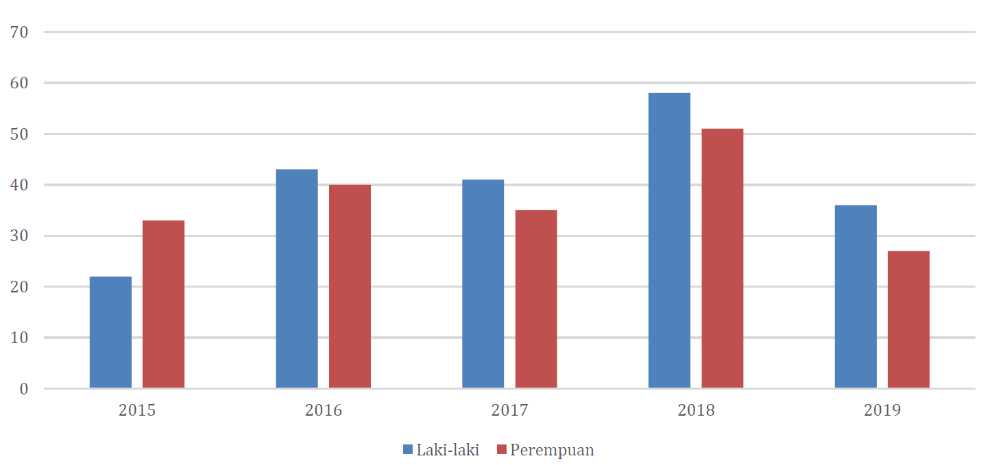
Of the 386 research subjects, there were 55 cases in 2015 (14%), 83 cases in 2016 (22%), 76 cases in 2017 (20%), 109 cases in 2018 (28%) and 63 cases in 2019(16%). (Table 1) (Figure 1).
Distribution by Gender
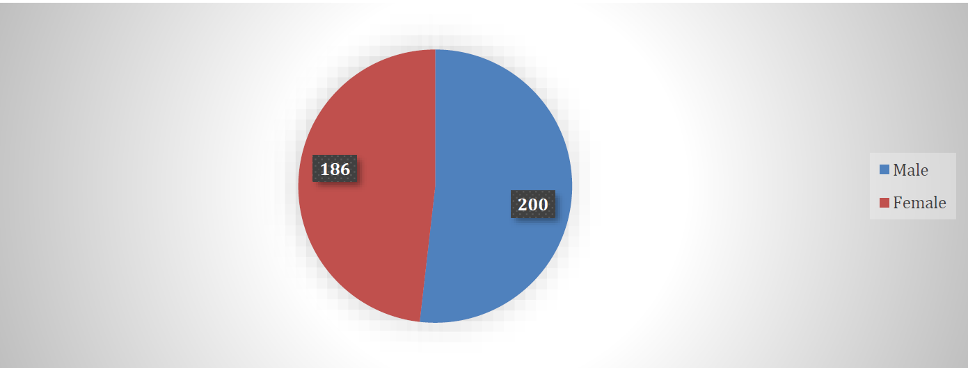
From the data in diagram 3 above, from 363 cases of sarcoma in the Surgery section, RSUD dr. Moewardi Surakarta186 female patients (48%) and 200 male patients (52%) (Figure 2,3). So that in this study, the number of male sarcoma patients was more than the number of female sarcoma patients (Table 2).
Distribution by Age
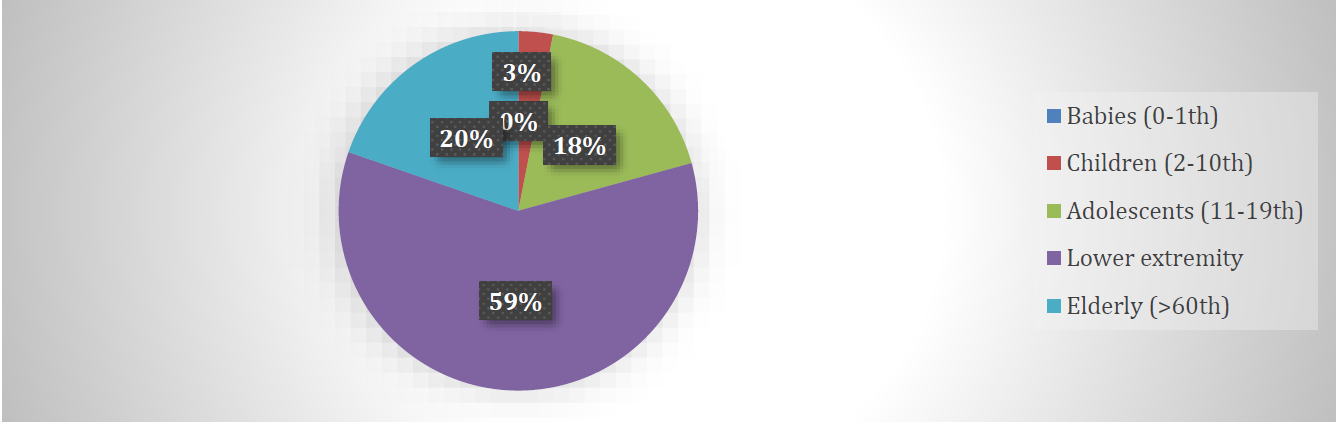
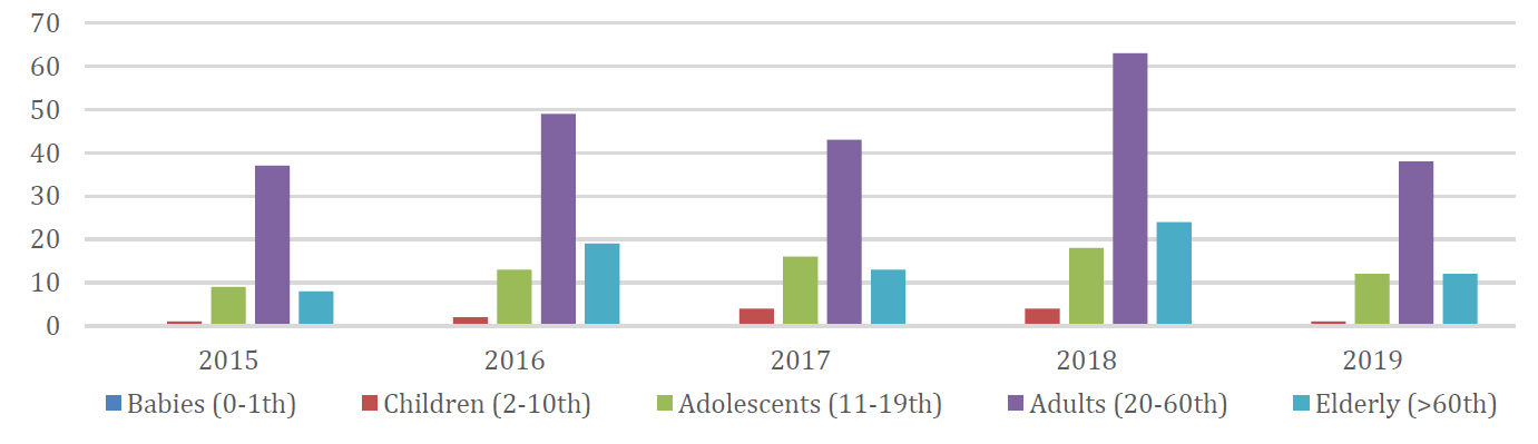
From 386 research subjects, there were 0 patients aged infancy (0-1 years) (0%), 12 patients aged children (2-10 years) (3%), 68 adolescent patients (11-19 years) (18%), 230 patients adults (20-60 years) (59%) and 76 elderly patients (>60 years) (20%) (Figure 4,5) (Table 3).
Distribution based on Anatomical Location
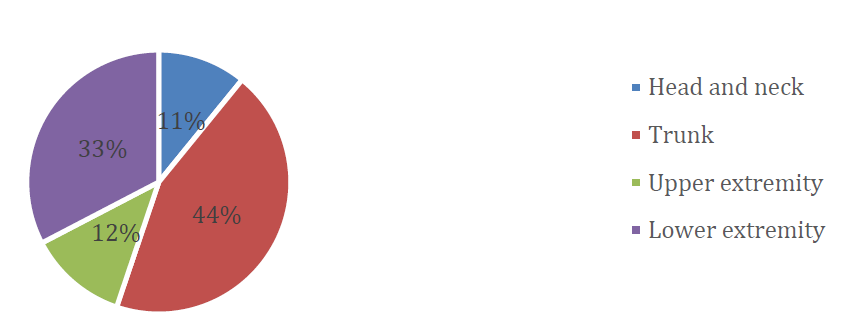
From Figure 6 of 363 cases of sarcoma in the Surgery section RSUD dr. Moewardi SurakartaThere were 42 cases of sarcoma in the head and neck (11%), 171 cases of sarcoma on the trunk (44%), 47 cases of sarcoma in the upper extremity (12%), 126 cases of sarcoma in the lower extremity (33%). From these data, the most anatomical locations of sarcomas were found in the trunk and the least in the head and neck.In 2015 there were 7 cases of sarcoma in the head and neck, 32 cases in the trunk, 5 cases in the superior extremities and 11 cases in the lower extremities. In 2016 there were 9 cases of sarcoma in the head and neck, 43 cases in the trunk, 14 cases in the superior extremities and 17 cases in the lower extremities. In 2017 there were 7 cases of sarcoma in the head and neck, 31 cases in the trunk, 6 cases in the superior extremities and 32 cases in the lower extremities. In 2018 there were 13 cases of sarcoma in the head and neck, 41 cases in the trunk, 11 cases in the superior extremities and 44 cases in the lower extremities. In 2019 there were 6 cases of sarcoma in the head and neck, 24 cases in the trunk, 11 cases in the superior extremities and 22 cases in the lower extremities.
Sarcoma is a rare malignancy with biological characteristics that are classified as aggressive13. This research was conducted by taking data in the anatomical pathology laboratory of RSUD dr. Moewardi Surakarta by collecting data on all sarcoma cases in the Surgery department from 2015 to 2019. In this study, data collection was carried out based on histopathological diagnosis, number of sarcomas, gender, age and anatomical location of sarcomas on the patient's body. From this study, it was found that the number of sarcoma cases in the Surgery section of RSUD dr. Moewardi from 2015 to 2019 rose and fell with an average of 77.2 cases per year.

Figure 1: Data by Year
Table 1: Distribution Histological Subtypes of Sarcomas each Year (2015-2019)
| Sarcoma | Year | Total | ||||
2015(%) | 2016(%) | 2017(%) | 2018(%) | 2019(%) | ||
Alveolar Soft Part Sarcoma. | 0 | 1 (1,2) | 0 | 3 (2.8) | 1 (1.6) | 5 (1,3) |
Angiosarcoma | 2 (3.6) | 9 (10.8) | 5 (6.6) | 2 (1.8) | 1 (1.6) | 19 (4.9) |
botryoid sarcoma | 0 | 0 | 1 (1,3) | 0 | 0 | 1 (0.3) |
Chordoid Sarcoma | 0 | 2 (2,4) | 1 (1,3) | 5 (4.6) | 2 (3,2) | 10 (2.6) |
Clear Cell Sarcoma | 0 | 1 (1,2) | 0 | 0 | 0 | 1 (0.3) |
dermatofibrosarcoma | 0 | 0 | 2 (2.6) | 4 (3,7) | 2 (3,2) | 8 (2) |
Ewing Sarcoma. | 0 | 4 (4.8) | 2 (2.6) | 3 (2.8) | 5 (7.9) | 14 (3.6) |
Fibrosarcoma | 4 (7.3) | 5 (6) | 8 (10.5) | 8 (7.3) | 4 (6.3) | 29 (7.5) |
Giant cell tumor | 3 (5.5) | 6 (7.2) | 3 (4) | 13 (11.9) | 10 (15.9) | 35 (9) |
GIST | 8 (14.4) | 8 (9.6) | 9 (11.8) | 17 (15.6) | 10 (15.9) | 52 (13.5) |
Leiomyosarcoma. | 3 (5.5) | 1 (1,2) | 1 (1,3) | 0 | 0 | 5 (1,3) |
Liposarcoma | 9 (16.4) | 9 (10.8) | 9 (11.8) | 19 (17.4) | 6 (9.5) | 52 (13.5) |
Malignant Fibrohystiocytoma | 3 (5.5) | 10 (12) | 3 (4) | 5 (4.6) | 7 (11,1) | 28 (7.3) |
Malignant Phylloides Tumor. | 4 (7.3) | 6 (7.2) | 3 (4) | 1 (0.9) | 1 (1.6) | 15 (3.9) |
Malignant Schwannoma. | 2 (3.6) | 0 | 0 | 0 | 0 | 2 (0.5) |
XMyxoinflammatory Fibroblastic Sarcoma | 0 | 0 | 0 | 0 | 1 (1.6) | 1 (0.3) |
Osteosarcoma. | 4 (7.3) | 3 (3.6) | 17 (22.4) | 16 (14.7) | 7 (11,1) | 47 (12.2) |
Rhabdomyosarcoma. | 7 (12.7) | 12 (14.5) | 8 (10.5) | 7 (6.4) | 4 (6.3) | 38 (9.8) |
Round Cell Sarcoma. | 2 (3.6) | 2 (2,4) | 3 (4) | 5 (4.6) | 1 (1.6) | 13 (3,4) |
Synovial Sarcoma | 3 (5.5) | 3 (3.6) | 1 (1,3) | 0 | 1 (1.6) | 8 (2) |
Epitheloid Sarcoma | 1 (1.8) | 1 (1,2) | 0 | 1 (0.9) | 0 | 3 (0.8) |
Total | 55(100) | 83(100) | 76(100) | 109(100) | 63(100) | 386 |

Figure 2: Data by Gender

Figure 3: Sarcoma Distribution by Sex in 2015-2019
Table 2: Distribution Sarcoma Subtype by Gender Year ( 2015 – 2019 )
| Sarcoma | Gender | |
Man (%) | Woman (%) | |
Alveolar Soft Part Sarcoma. | 2 (1) | 3 (1.6) |
Angiosarcoma | 11 (5.5) | 8 (4,3) |
botryoid sarcoma | 0 | 1 (0.5) |
Chordoid Sarcoma | 4 (2) | 6 (3,2) |
Clear Cell Sarcoma | 1 (0.5) | 0 |
Dermatofibrosarcoma | 5 (2.5) | 3 (1.6) |
Ewing Sarcoma. | 8 (4) | 6 (3,2) |
Fibrosarcoma | 13 (6.5) | 16 (8.6) |
Giant cell tumor | 20 (10) | 15 (8,1) |
GIST | 29 (14.5) | 23 (12,4) |
Leiomyosarcoma. | 2 (1) | 3 (1.6) |
Liposarcoma | 32 (16) | 20 (10.8) |
Malignant Fibrohystiocytoma | 16 (8) | 12 (6.4) |
Malignant Phylloides Tumor. | 0 | 15 (8,1) |
Malignant Schwannoma. | 0 | 2 (1,1) |
Myxoinflammatory Fibroblastic Sarcoma | 1 (0.5) | 0 |
Osteosarcoma. | 25 (12.5) | 22 (11.8) |
Rhabdomyosarcoma. | 21 (10.5) | 17 (9.1) |
Round Cell Sarcoma. | 6 (3) | 7 (3.8) |
Synovial Sarcoma | 3 (1.5) | 5 (2.7) |
Epitheloid Sarcoma | 1 (0.5) | 2 (1,1) |
Total | 200 (100) |
|

Figure 4: Data by Age

Figure 5: Sarcoma Distribution by Age 2015-2019

Figure 6: Data based on Anatomical Location
Table 3: Distribution of Sarcoma Subtypes by Age (2015 – 2019)
| Sarcoma | Baby | Child | Teenager | Mature | Seniors |
0-1th (Percentage) | 2 – 10th (Percentage) | 11 - 19th (Percentage) | 20 - 60th (Percentage) | > 60 Years Old (%) | |
Alveolar Soft Part Sarcoma | 0 | 0 | 1 (1.5) | 4 (1.7) | 0 |
Angiosarcoma | 0 | 0 | 2 (2.9) | 12 (5,2) | 5 (6.6) |
botryoid sarcoma | 0 | 0 | 0 | 1 (0.4) | 0 |
Chordoid Sarcoma | 0 | 0 | 0 | 8 (3.5) | 2 (2.7) |
Clear Cell Sarcoma | 0 | 0 | 0 | 1 (0.4) | 0 |
Dermatofibrosarcoma | 0 | 1 (8.3) | 0 | 6 (2.7) | 1 (1,3) |
Ewing Sarcoma. | 0 | 3 (25) | 6 (8.8) | 4 (1.7) | 1 (1,3) |
Fibrosarcoma | 0 | 0 | 1 (1.5) | 23 (10) | 5 (6.6) |
Giant cell tumor | 0 | 0 | 4 (5,9) | 30 (13) | 1 (1,3) |
GIST | 0 | 1 (8.3) | 0 | 32 (14) | 19 (25) |
Leiomyosarcoma. | 0 | 0 | 0 | 4 (1.7) | 1 (1,3) |
Liposarcoma | 0 | 0 | 1 (1.5) | 30 (13) | 21 (27.6) |
Malignant Fibrohystiocytoma | 0 | 0 | 2 (2.9) | 18 (7,8) | 8 (10.5) |
Malignant Phylloides Tumor | 0 | 0 | 0 | 15 (6.5) | 0 |
Malignant Schwannoma. | 0 | 0 | 0 | 1 (0.4) | 1 (1,3) |
Myxoinflammatory Fibroblastic Sarcoma | 0 | 0 | 0 | 0 | 1 (1,3) |
Osteosarcoma. | 0 | 3 (25) | 37 (54.4) | 6 (2.7) | 1 (1,3) |
Rhabdomyosarcoma. | 0 | 2 (16.7) | 7 (10.3) | 22 (9.6) | 7 (9.2) |
Round Cell Sarcoma. | 0 | 2 (16.7) | 3 (4,4) | 6 (2.7) | 2 (2.7) |
Synovial Sarcoma | 0 | 0 | 4 (5,9) | 4 (1.7) | 0 |
Epitheloid Sarcoma | 0 | 0 | 0 | 3 (1,3) | 0 |
- | 0 | 12 (100) | 68 (100) | 230 (100) | 386 (100) |
In this study data, there were 21 subtypes of sarcoma based on histological diagnosis, sorted from highest to lowest number, namely liposarcoma, GIST (Gastro-Intestinal Stromal Tumor), osteosarcoma, rhabdomyosarcoma, giant cell tumor, fibrosarcoma, malignant fibrohystiocytoma, angiosarcoma, malignant phylloides tumor, Ewing. sarcoma, round cell sarcoma, chordoid sarcoma, dermatofibrosarcoma, synovial sarcoma, alveolar soft part sarcoma, leiomyosarcoma, epithelioid sarcoma, malignant schwannoma, botryoid sarcoma, myxoinflammatory fibroblastic sarcoma, clear cell sarcoma. The most diagnoses were liposarcoma and GIST, namely 52 cases with an average of 10.4 cases per year, while the least diagnoses were botryoid sarcoma, myxoinflammatory fibroblastic sarcomaandclear cell sarcoma obtained 1 case each in 5 years. This is consistent with a study conducted in Italy at 15 cancer centers between 2009-2012 where liposarcoma also ranked first in the most frequent diagnosis of sarcoma, followed by fibrosarcoma and leiomyosarcoma [11]. The increase in liposarcoma in America from 2001-2016 was reported by Bock et al. [12], with the analysis that obesity and an unhealthy lifestyle are two possible factors that increase the risk of developing liposarcoma.
From the above study, it was found that the number of male patients was more than female. With an average number of male patients 40 patients per year and female patients 37.2 patients per year. This is in accordance with a previous study in Pekanbaru Indonesia in 2009-2013 that found the incidence of sarcoma in male patients was more than female, namely 66.7% in males and 51.4% in females. Age 65 and over found that the prevalence of sarcoma is increasing in men which can be doubled compared to women11 and the same thing also happened in Iran [13].
In this study, the most sarcomas were found in adult patients (20-60 years) i.e. 46 patients per year and the least in infants (0-1 years) i.e. 0 patients for 5 years. Almost all types of sarcomas in this study were most common in adults, insynovial sarcoma the number of patients aged adolescents and adults is the same,only in osteosarcoma the highest incidence is in adolescents (11-19 years). This data is different from what happened in Europe between 2003-2007 conducted by Burnham et al, where age >65 years showed an increase in prevalence and incidence that was directly proportional to the age group. Research in general in Europe (2003-2007), Germany (2003-2012) and Italy (2009-2012) at the age of >65 years with a prevalence of 45%, 26% and 44.5% while in this study it was 20%.6 ,9 These data are in line with research from Arfiana W et al that more soft tissue sarcomas are obtained at the age of 40-49 years. 16 Meanwhile, according to WHO in 2017, soft tissue sarcomas are increasing with increasing age, the median is 65 years.
From the sarcoma patient data above, the most anatomical locations of sarcomas are on the trunk with an average value of 34.2 cases per year (44%) and the least in the upper extremities, namely 8.4 cases per year (11%). This is different from the study conducted by Handawi in 15 cancer centers in Italy which showed that the upper extremity (35.6%) followed by the lower (33.2%) was the most common location. conducted in Medan 2006-2007 on dermatofibrosarcoma protuberans predilection showed extremity location of 66%.16 DeVita (2001) explained that soft tissue sarcoma mostly originates from the upper and lower extremities (59%), trunk (19%), retroperitoneum ( 15%) and head and neck (9%).
The number of sarcoma cases in the Surgery section of RSUD Dr. Moewardi Surakarta from 2015 to 2019 was 386 cases, with the most cases in 2018 being 109 cases and the lowest in 2015 being 55 cases
Sarcoma diagnosis based on histopathology in the Surgery section of RSUD dr. Moewardi Surakarta from 2015 to 2019 obtained 21 diagnoses with the most cases of liposarcoma and GIST, each with 52 cases (10.4%)
The distribution of sarcomas based on gender in the Surgery section of RSUD dr. Moewardi Surakarta from 2015 to 2019 found that there were more male patients than female, with 200 male cases (40%) and 186 female patients (37.2%)
Distribution of sarcomas by age in the Surgery department of dr. Moewardi Surakarta from 2015 to 2019 found the most cases in adult patients (20-60 years old), namely 230 cases (46%) and there were no cases of infants aged (0-1 years)
The distribution of sarcomas based on anatomical location in the Surgery section of RSUD dr. Moewardi Surakarta from 2015 to 2019 found the most cases in the trunk, namely 171 cases (34.2%) and the least in the head and neck, namely 42 cases (8.4%)
Arfiana, W. et al. “The distribution of soft tissue sarcoma based on histopathology’s check in pekanbaru’s hospital between 2009–2013.” Let’s FK, vol. 3, no. 1, February 2016, pp. 1–22.
Fabiano, S. et al. “Epidemiology of soft tissue sarcoma and bone sarcoma in Italy: Analysis of data from 15 population-based cancer registries.” Sarcoma, vol. 2020, 2020, Article ID 6142613. https://doi.org/10.1155/2020/6142613.
American Cancer Society. Cancer Facts and Figures 2015. American Cancer Society, 2015.
Gatta, G. et al. “Rare cancers are not so rare: The rare cancer burden in Europe.” European Journal of Cancer, vol. 47, no. 17, 2011, pp. 2493–2511.
Ducimetière, F. et al. “Incidence of sarcoma histotypes and molecular subtypes in a prospective epidemiological study with central pathology review and molecular testing.” PLoS ONE, vol. 6, no. 8, 2011, e20294, pp. 1–14.
Burningham, Z. et al. “The epidemiology of sarcoma.” Clinical Sarcoma Research, vol. 2, 2012, pp. 14. https://doi.org/10.1186/2045-3329-2-14.
Alwan, A. et al. Noncommunicable Diseases Country Profiles 2011. World Health Organization, 2011.
Ministry of Health of the Republic of Indonesia. Cancer Situation. Center for Data and Information, Ministry of Health of the Republic of Indonesia, 2015.
Saltus, C.W. et al. “Epidemiology of adult soft-tissue sarcomas in Germany.” Sarcoma, vol. 2018, 2018, Article ID 5671926, pp. 1–11. https://doi.org/10.1155/2018/5671926.
Ducimetière, F. et al. “Global cancer statistics 2018: GLOBOCAN estimates of incidence and mortality worldwide for 36 cancers in 185 countries.” CA: A Cancer Journal for Clinicians, vol. 68, 2018, pp. 394–424.
Younger, E. et al. “Age-related sarcoma patient experience: results from a national survey in England.” BMC Cancer, vol. 18, 2018, pp. 991. https://doi.org/10.1186/S12885-018-4866-8.
Bock, S. et al. “Increasing incidence of liposarcoma: A population-based study of national surveillance databases, 2001–2016.” International Journal of Environmental Research and Public Health, vol. 17, no. 8, 2020, pp. 2710. https://doi.org/10.3390/ijerph17082710.
Acem, I. et al. “Age-related differences of oncological outcomes in primary extremity soft tissue sarcoma: A multistate model including 6260 patients.” European Journal of Cancer, vol. 141, 2020, pp. 128–136. https://doi.org/10.1016/j.ejca.2020.09.021.