Myringoplasty is a surgical procedure performed to repair tympanic perforation. Different techniques have been developed and improved over time using various materials. The technique we describe in this article uses fat graft under endoscopic guidance. This minimally invasive and simple surgical procedure allows excellent results. It presents itself as a good alternative in poor countries where strong demand of tympanic repairs faces few numbers of otologists and deficient technical platforms.
Myringoplasty is a commonly used procedure in ear surgery [1]. It is conventionally performed under microscopy [2]. Different techniques and various graft materials are used [2, 3, 4].
Recently, the use of endoscopy in otologic surgery allowed evolution of these techniques [2, 4, 5, 6].
The myringoplasty procedure we describe hereby derives from coupling endoscopy with fat graft material.
Surgical Technique
Surgical Team
The surgeon operates alone; a circulating nurse will take care of dressing and serving instruments.
Instrumentation
A 0o or 30o rigid endoscope (2.7- or 3-mm diameter, 10 to 18 cm length) is connected to video system (camera, monitor and light source).
Classic myringoplasty instruments, suction device with fine cannulas are needed.
Installation
The patient is in supine position with head slightly turned towards tympanic perforation opposite side. The surgeon stands on operated side, video monitor in front and instrument table at patient's head.
Anesthesia
For adults, the procedure is most often performed under local anesthesia. For children, pusillanimous adults, or on request, the intervention is done under general anesthesia.
Procedure
Otoendoscopic Examination: After external acoustic meatus cleaning, initial otoendoscopy visualizes all eardrum parts, especially anterior angle; it permits to precise perforation seat and size, and also appreciates tympanic remains.
Fat Graft Sampling
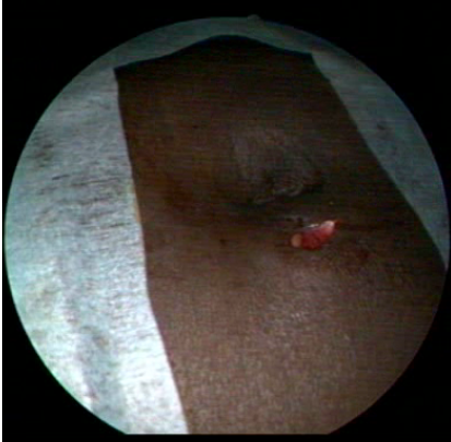
A piece of fat, slightly larger than perforation diameter is taken in abdominal peri-umbilical area through a 5 to 10 mm incision (Figure 1).
Myringoplasty
The endoscope held by left hand (for a right-handed operator) slowly goes down through external auditory meatus. Endoscope trip is followed on the monitor.

Figure 1: Abdominal Peri-Umbilical Fat Graft Collection Site

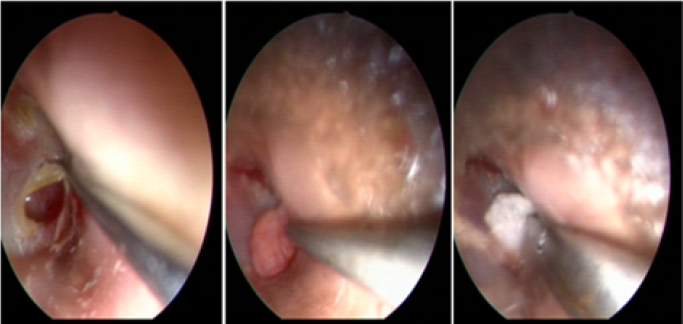
Figure 2: (A) Tympanic Perforation Margins Reviving, (B) Fat Graft Set Up, (C) Gelfoam on the Graft.

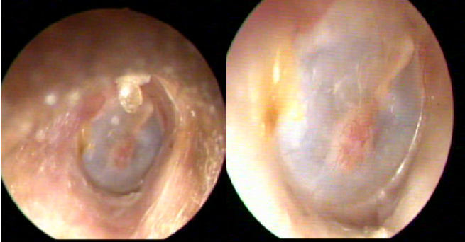
Figure 3: Otoscopic View One Month Postoperative (A), three Months Postoperative (B)
Desepidermisation of Perforation Margins
The tympanic perforation is revitalized by making small circumferential holes at 1 mm from the banks using a curved tip. The holes are then connected by a scanning movement of the tip. The scar tissue is then extracted using a micro-clamp (Figure 2A).
Graft Implementation
The fat graft is inserted by "champagne cork" technique through the perforation allowing the fatty tissue to cover medial and lateral faces of the perforation (Fig. 2B). A piece of gelfoam is placed on the graft, and wedged by a cotton ball or a pope (Figure 2C).
Post-Operative Care
Antibiotic drops are prescribed for a week. Removal of the cotton ball is done on 7th day. The patient is seen one month, and three months postoperative (Figure 3A and 3B).
Recently, endoscopy is increasingly used in otologic surgery [2,4,5,6]. In myringoplasty, the endoscopic approach permits to obtain a wider field of view [4,5,6], especially in anterior perforations, but also in cases of narrow or tortuous external acoustic meatus and in presence of exostosis.
The transmeatal pathway thus avoids endaural and retroauricular incisions [3,4,7,8]. It also avoids meatoplasty [5,7,8]. Use of endoscopy reduces the operative time [4,7,9].
Anatomical and functional results are similar for conventional myringoplasty under microscope and endoscopic myringoplasty [2,4,8,9].
The main disadvantage of using endoscopy is that surgeon works with only one free hand, when the other is holding the endoscope [4,5].
The use of fat graft gives same results as other conventional materials with success rate higher than 92% [1,10]. However, it should be reserved for dry, non-marginal, small or medium-sized (-50%) perforations without associated myringosclerosis [9].
Perforation seat does not affect failure rate [9], and no more complication has been described when using fat graft in myringoplasty [1,9,10]. This material can be used in first or seconde intention [9].
Endoscopic myringoplasty with fat graft is a simple and effective surgical procedure. It may be indicated in case of tympanic perforation in a rigorously selected patient.
It is thus an alternative in developing countries; where, despite of a very strong demand for tympanic repairs, otologists are still rare and technical platform deficient.
Conflict of Interest
The authors declare that they have no competing interest
Kwong, K.M. et al. “Fat graft myringoplasty using umbilical fat.” International Journal of Pediatric Otorhinolaryngology, vol. 76, 2012, pp. 1098–1101.
Akyigit, A. et al. “Endoscopic tympanoplasty.” Journal of Otology, vol. 12, 2017, pp. 62–67.
Kiringoda, E.R. and M.S. Cohen. “Endoscopic tympanoplasty.” Operative Techniques in Otolaryngology, vol. 28, 2017, pp. 17–22.
Min, J. and S.H. Kim. “Comparison of transcanal endoscopic tympanoplasty with sterile acellular dermal allograft to conventional endaural microscopic tympanoplasty with tragal perichondrium.” American Journal of Otolaryngology, vol. 39, 2018, pp. 167–170.
Mohanty, S. et al. “Endoscopic cartilage versus temporalis fascia grafting for anterior quadrant tympanic perforations: a prospective study in a tertiary care hospital.” Auris Nasus Larynx, vol. 45, 2018, pp. 936–942.
Tseng, C.C. et al. “Comparison of endoscopic transcanal myringoplasty and endoscopic type i tympanoplasty in repairing medium-sized tympanic perforations.” Auris Nasus Larynx, vol. 44, 2017, pp. 672–677.
Daneshi, A. et al. “Bilateral same-day endoscopic transcanal cartilage tympanoplasty: Initial results.” Brazilian Journal of Otorhinolaryngology, vol. 83, 2017, pp. 411–415.
Isaacson, G. and J.A. Harounian. “Results of pediatric endoscopic and endoscopically assisted tympanoplasty.” World Journal of Otorhinolaryngology–Head and Neck Surgery, vol. 3, 2017, pp. 136–141.
Thomassin, J.M. et al. “Intérêt de l’Oto-Endoscopie dans la myringoplastie par greffon adipocytaire.” Annales d’Otolaryngologie et de Chirurgie Cervico-Faciale, vol. 121, 2004, pp. 346–349.
Tringali, S. et al. “Les perforations tympaniques et les greffes de tympan.” Annales d’Otolaryngologie et de Chirurgie Cervico-Faciale, vol. 125, 2008, pp. 261–272.