Introduction: Hoffa in 1904 first described fractures of the femoral condyle in tangential plane and are now known as Hoffa fractures. Approach to isolated medial Hoffa fracture is, however, confusing. On reviewing literature, majority cases are operated using parapatellar arthrotomy, some through a medial (subvastus) approach, and few was fixed percutaneously.Materials and Methods: We studied 6 patients of medial Hoffa fracture presenting in Orthopedics department of Kalpana Chawala Government Medical College, Karnal. They were evaluated radiologically with X-rays of knee anteroposterior and lateral view. All patients were approached through subvastus approach, and were treated with screws and buttress plating. The follow up of patients was done after 2 weeks, 1 month, 3 months and 6 months post operatively. Results: The age of the patients ranged from 27-31 (Mean 29 years). Mean range of motion was 125.83(range 120-130). All our patients had radiological bony union at 6 months. Functional outcome was calculated by Knee society score which was average 84.16 (range 80-88), which comes under excellent category (80-100). Conclusion: We concluded that outcome results of operated case of isolated medial Hoffa’s fracture using medial subvastus approach and fixation of fracture with screws and buttress recon plate are excellent (as per knee society score).
Hoffa in 1904 first described fractures of the femoral condyle in tangential plane and are now known as Hoffa fractures [1]. They are classified as 33-B3.2 as per AO/OTA classification [2].
They can be described as intra-articular lateral or medial or bicondylar fracture that occurs in the coronal plane of the distal femur hence immediate needing the anatomical reduction with internal fixation as early as possible [1].
Lateral or bicondylar Hoffa fractures and well described in literature in respect to classification, mechanism of injury, surgical approach, reduction and fixation methods but Isolated coronal fracture of medial femoral condyle, with intact lateral femoral condyle, is extremely rare and barring a few isolated case reports [3].
This is likely because of the often-associated articular comminution, associated injuries, the relative inaccessibility of the medial femoral condyle articular surface, the difficulty associated with hardware placement, and the magnitude of shear forces that must be resisted with the fixation to avoid loss of fracture reduction [4].
The mechanism of injury for isolated medial Hoffa fracture is controversial. Some authors described direct impact with the knee in a flexed position as the mechanism of injury, while others have attributed the fracture to simultaneous vertical shear and twisting forces(2). Majority of these injuries are a result of accidents two-wheeler riding. The normal riding posture of the motor cyclist involves sitting with the knee flexed at or beyond 90. In this position with slight abduction, lateral femoral condyle is the leading part of the knee to receive a direct impact. Medial impact directly to the medial femoral condyle with the knee in greater than 90 of flexion and an element of adduction and internal rotation can lead to medial Hoffa’s fracture [5].
Diagnosis of this fracture is challenging and needs strong suspicion as this type of fracture is rare and misdiagnosis is common. In cases of isolated fracture without any associated fractures of the knee, diagnosis should be based on physical examination of skin lesions, oedema, effusion and careful radiological evaluation especially oblique views However, in cases with associated distal femoral fractures, diagnosis of coronal fractures is quiet difficult unless a strong index of suspicion is kept for the same [6].
The imaging evaluation of distal femoral fractures is based primarily on radiographs. Supracondylar distal femoral fractures may be classified as extraarticular, unicondylar, or bicondylar, and the fractures may have an intercondylar extension [7].
Nork et al., strongly recommended preoperative CT scan in cases of supracondylar-intercondylar fractures of the distal femur because of misdiagnosis on conventional radiographs [8].
Approach to isolated medial Hoffa fracture is, however, confusing. On reviewing literature, majority cases are operated using parapatellar arthrotomy, some through a medial (subvastus) approach, and few was fixed percutaneously [4,9].

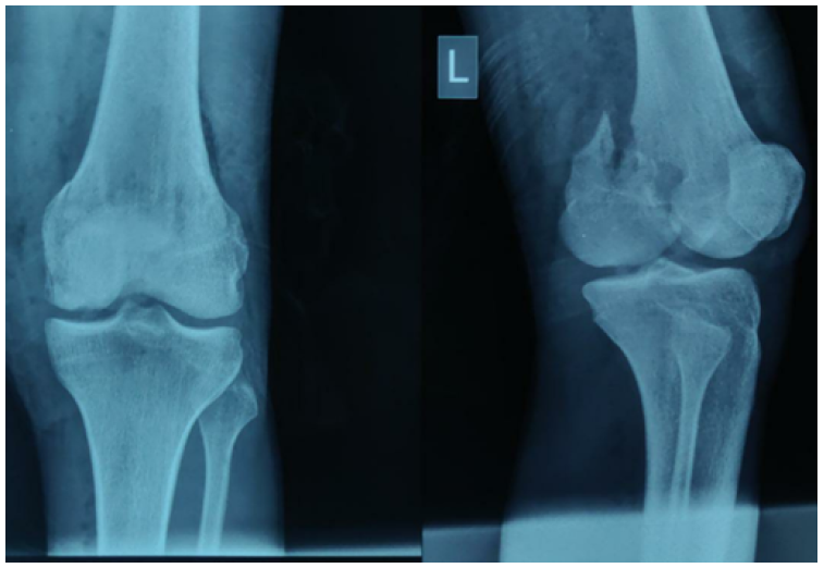
We studied 6 patients of medial Hoffa fracture presenting in Orthopedics department of Kalpana chawala government medical college, Karnal, Haryana. They were evaluated radiologically with X-rays of knee anteroposterior and lateral view (Figure- 3). Due to financial constraints of patients CT scan couldn’t be done. Of 6 patients 5 were male and one was female. The average age of patients was 29 years (range 27-31 years).
After PAC and obtaining clearance for surgery patients were taken up for surgery.
The operation was conducted under the guidance of professional surgeons. The patient was placed supine on a radiolucent table. Preoperative antibiotics and a general or spinal anaesthetic were administered. A tourniquet was placed around the ipsilateral thigh and inflated at surgeon discretion. The knee was initially placed in 20° of flexion over a towels and sheets.
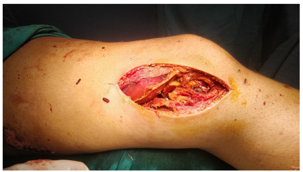
All the fractures were treated by open reduction through the medial subvastus approach is used through an extensile longitudinal anterior skin incision approximately 25 cm in length. (Figure 1).
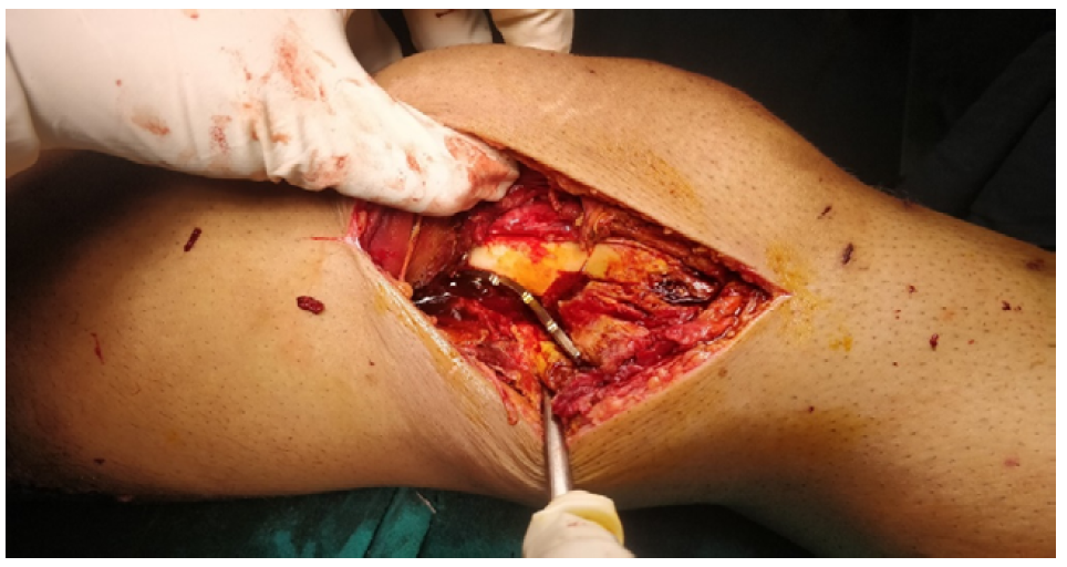
And after exposure done the vastus medialis muscle, sartorius muscle and saphenous nerve, which were retracted forward to the anterior to explore the gracilis, semimembranosus and semitendinosus muscles. After that, the semimembranosus and semitendinosus were retraced to the posterior and we could go through the interval space between the gracilis muscle and caput medial of the gastrocnemius to expose the fragment, and then the origin of the medial collateral ligament could be clearly exposed and protected. The intact fragment of Hoffa fracture was reduced and fixed with one or two cannulated or lag screws and meanwhile a recon plate was anatomically contoured and used (Figure 2) for fixing with angular stability and also preventing from the vertical gliding of the fragment (for buttressing) (Figure 4). Knee immobilizer was given to the patients post operatively.
The follow up of patients was done after 2 weeks, 1 month, 3 months and 6 months post operatively.
For 12 weeks after surgery, range-of-motion exercises were unlimited, and weight-bearing was restricted. All fractures healed, and reductions were maintained. One patient has superficial wound infection which was treated with intravenous antibiotics and antiseptic dressings.
On follow up we measured functional out come by checking for knee range of motion, knee society score [10] and bony union time.
Mean Range of motion of patients was 125.83 (range 120-130). Average knee society score was 84.16 (range 80-88) which comes under excellent category (80-100). All our patients had bony union at 6 months.
Table 1: Observation at 6 months of follow up
Sr. no | Range of motion | Knee society score | Bony union (confirmed radiologically) |
1 | 0-125 | 84 | Yes |
2 | 0-120 | 80 | Yes |
3 | 0-130 | 86 | Yes |
4 | 0-125 | 84 | Yes |
5 | 0-130 | 88 | Yes |
6 | 0-125 | 83 | Yes |

Figure 1: Intra Operative Picture Showing Subvastus Approach

Figure 2: Intra Operative Picture Showing Anatomical Reduction of Fracture and Buttressed with Recon Plate

Figure 3: Antero Posterior and Lateral Radiograph of Left knee Showing Isolated Medial Hoffas Fracture
An isolated coronal plane fracture of the posteromedial distal femoral condylar was originally described by Hoffa in 1904 [1].
It is assumed to occur secondary to a high-energy blunt force trauma that, applied to a flexed knee, causes shearing of the posterior condyle [11].
Associated injuries, which are common, occur in the ipsilateral knee, lower extremity and more distant locations [4].
Unicondylar Hoffa fracture has been reported in several studies [12,13]. However, few reports about medial Hoffa fracture were shown in the literature [14,15,2].
Hoffa fractures are usually caused by motor vehicular accidents. Until now, the injury mechanisms of this fracture pattern have not been clearly described. To detect the coronal fractures more accurately, CT scanning is necessary to these fractures that might be easily overlooked on plain radiographs. On the other hand, CT scanning or MRI examination is essential to find out a potential fracture and other accompanying structure injuries [8]. Ozturk [16] recorded a case of neglected medial Hoffa fracture that was initially missed because of seemingly normal anteroposterior (AP) radiographs of the knee.
The medial Hoffa fracture is a kind of intra-articular fracture, and most of these cases need operation [2,13]. Conservative management often leads to non-union or loss of knee function [16]. The surgical approaches are usually parapatellar approaches (anteromedial), or combined with posterior approaches, depending on the fracture classification and fixation methods.
Several approaches and fixation methods for distal femoral coronal fractures have been described, but most descriptions involve treatment of lateral Hoffa fragments. Previous descriptions of medial Hoffa fracture surgical fixation are limited. Attempts at fixation with a limited medial approach or a medial parapatellar approach with the knee in extension can be frustrating. The MCL lies over the typical coronal intra-articular fracture plane and limits exposure and reduction. Femoral distraction with the knee in extension is limited in increasing visualization. In a case report, Holmes and colleagues [12] described using an anterior midline incision with a medial parapatellar approach. Although the parapatellar approach allows access for reduction anteriorly, visualization of posterior comminution and ease of reduction may be limited. A medial subvastus approach allows extensile exposure of the articular surface and, if needed, access to the posterior femoral condyle— for placement of fixation in multiple planes. The subvastus approach has been popularized by Hofmann and colleagues [17].
As far as we know, there was no clinical comparison of different fixation methods on Hoffa fracture in the literatures. Most of Hoffa fractures were treated with cannulated screws or cancellous screws from anterior to posterior or the opposite direction, and the clinical outcomes were different [12,15,16,19]. It is generally accepted that the screw heads are recessed beneath the articular surface.
A biomechanical study showed that cannulated screws placed from posterior to anterior provided more stable fixation of Hoffa fractures in embalmed femurs than anteroposteriorly placed cannulated screws [20].
We concluded that outcome results of operated case of isolated medial Hoffa’s fracture using medial subvastus approach and fixation of fracture with 6.5 cancellous cannulated screws and buttress recon plate are excellent (as per knee society score). We obtained very good range of motion (average 125.83) at 6 months follow up. We obtained radiological bony union in 100% of our cases (Figure 5).
Although conclusions about the long-term outcomes of these injuries cannot be drawn, reduction and fixation of these high-energy intra-articular injuries using this surgical approach have been favourable and may be useful for the medial Hoffa fracture.
Karim, A., and N. Rossiter. “Isolated Medial Uni-Condylar Hoffa Fracture Following Traumatic Knee Dislocation.” Injury Extra, vol. 37, no. 1, 2006, pp. 12–14.
Dhillon, M.S. et al. “Coronal Fractures of the Medial Femoral Condyle: A Series of 6 Cases and Review of Literature.” Musculoskeletal Surgery, vol. 96, no. 1, 2012, pp. 49–54.
Ocguder, A. et al. “Hoffa Fracture, Eminentia Fracture and Posterior Cruciate Ligament Damage: An Unusual Knee Injury.” Injury Extra, vol. 39, no. 3, 2008, pp. 88–91.
Viskontas, D.G. et al. “Technique of Reduction and Fixation of Unicondylar Medial Hoffa Fracture.” American Journal of Orthopedics, vol. 39, no. 9, 2010, pp. 424–428.
Lewis, S.L. “Femoral Accident.” Surgery, 1989, pp. 118–120.
Marzouki, A. et al. “A Rare Case of Unicondylar Medial Hoffa Fracture Associated with Ipsilateral Vertical Patella Fracture.” Journal of Clinical Orthopaedics and Trauma, vol. 4, no. 2, 2013, pp. 102–105. https://doi.org/10.1016/j.jcot.2013.01.003
Skaggs, D.L., and J.M. Flynn. “Supracondylar Fractures of the Distal Humerus.” Rockwood and Wilkins’ Fractures in Children, 8th ed., 2014, pp. 2002–2004.
Fractures, C.P. “Distal Femoral Fractures and Coronal Plane Fractures.” 2005, pp. 564–569.
Gao, M. et al. “Surgical Treatment and Rehabilitation of Medial Hoffa Fracture Fixed by Locking Plate and Additional Screws: A Retrospective Cohort Study.” International Journal of Surgery, vol. 19, 2015, pp. 95–102. https://doi.org/10.1016/j.ijsu.2015.05.027
Liow, R.Y. et al. “The Reliability of the American Knee Society Score.” Acta Orthopaedica Scandinavica, vol. 71, no. 6, 2000, pp. 603–608.
HAL. Der Frakturen und L für Ä und SE. 1904.
Holmes, S.M. et al. “Coronal Fractures of the Femoral Condyle: A Brief Report of Five Cases.” Journal of Orthopaedic Trauma, vol. 18, no. 5, 2004, pp. 316–319.
Ostermann, P.A. et al. “Long Term Results of Unicondylar Fractures of the Femur.” Journal of Orthopaedic Trauma, vol. 8, no. 2, 1994, pp. 142–146.
Bali, K. et al. “Isolated Hoffa Fracture of the Medial Femoral Condyle in a Skeletally Immature Patient.” Bulletin of the NYU Hospital for Joint Diseases, vol. 69, no. 4, 2011, pp. 335–358.
Chang, J.J.H.T. et al. “Treatment of an Osteoporotic Hoffa Fracture.” Knee Surgery, Sports Traumatology, Arthroscopy, vol. 18, no. 6, 2010, pp. 784–786.
Oztürk, A. et al. “Nonunion of a Hoffa Fracture in an Adult.” La Chirurgia degli Organi di Movimento, vol. 93, no. 3, 2009, pp. 183–185.
Hofmann, A.A. et al. “Subvastus (Southern) Approach for Primary Total Knee Arthroplasty.” Clinical Orthopaedics and Related Research, no. 269, 1991, pp. 70–77.
Gavaskar, A.S. et al. “Operative Management of Hoffa Fractures—A Prospective Review of 18 Patients.” Injury, vol. 42, no. 12, 2011, pp. 1495–1498.
Jarit, G.J. et al. “A Mechanical Evaluation of Two Fixation Methods Using Cancellous Screws for Coronal Fractures of the Lateral Condyle of the Distal Femur (OTA Type 33B).” Journal of Orthopaedic Trauma, vol. 20, no. 4, 2006, pp. 273–276.