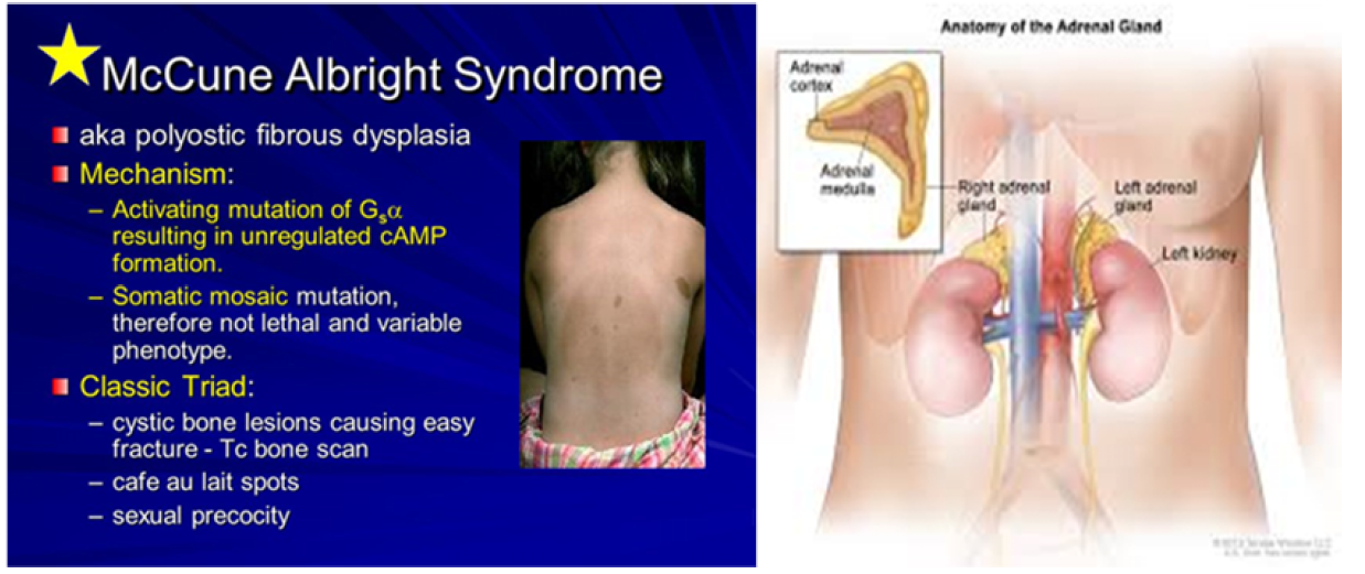
McCune-Albright syndrome (MAS) is classically defined by the clinical triad of fibrous dysplasia of bone (FD), café-au-lait skin spots, and precocious puberty (PP). It is a rare disease with estimated prevalence between 1/100,000 and 1/1,000,000. FD can involve a single or multiple skeletal sites and presents with a limp and/or pain, and, occasionally, a pathologic fracture. Scoliosis is common and may be progressive. In addition to PP (vaginal bleeding or spotting and development of breast tissue in girls, testicular and penile enlargement and precocious sexual behavior in boys), other hyperfunctioning endocrinopathies may be involved including hyperthyroidism, growth hormone excess, Cushing syndrome, and renal phosphate wasting. Café-au-lait spots usually appear in the neonatal period, but it is most often PP or FD that brings the child to medical attention. Renal involvement is seen in approximately 50% of the patients with MAS. The disease results from somatic mutations of the GNAS gene, specifically mutations in the cAMP regulating protein, Gs alpha. The extent of the disease is determined by the proliferation, migration and survival of the cell in which the mutation spontaneously occurs during embryonic development. Diagnosis of MAS is usually established on clinical grounds. Plain radiographs are often sufficient to make the diagnosis of FD and biopsy of FD lesions can confirm the diagnosis. The evaluation of patients with MAS should be guided by knowledge of the spectrum of tissues that may be involved, with specific testing for each. Genetic testing is possible, but is not routinely available. Genetic counseling, however, should be offered. Differential diagnoses include neurofibromatosis, osteofibrous dysplasia, non-ossifying fibromas, idiopathic central precocious puberty, and ovarian neoplasm. Treatment is dictated by the tissues affected, and the extent to which they are affected. Generally, some form of surgical intervention is recommended. Bisphosphonates are frequently used in the treatment of FD. Strengthening exercises are recommended to help maintaining the musculature around the FD bone and minimize the risk for fracture. Treatment of all endocrinopathies is required. Malignancies associated with MAS are distinctly rare occurrences. Malignant transformation of FD lesions occurs in probably less than 1% of the cases of MAS.
Keywords
McCune-Albright Syndrome (MAS)
CAMP
FD
INTRODUCTION
Originally, the McCune-Albright syndrome (MAS) was defined by the triad of polyostotic fibrous dysplasia of bone (FD), café-au-lait skin pigmentation, and precocious puberty (PP) [1,2]. It was later recognized that other endocrinopathies, including hyperthyroidism (reviewed in [3], growth hormone (GH) excess [4,5] renal phosphate wasting with or without rickets/osteomalacia [6] and Cushing syndrome could be found in association with the original triad [6,7,8]. Rarely, other organ systems may be involved (liver, cardiac, parathyroid, pancreas) [9]. While MAS is rare, FD is not. FD can involve a single skeletal site (monostotic FD, MFD), or multiple sites (polyostotic FD, PFD) [10,11]. Very rarely PP can be found in association with café-au-lait skin pigmentation in the absence of FD (about 1% of the cases), but in general, FD seems to be the most common component of MAS. Therefore, a more clinically relevant definition of MAS, broader than the original triad of FD + PP + café-au-lait is: MAS = FD + at least one of the typical hyperfunctioning endocrinopathies and/or café-au-lait spots, with almost any combination possible [4].
CLINICAL DESCRIPTION
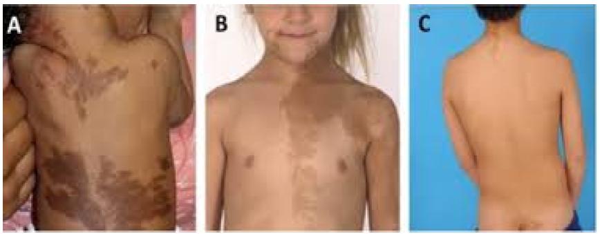
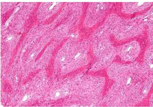
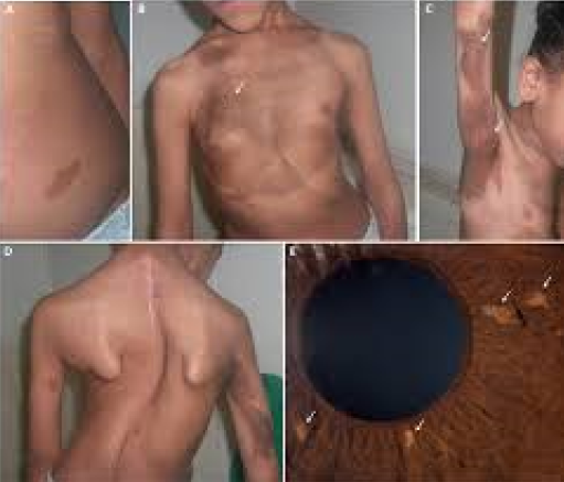
Typically, the signs and symptoms of either PP or FD usually account for the initial presentation. In girls with PP, it is usually vaginal bleeding or spotting, accompanied by development of breast tissue, usually without the development of pubic hair. In boys, it can be bilateral (or unilateral) testicular enlargement with penile enlargement, scrotal rugae, body odor, pubic and axillary hair, and precocious sexual behavior. In retrospect, café-au-lait spots (Figure 1), which are usually present at birth or shortly thereafter, are the most common but unappreciated "presenting" sign. Fibrous dysplasia in the appendicular skeleton usually presents with a limp and/or pain (sometimes reported by children as being "tired"), but occasionally a pathologic fracture may be the presenting sign. Radiographs will demonstrate typical expansile lesions with endosteal scalloping and thinning of the cortex with the matrix of the intramedullary tissue demonstrating a "ground glass" appearance (Figure 2 and 3). FD in the craniofacial bones usually presents as a painless "lump" or facial asymmetry. Representative radiographic findings and the histological appearance of FD are shown in Figures 2 and 3. The areas most commonly involved are the proximal femora and skull base. The sites of FD involvement are established early; 90% of the total body skeletal disease burden is usually established by age 15 [10]. Hart et al. found that lesions in the craniofacial region were established earliest, with 90% of the lesions present by 3.4 years of age. In the extremities, 90% were present by 13.7 yr, and in the axial skeleton, 90% were present by 15.5 yr. The appearance of new lesions later in life is a very uncommon occurrence in FD. The incidence of fractures is greatest in childhood, between the age of 6 and 10 yr, but due to the intrinsic abnormalities in FD bone, some fractures continue to occur into adulthood (Figure 4).
Etiology
The observation that the G protein/cAMP/adenylate cyclase signaling pathway was central to all of the tissues involved in MAS eventually led to the discovery that mutations in the regulatory Gsα protein (encoded by the GNAS gene) were the underlying molecular etiology of MAS (1) (Figure 5) In all published cases of MAS, PFD, and even MFD, activating mutations of Gsα at the R201 position have been identified [6]. More recently, mutations at the Q227 position have been found in association with FD [7]. The lack of vertical transmission of the disease, along with the observation that skin and bone lesions tend to respect the midline and be on one or the other side of the body, has led to the unproven, but accepted, concept that the disease is the result of postzygotic mutations, and that patients are therefore somatic mosaics. The point in time in development at which the mutation occurs, the specific cell in which it occurs, and to where its progeny migrate, determines what tissues will be affected, and thus the phenotype. Therefore, in cases in which tissues of endodermal, mesodermal, and ectodermal origin are involved, it would appear that the mutation occurred at the inner cell mass stage (Figure 5).
Figure 2: Clinical Features of Mccune-Albright Syndrome Data
DIAGNOSIS
Diagnosis of MAS is usually established on clinical grounds. Plain radiographs are often sufficient to make the diagnosis of FD (Figure12). Isotopic bone scans are the most sensitive tool for detecting the presence of FD lesions, and are often useful, especially at the initial evaluation, for determining the extent of the disease and predicting functional outcome (Figure 4) FD has a typical appearance on radiographs described as "ground glass." In general, lesions in the long bones have a "lytic" appearance. The lesions usually arise in the medullary cavity and expand outward replacing normal bone, which results in thinning of the cortex (Figure 5). It is usually the metaphysis and/or the diaphysis that are involved, with sparing of the epiphysis. It is possible for any bone to be involved, but the skull base and the proximal femur are the sites most commonly involved [4,5]. Due to the fact that these lesions are undermineralized [2], the bones are "soft" and prone to deformation, as exemplified by the classic "shepherd's crook" deformity of the proximal femur (Figure 2).
FD in the craniofacial bones tends to have a "sclerotic" appearance on plain radiographs. This is due to the relatively greater degree of mineralization of FD tissue in the craniofacial bones (Figure 4} Computed tomography (CT) scanning is the best technique for imaging FD lesions in the skull, revealing a "ground glass" appearance. In children and young adults, the lesions appear homogeneous on CT, but in older patients the appearance is mixed, with the development of "cystic" lesions in some areas. The density of these areas is that of soft tissue, so while they may have a cystic appearance they are not true cysts. That said, it is possible for true cysts to develop in FD, both in the long bones, but more often in the craniofacial bones (Figure 5). This has occurred in about 5% of the patients with FD in the NIH cohort (unpublished data). If needed, bone cysts may be diagnosed using magnetic resonance imaging (MRI). The cysts tend to have a more aggressive course. They can expand rapidly and produce symptoms which vary, depending on the location. One of the complications can be the fracture through a cyst. This usually requires surgical intervention. Biopsy of FD lesions can confirm the diagnosis if doubt remains after review of the radiographs. One characteristic of FD bone is the absence of the lamellation pattern seen in normal bone under polarized light. This indicates that the matrix produced in the lesions is of the woven type. The histopathological description of FD is often described as a "Chinese writing" pattern, and with special preparation and stains used to detect mineralized and unmineralized tissue, extensive areas of unmineralized osteoid are evident (Figure 3) For an extensive description of the histopathological changes that can be observed in FD, the reader is referred to Riminucci et al., and Corsi et al., [3,4].
Clinical Features
Fibrous Dysplasia: Clinical sequelae of FD in the appendicular skeleton arises due to FD’s tendency to fracture and deform under weight-bearing forces. Patients frequently present to care due to limp or pain [2]. The proximal femur is one of the most commonly involved sites and may develop a characteristic coxa vara (“shepherd’s crook”) deformity [7] (Figure 4). Craniofacial FD typically presents with a slow-growing, painless swelling, which may result in facial asymmetry. Mild, asymptomatic craniofacial FD is often found incidentally on imaging studies such as dental radiographs and posttraumatic computed tomography scans. In rare and severe cases, patients may experience pain, paresthesia, or functional deficits, such as malocclusion, hearing impairment, and/or visual disturbances [10] (Figure 2). Rarely, compression of the cerebellum and brainstem can develop in patients with skull base FD [11]. The natural history of FD includes typical age-related changes in disease progression and activity. In utero skeletal development appears to occur relatively normally, without obvious signs of FD at birth. FD lesions become apparent during early childhood and tend to progress in number and size until final skeletal burden is established by age 15 years [7]. In vitro studies suggest that lesions eventually “burn out” during adulthood as the population of mutated skeletal stem cells depletes. Age-related changes in FD are also observed radiographically. Overproduction of fibroblast growth factor 23 (FGF23) from mutation-bearing skeletal stem cells is a key feature of FD [11]. FGF23 is potent phosphate regulator, acting at the proximal renal tubule to decrease 1-α-hydroxylase activity and increase urinary phosphate excretion.
Skin
Café-au-lait macules are often the first clinically apparent manifestations of MAS, presenting at or shortly after birth [2]. However, their significance is often noted only in retrospect, after other symptoms have developed. Café-au-lait macules have a characteristic appearance that includes jagged, irregular borders (often described as resembling the “coast of Maine”) and location respecting the midline of the body [4].
Gonadal Involvement
Although MAS gonadal involvement occurs equally in girls and boys, overproduction of sex steroids is far more common in girls. In one large series of patients seen at the NIH, GNAS activation in ovarian tissue resulted in recurrent estrogen-producing cysts in approximately 85% of girls [4,5] (Figure 4). Patients develop acute onset of pubertal signs, including breast development and growth acceleration. Labs show elevated estradiol levels with suppressed gonadotropins, and ultrasonography typically shows uterine enlargement with single or multiple ovarian cysts. Cyst resolution is associated with an acute drop in estradiol, which triggers vaginal bleeding. Between episodes, girls are often clinically asymptomatic with undetectable estradiol levels and normal ultrasonographic findings, which may lead to delayed diagnoses.
Thyroid
Thyroid abnormalities have been reported in approximately ∼50% of patients with MAS, about half of whom develop frank hyperthyroidism [3,4] (Fig. 3). Clinical and ultrasonographic findings include diffuse enlargement, heterogeneity, and discrete cystic and solid nodules [6,7]. Biochemically, GNAS mutations result in constitutive 5′-deiodinase activity, resulting in increased conversion of T4 to T3 and a primary T3 toxicosis [11]. Hyperthyroidism typically develops during childhood and persists throughout adulthood.
Pituitary
GNAS activation in the pituitary results in somatolactroph cell hyperplasia, leading to constitutive growth hormone (GH) and prolactin production in approximately 15% of patients [3,4]. The most common clinical sign is expansion of craniofacial FD, which is particularly sensitive to the effects of GH [7,8] (Fig. 2). Symptoms may include progressive macrocephaly, vision loss, and hearing loss, all of which may signal the presence of GH secretion abnormalities, even in the absence of frankly elevated IGF-1 levels [8,9]. Other manifestations include acromegalic features and the development of secondary pituitary hormone insufficiencies [9]. While growth acceleration and tall stature may suggest the presence of GH excess, this is not a consistent finding in patients with MAS because linear growth is often confounded by skeletal deformities and other endocrinopathies. GH excess is diagnosed by IGF-1, oral glucose tolerance test, and/or overnight GH sampling; most patients also demonstrate concomitant mild elevations in prolactin.
Gastrointestinal
MAS may be associated with a broad spectrum of gastrointestinal disease. Neonatal cholestasis and hepatitis have been reported in infants [5], and a variety of hepatobiliary abnormalities have been reported in adults, including hepatocellular adenomas, choledochal cysts, and inflammatory adenomas [7,8]. Gastric polyps may be asymptomatic or associated with symptoms of reflux [7]. Activating GNAS mutations are known drivers for the development of intraductal papillary mucinous neoplasms (IPMNs) [2]. These pancreatic cysts have been reported in up to 40% of patients with MAS and have rarely been associated with obstructive pancreatitis and diabetes [5] Gastrointestinal evaluation, including pancreatic imaging, should therefore be considered in all symptomatic patients [2,3]. Although IPMNs are considered a potentially premalignant lesion in the general population, the risk of pancreatic adenocarcinoma in patients with MAS appears to be low, with only one reported case [4]. It is unknown if IPMNs associated with MAS are truly at lower risk compared to the general population, or if the malignant potential of IPMNs is lower than previously thought [6].
Bone Marrow and Hematologic
Bone marrow failure with pancytopenia and extramedullary hematopoiesis has been rarely reported in patients with severe FD. The etiology is not well-established but may be related to reduce marrow capacity in conjunction with splenic sequestration, and patients have clinically improved after splenectomy [6]. Platelet dysfunction has also been reported in association with MAS [7], which together with the hypervascularity of FD lesions may contribute to blood loss with orthopedic procedures.
Queries
While bisphosphonates are usually effective in relieving FD-related pain, whether or not the treatment of FD with bisphosphonates changes the natural history of the disease remains an open question. The most recent, and strongest data to date, suggests that they do not [5]. Ongoing placebo-controlled trials in the US and Europe, should help to resolve this open question. More effective treatment for FD is needed. The development of cell based and/or GSAdirected therapies may hold promise.
The best treatment for PP is also not clear. Letrozole, a potent third generation aromatase inhibitor, has recently been shown to be an effective therapy in some girls with MAS [6]. Studies suggest tamoxifen, the estrogen agonist/antagonist, may also be effective [8]. However, all trials for the treatment of PP have been uncontrolled, and this fact, combined with the extreme variability in the clinical course of the disease, makes conclusions about relative efficacy very difficult. A trial with the pure anti-estrogen, fulvestrant, in girls with MAS is underway. Controlled and head-to-head comparison trials are needed to establish the best treatment for PP in MAS.
Figure 4:Fibrous Dysplasia of Bone
Figure 5: C/F of Mccune-Albright Syndrome
Figure 6: Involvement of Bone As Clinical Features
REFERENCE
McCune, D.J. “Osteitis Fibrosa Cystica: The Case of a Nine Year Old Girl Who Also Exhibits Precocious Puberty, Multiple Pigmentation of the Skin and Hyperthyroidism.” American Journal of Diseases of Children, vol. 52, 1936, pp. 743–744.
Albright, F. et al. “Syndrome Characterized by Osteitis Fibrosa Disseminata, Areas of Pigmentation and Endocrine Dysfunction, with Precocious Puberty in Females: Report of Five Cases.” New England Journal of Medicine, vol. 216, no. 17, 1937, pp. 727–746.
Mastorakos, G. et al. “Hyperthyroidism in McCune-Albright Syndrome with a Review of Thyroid Abnormalities Sixty Years after the First Report.” Thyroid, vol. 7, no. 3, 1997, pp. 433–439.
Sherman, S.I., and P.W. Ladenson. “Octreotide Therapy of Growth Hormone Excess in the McCune-Albright Syndrome.” Journal of Endocrinological Investigation, vol. 15, no. 3, 1992, pp. 185–190.
Akintoye, S.O. et al. “Characterization of gsp-Mediated Growth Hormone Excess in the Context of McCune-Albright Syndrome.” The Journal of Clinical Endocrinology & Metabolism, vol. 87, no. 11, 2002, pp. 5104–5112. https://doi.org/10.1210/jc.2001-012022.
Danon, M., and J.D. Crawford. “The McCune-Albright Syndrome.” Ergebnisse der Inneren Medizin und Kinderheilkunde, vol. 55, 1987, pp. 81–115.
Diaz, A. et al. “McCune-Albright Syndrome and Disorders Due to Activating Mutations of GNAS1.” Journal of Pediatric Endocrinology and Metabolism, vol. 20, no. 8, 2007, pp. 853–880.
Kirk, J.M. et al. “Cushing’s Syndrome Caused by Nodular Adrenal Hyperplasia in Children with McCune-Albright Syndrome.” The Journal of Pediatrics, vol. 134, no. 6, 1999, pp. 789–792. https://doi.org/10.1016/S0022-3476(99)70302-1.
Shenker, A. et al. “Severe Endocrine and Nonendocrine Manifestations of the McCune-Albright Syndrome Associated with Activating Mutations of Stimulatory G Protein GS.” The Journal of Pediatrics, vol. 123, no. 4, 1993, pp. 509–518. https://doi.org/10.1016/S0022-3476(05)80943-6.
Liechtenstein, L., and H.L. Jaffee. “Fibrous Dysplasia of Bone: Conditions Affecting One, Several or Many Bones, Graver Cases of Which May Present Abnormal Pigmentation of Skin, Premature Sexual Development, Hyperthyroidism, and Still Other Extra-Skeletal Abnormalities.” Archives of Pathology, vol. 33, 1942, pp. 777–816.
Bianco, P. et al. “Fibrous Dysplasia.” Pediatric Bone: Biology and Disease, edited by F.H. Glorieux et al., Academic Press, Elsevier, 2003, pp. 509–539.
Collins, M.T. et al. “Renal Phosphate Wasting in Fibrous Dysplasia of Bone Is Part of a Generalized Renal Tubular Dysfunction Similar to That Seen in Tumor-Induced Osteomalacia.” Journal of Bone and Mineral Research, vol. 16, no. 5, 2001, pp. 806–813. https://doi.org/10.1359/jbmr.2001.16.5.806.
Recommended Articles
Research Article
A study on the feasibility of online classes for medical education
Seema Bose,
Atindra Krishna Ghosh
Published: 15/02/2021
Download PDF
Research Article
Amyand’s Hernia: A Rare form of Common Entity
Download PDF
Research Article
Prevalence of Typhoid Fever among Patients Attending General Sani Abatcha Specialist Hospital Damaturu, Nigeria
Ahmed Idriss Jajere,
...
Usman Umar Faruk
Published: 10/10/2020
Download PDF
Research Article
A case report of management of a Case of Hyperalgesia by Ultrasound guided Fluoroscopically Verified Transforaminal Nerve Root Block
Sunil Dutt,
...
Anil Kumar
Published: 29/06/2024
Download PDF
PO Box 101, Nakuru
Kenya.
Email: office@iarconsortium.org
Editorial Office:
J.L Bhavan, Near Radison Blu Hotel,
Jalukbari, Guwahati-India



 Download PDF
 Download XML