Contents
Download PDF
pdf Download XML
419 Views
282 Downloads
Share this article
Case Series | Volume 1 Issue 1 (Jul-Dec, 2020) | Pages 1 - 4
Herniorrhaphy mesh in Orbital Implants of Enucleated eyes for post-operative functionality – A Case Series
 ,
 ,
Under a Creative Commons license
Open Access
Received
Oct. 2, 2020
Revised
Oct. 23, 2020
Accepted
Nov. 19, 2020
Published
Dec. 30, 2020
Abstract

Imparting postoperative functionality and cosmesis is a major challenge in enucleated eyes. Orbital implants are used to replace the volume lost by enucleated eye and maintain cosmetic symmetry with the fellow eye.A variety ofnon-integrated, synthetic semi-integrated, integrated, Bio-integrated & Biogenic varieties have been tried employing different materials. The favoured hydroxyapatite (bio-integrated) implant, due to its rough surface needs to be wrapped in donor sclera or other wrapping materials (like polyglactin-910 mesh, polytetrafluoroethylene sheet, etc.) to allow muscle attachment and integration. Monofilament polypropylene mesh used in herniorrhaphy is a bio-integrable material with little rejection hitherto reported. A series of 7 eyes of 7 patients were implanted with hydroxyapatite balls of sizes ranging from 12 to 16 mm. Each of such implants were given polypropylene mesh wrappings using 7- 0 prolene sutures. 6 of such implants were performed in the primary setting while 1 implant was undertaken as a secondary surgery, 8 weeks following the primary procedure of globe excision. 5 of these surgeries were done under local anaesthesia of a peribulbar block while 2 of these surgeries were performed under general anaesthesia. Post operative evaluation was done after 1 day, 1 week, 1 month, 3 months and 6 months in each of these cases. 1 case was lost to follow-up after 3 months. Surgical evaluation was rated on basis of intra operative ease of implantation and muscle attachment, early and late post operative complications, implant mobility and cosmesis achieved. Early complications included peri-orbital surgical edema, discharge, watering and foreign body sensation persisting upto 2 weeks post-operatively. Implant motility as judged 6 months post operatively was satisfactory in all cases. Sulcus deformity was noted in 1 case. No case presented with implant migration or exposure.

Keywords
INTRODUCTION

Following enucleation or evisceration, there is a reduction in the volume of the orbital contents, which needs to be replaced by an orbital implant. A prosthetic eye without an implant cause stretching of the lower lid under its weight and has poor motility. Thus, an orbital implant is inserted for achieving satisfactory prosthetic motility and better cosmetic results. The implant can be inserted at the time of surgery (enucleation or evisceration) or later.               


On the basis of integrability of implant material with orbital tissue, implants are classified as Non-integrated, semi-integrated, Integrated, Bio-integrated & Biogenic [1]. Since their introduction in 1989, the Integrated Hydroxyapatite implant has gained popularity due to its porous structure allowing for fibrovascular ingrowth and better cosmetic outcomes [2,3]. The need for a smooth wrapping allowing for extra-ocular muscle attachments adds to the cost and constriction of resource in an emergent setting. The most preferred wrapping materials like Donor Sclera, Processed pericardium or harvested facia lata are either not readily available or their procurement cumbersome. The semi-synthetic material of Polyglactin used in Herniorrhaphy mesh is readily available and is a novel method to wrap implants allowing for easier attachment of extra-ocular muscles [4]. Even though its smoothness is inferior to other wrapping materials, Polypropylene mesh serves the purpose of volume augmentation and reduces the risk of implant exposure with easier implant insertion. It is also suitable in a multi-speciality surgical setting with facio-maxillary and Neurosurgical teams.


Experimental Section

A series of 7 patients that underwent globe excision for purposes ranging from Ruptured globe in Road traffic Accidents, Painful Blind eye and Choroidal Melanoma, were implanted with Hydroxyapatite implants using the Herniorrhaphy mesh of monofilament Polyglactin. The patient with Choroidal Melanoma underwent the implant after 3 cycles of chemotherapy as a secondary procedure following initial Globe Excision. One case with Ruptured globe along with multiple facial injuries that included fracture of Zygomatic arch, lateral wall of orbit & Lamina papryciaunderwent a combined procedure of globe excision with ORIF [5].


The sizing of implant was done prior to the procedure using the Axial length of fellow eye as a guide. Implant sizes were kept 2 mm less to allow for the dimensions of the wrapping. Orbital implant was wrapped in the monofilament polyglactin mesh used in herniorrhaohy and sutured with 50 prolene suture. The wrapped implant was placed in normal saline for 5 minutes to loosen the stiffened fibres of the mesh. Meticulous muscle attachment with wrapping using 6–0 prolene suture and conjunctival wound closure with 6 0 Vicryl in a horizontal mattress fashion were done. Prosthesis was fitted following resolution of orbital edema.


Post operative results were evaluated for early complications like wound edema, chemosis, discharge and watering from eye at intervals of 1 day, 1 week and 1 month. Late complications and Implant status were evaluated for sulcus deformity, implant mobility and migration or wound gape with extrusion of implant.

RESULTS

Of the 7 patients implanted, all had lid oedema, watering and foreign body sensation lasting for a maximum of 2 weeks and responding to the conventional therapy of broad spectrum systemic and topical antibiotics along with anti-inflammatory drugs. Significant mucopurulent discharge was noted in 3 of the patients that required fortified preparations of topical antibiotics (Gatifloxacin 0.5% and Tobramycin 1%). Ocular motility gained subsequent to the resolution of chemosis and edema.


Figure 1: (A and B) Presentation of the Patient (C)  NCCT Orbit Showing Multiple Fractures


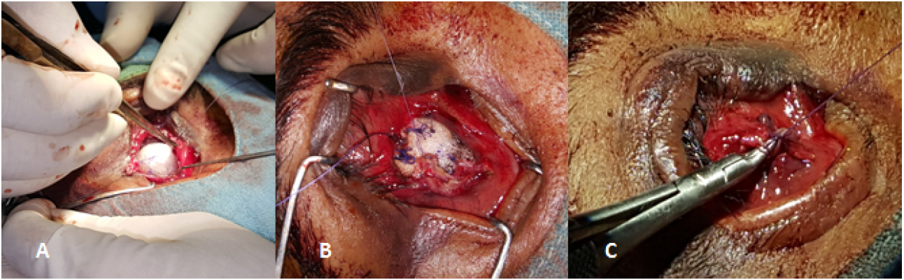
Figure 2: Wrapping of the Implant with the Prolene Mesh


Figure 3: A Placing the Implant in the Socket B Suturing the Extraocular Muscles to the Mesh C Closing the Socket


Figure 4: A Post-Op Day 1 B Post - Op Day 7 C After 6 Months 


Good cosmesis and globe functionality was achieved 3 months following the surgery and was maintained at the 6 month follow up visit. One patient did not report for the 6 monthly follow up. A superior implant migration was noted in the patient with Choroidal melanoma.

DISCUSSION

Globe rupture due to road traffic accidents is a common accompaniment of facio-maxillary surgeries. It entails surgical excision of globe that may leave a cosmetically compromised face. Other settings requiring Enucleation include Ocular malignancies and painful blind eye where a cosmetically unacceptable eye require orbital volume replacement employing orbital implant. This is particularly essential for eyes in pediatric age group since an empty socket can lead to facial disfigurement and asymmetry.


A variety of materials have been used for socket reconstruction. The previous approach to use inert materials like Poly methyl methacrylate (PMMA) and silicone were inexpensive, well tolerated but were non-integrated in the adjoining tissues [1,2]. Such implants met with the fate of high rate of implant extrusion, migration and inability to allow for muscle attachments2. This led to poor cosmetic outcomes and unpredictable stability of implant.


Semi-integrated implants like the Allen implant had an indirect, mechanical integration with orbital structures but not with the prosthesis.


The porous coralline hydroxyapatite implant had the property of good integration with the orbital tissues, being a component of human bone. Its porous system provided for a framework for fibrovascular ingrowth. This allowed for a firm stability of implant giving long term cosmetically acceptable outcomes. However, its rough surface is a hindrance to smooth implantation and muscle attachment. Different wrapping materials have been used for such implants like donor sclera, facia lata or processed pericardium. Their procurement however is difficult, and often time consuming.


Monofilament polyglactin mesh used in herniorrhapy is a readily available material and allows for easy attachment of extra ocular muscles. It facilitates smooth placement of implant and its inert properties prevent extrusion or post-operative infections and easier incorporation into the adjoining orbital tissues. Implant exposure seems to be a major complication with hydroxyapatite implant (1-15%) [6]. The results vary vastly due to variations in surgical procedure. Proper implant size and meticulous wound closure minimize the risk of implant exposure [7].


Polyglactin mesh wrapping also facilitates pegging of implant at a later date to allow for prosthetic placement.A case series of 200 recontructive surgeries has been reported by Jordan,et al using the wrapping material of a vicryl mesh in porous orbital implants [8,9]. The series showed few exposures and suggested such mesh as a viable material for implant wrappings. The material used is similar to the monofilament polyglactin mesh used in our series of cases and substantiates its utility in orbital reconstructive surgeries. 

CONCLUSION

In surgeries involving orbital implants, a monofilament polypropylene mesh can be used safely as an alternative to other synthetic orbital implant wrapping material. In addition to the advantage of using a biointegrated implant, the newly tried wrapping material, economizes the implant procedure.Present work had tried a monofilament polypropylene surgical mesh as an alternative implant wrapping material, which is used in herniorrhaphy procedures, with satisfactory post-operative results. Key advantages were easy availability of the mesh and minimizing the cost of the implant.

REFERENCE
  1. Acker, E., and P. De Potter. “[Porous Polyethylene (Medpor) Orbital Implant: Prospective Study of 75 Primary Implantations].” Journal Français d’Ophtalmologie, vol. 24, 2001, pp. 1067–1073.

  2. Trichopoulos, N., and J.J. Augsburger. “Enucleation with Unwrapped Porous and Nonporous Orbital Implants: A 15-Year Experience.” Ophthalmic Plastic and Reconstructive Surgery, vol. 21, 2005, pp. 331–336.

  3. Ashworth, J. et al. “A Study of the Hydroxyapatite Orbital Implant Drilling Procedure.” Eye, vol. 12, 1998, pp. 37–42.

  4. Klapper, S.R. et al. “Hydroxyapatite Orbital Implant Vascularization Assessed by Magnetic Resonance Imaging.” Ophthalmic Plastic and Reconstructive Surgery, vol. 19, 2003, pp. 46–52.

  5. Jordan, D.R. et al. “Complications Associated with Pegging Hydroxyapatite Orbital Implants.” Ophthalmology, vol. 106, 1999, pp. 505–512.

  6. Lin, C.J. et al. “Complications of Motility Peg Placement for Porous Hydroxyapatite Orbital Implants.” British Journal of Ophthalmology, vol. 86, 2002, pp. 394–396.

  7. Kaltreider, S.A. “The Ideal Ocular Prosthesis: Analysis of Prosthetic Volume.” Ophthalmic Plastic and Reconstructive Surgery, vol. 16, 2000, pp. 388–392.

  8. Arat, Y.O. et al. “Bovine Pericardium versus Homologous Sclera as a Wrapping for Hydroxyapatite Orbital Implants.” Ophthalmic Plastic and Reconstructive Surgery, vol. 19, 2003, pp. 189–193.

  9. Jordan, D.R. et al. “The Use of Vicryl Mesh in 200 Porous Orbital Implants: A Technique with Few Exposures.” Ophthalmic Plastic and Reconstructive Surgery, vol. 19, 2003, pp. 53–61.

Recommended Articles
Research Article
A study on the feasibility of online classes for medical education
Published: 15/02/2021
Download PDF
Research Article
Amyand’s Hernia: A Rare form of Common Entity
Download PDF
Research Article
Prevalence of Typhoid Fever among Patients Attending General Sani Abatcha Specialist Hospital Damaturu, Nigeria
...
Published: 10/10/2020
Download PDF
Research Article
A case report of management of a Case of Hyperalgesia by Ultrasound guided Fluoroscopically Verified Transforaminal Nerve Root Block
...
Published: 29/06/2024
Download PDF
Chat on WhatsApp
Flowbite Logo
PO Box 101, Nakuru
Kenya.
Email: office@iarconsortium.org

Editorial Office:
J.L Bhavan, Near Radison Blu Hotel,
Jalukbari, Guwahati-India
Useful Links
Order Hard Copy
Privacy policy
Terms and Conditions
Refund Policy
Shipping Policy
Others
About Us
Team Members
Contact Us
Online Payments
Join as Editor
Join as Reviewer
Subscribe to our Newsletter
+91 60029-93949
Follow us
MOST SEARCHED KEYWORDS
Copyright © iARCON International LLP . All Rights Reserved.