Brain abscess constitutes a rare medical condition in the newborn period. This condition if present in the majority of cases is a complication of meningitis with high morbidity and mortality at short and long term. Many unusual bacterial pathogens were implicated in this condition like Proteus, Klebsiella, Citrobacter, Staphylococcus Aureus, MRSA, StaphylococcusEpidermidis and Enterococcus (1, 2, 3). In this article, we report a case of a 28 days old late preterm baby presented with a history of poor feeding and low grade fever, who was found to have StaphylococcusEpidermidis meningitis without any focal neurologic signs with a huge brain abscess of 10 cm without neither midline shift nor hydrocephalus. The baby received a full course of antibiotherapy for total 4 weeks post drainage of abscess and was discharged home after documentation of radiological clearance in a repeated MRI without any neurologic sequalae.
Keywords
Brain abscess
Neonates
Coagulase negative Staphylococcus.
INTRODUCTION
During the neonatal period, meningitis mainly that of bacterial origin is still considered one of the most devastating conditions, with a high morbidity rate ranging from 20% to 60% [1]. Brain abscess is a rare and uncommon complication of neonatal meningitis that occurs in 1%–4% of all cases [2]. The first report of brain abscess in a neonate was published 100 years ago in the literature [1]. Brain abscess is an uncommon intracranial suppuration that usually occurs as a complication of bacterial meningitis or septicemia. Many pathogens have been implicated in the pathogenesis of brain abscess, ranging from the most common ones like Citrobacter species., Pseudomonas Aeruginosa, Klebsiella Pneumonia and Enterobacteria such as Proteus Mirabilis and Serratia Marcescens; to the less common like Staphylococcus Aureus and Staphylococcus Epidermidis which has been described as the leading species of coagulase-negative Staphylococci (CONS), and the predominant pathogen of sepsis in preterm infants [3,2,4]. We report in this article a case of a huge brain abscess status post drainage and full course of 4 weeks antibiotherapy due to coagulase negative staphylococcus meningitis with absent neurological deficit in a 28 days ex-late preterm baby who presented with poor feeding and low-grade fever.
Case Description
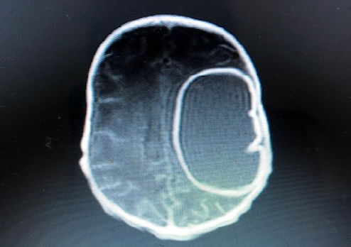
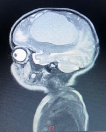
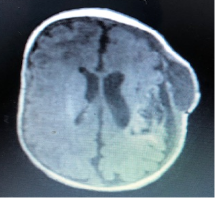
Our case is a 28 days old baby girl second member of twins, product of IVF and 34 weeks prematurity, born by cesarean section for scar pain to 38 years old mother G1P2A0 of blood group B positive, B positive father, no consanguinity between parents. The mother TORCH status was negative, GBS negative, history of recurrent UTIs at the last trimester treated at home with Cefixime. At birth the baby was 1920 grams, 50 cm length and 35cm head circumference with an Apgar score of 8/1min, 9/5mins, and no neonatal resuscitation was done. Immediately after birth the evolution was marked by progressive respiratory distress beginning at 30 minutes post-delivery (subcostal retractions with grunting and nasal flaring), the baby was admitted to the ICN for respiratory distress, got intubated for a total of 3 days and received a total course of 7 days triple antibiotherapy for early onset neonatal sepsis via a central access (umbilical venous line), then she was discharged home after documentation of negative blood culture with weight of 1900 grams. Newborn was referred to our hospital for history of poor feeding and low-grade fever at day 28 of life, no lethargy or irritability, no abnormal movements or seizure. On physical examination, the baby was tonic, active, had normal reflexes, good sucking reflex, normotensive non bulging open anterior fontanel, no signs of respiratory distress, she had good oxygen saturation on room air, regular S1 and S2, no murmur, positive femoral pulses, and good peripheral perfusion, mildly dehydrated (delayed skin turgor). Weight 1900g, HC 36cm, length 50cm. Full sepsis work-up done showed WBC 13000, Neutrophils 41%, Lymphocytes 36%, with high CRP 42, CSF analysis showed WBC 308 (neutrophils 70%, lymphocytes 30%), RBC 2, glucose 54mg/dl, Protein 0.8g/L. PCR viral panel meningitis (Neuro9) were also ordered with urine analysis, culture and blood culture. The baby started on Ampicillin, Gentamycin and Cefotaxime as meningeal doses with IV hydration. 72 hours later, the CSF culture showed coagulase negative Staphylococcus sensitive to Clindamycin, Vancomycin, and Erythromycin and resistant to Benzyl penicillin, so the baby was shifted to Meropenem and Vancomycin, also meningeal doses, and MRI brain was requested. MRI brain showed cyst-like area (6.4 cm x 4cm x 3,6cm) with inflamed /infected thick contour and thick fluid content just above the left Sylvian fissure involving the left fronto-praietal region with peripheral contrast enhancement without neither midline shift nor hydrocephalus Figure 1 and Figure 2.
Figure1: MRI Brain Showing Cyst-Like Area with Inflamed /Infected Thick Contour and Thick Fluid Content with Peripheral Contrast Enhancement without Neither Midline Shift nor Hydrocephalus
Figure2: MRI Brain Showing Cyst-Like Area Just Above the Left Sylvian Fissure Involving the Left Fronto-Praietal Region with Peripheral Contrast Enhancement
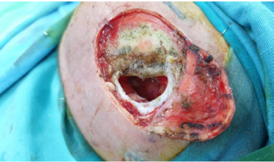
Neurosurgeon was consulted and the decision was to prepare the patient for craniotomy for drainage of abscess figure3.
Cardiac echocardiography showed normal findings. Pre-operative laboratory data showed: CRP 13; WBC 8000 with Neutrophils 25% and Lymphocytes 45%; Hbg 8, 3; thus 2 units of PRBCs were prepared. PCR viral meningitis panel revealed negative, blood culture and urine culture negative. Four weeks of Meropenem and Vancomycin post-op were considered according to the opinion of neurosurgeon and infectious diseases specialist.
Figure 3: Craniotomy for Drainage of Abscess
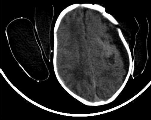
Pus drain specimen showed many neutrophils with no growth after 48 hrs, sections from soft gray tissue showed benign brain glial tissue with focal significant acute inflammation, no granuloma nor malignancy. Repeated CT scan of brain 1-week post-op showed significantly decreased collection/pus with few residual pockets, a left subdural collection with max thickness of 8 mm causing mild compressive effect with absence of neither midline shift nor hydrocephalus Figure 4.
Figure 4: CT Scan of Brain One Week Post Craniotomy Showed Residual Pockets, Left Subdural Collection of 8mm Thickness with Compressive Effect and Absence of Midline Shift
CT scan of brain was followed by MRI brain 2 weeks later which showed a subcutaneous fluid collection (4cm*4cm*1.7cm) overlying the left craniotomy site with enhancing rim Figure 5.
Antibiotherapy were completed for more 2 weeks to achieve a total of 5 weeks post-operatively mainly in correlation with the subcutaneous collection which was decreased remarkably on physical examination of the skull. During her hospital stay, the baby retained a good PO intake, weight gain and a normal level of activity and tonicity, she had normal primitive reflexes, no focal deficit and follow-up labs showed negative CRP < 0.3 and WBC 9000, Neutrophils 40%, Hgb 12. The baby was discharged home with complete normal neurological exam and presented for follow-up at outside clinic.
Figure 5: MRI Brain 2 Weeks Post Craniotomy Showed Left Subcutaneous Fluid Collection with Enhancing Rim
DISCUSSION
Despite the advances in the neuroimaging, neurosurgical and microbiological techniques, brain abscesses remains uncommon in infants and neonates with a prevalence ranging from 1.3 to 4% and with a significant rate of mortality and morbidity [3,5]. Brain abscess in newborns constitutes an uncommon central nervous disease. It is described as a focal, intracerebral infection that begins by a localized area of cerebritis and develops into a collection of pus surrounded by a well-vascularized capsule [6]. Neonatal cerebral abscesses usually occur as a complication of bacterial meningitis or septicemia [5]. Rarely they can be a complication of head trauma, congenital heart disease, or shunt surgery. Multiple abscesses are more frequently reported in immunocompromised patients [3]. Despite the initiation of intrapartum prophylaxis against GBS, it remains the most common cause of both meningitis and neonatal sepsis, with a high incidence of more than 40%, followed by E. coli especially in very low birth weight infants [7]. For the late onset one, coagulase-negative Staphylococci and Staphylococcus Aureus followed by E. coli and Klebsiella, Listeria, Pseudomonas Aeruginosa and methicillin-resistant Staphylococcus Aureus [7,8] are more common. The neonatal brain abscess is mainly caused by gram-negative micro-organisms commonly Citrobacter Diversus, Proteus and Pseudomonas species which are characterized by their ability to invade the nervous tissue and cause necrotizing vasculitis. Klebsiella Pneumonia was isolated in a reported cases of brain abscess in the literature and was been described as the most common organism causing neonatal sepsis in developing countries [8,1,2]. Staphylococcus Aureus and MRSA are described in the literature as rare causes of brain abscesses during the first months of life [3,9]. The blood stream infection with invasion of the brain parenchyma constitutes the most common mechanism of formation of brain abscess followed by a direct invasion of CSF through a right to left shunt especially in premature newborns [9,1,7]. An intrauterine infection was reported in a case of an E. Coli large brain abscess in 7 days old boy in the literature [6]. The clinical presentation was nonspecific and widely variable ranging from fever, poor feeding to irritability, variable ranging from fever, poor feeding to irritability, vomiting, focal/multifocal seizures and hydrocephalus [8,9,2]. Our case constitutes an unusual presentation of a large brain abscess measuring 10 cm approximately in a neonate with a rare and uncommon organism (coagulase negative Staphylococcus) treated with craniotomy for drainage associated with 5 weeks antibiotherapy post craniotomy without any neurologic sequalae.
CONCLUSION
More attention must be paid to the silent sometimes uncharacteristic, silent pathway to a potentially devastating outcome in the late preterm and preterm babies which constitutes a vulnerable population. The development of a brain abscess in a well appearing premature newborn may be only detected on a routine ultrasound of brain or may be a result of colonization by an infrequent germ of the CSF or the bloodstream.
REFERENCE
Masand, R. et al. “Neonatal Brain Abscess: An Atypical Presentation.” Journal of Pediatric Neurosciences, vol. 10, no. 3, 2015, p. 282.
Singh, A. et al. “Neonatal Brain Abscess: Clinical Report and Review of Indian Cases.” Journal of Clinical Neonatology, vol. 5, no. 3, 2016, p. 213.
Suárez, M. et al. “Combination of Drainage and Chemotherapy for Treatment of a Staphylococcus aureus Brain Abscess in a Pre-Term Infant.” JMM Case Reports, vol. 2, no. 5, 2015, e000100.
Dong, Y. et al. “Beyond Sepsis: Staphylococcus epidermidis Is an Underestimated but Significant Contributor to Neonatal Morbidity.” Virulence, vol. 9, no. 1, 2018, pp. 621–633.
Anca, I.A. et al. “Transfontanelar Ultrasound Diagnosis of Brain Abscesses in Two Neonates.” Medical Ultrasonography, vol. 11, no. 4, 2009, pp. 77–82.
Bundy, L.M., and A. Noor. “Neonatal Meningitis.” StatPearls, StatPearls Publishing, 2019.
Senapati, S. et al. “Large Brain Abscess in a Newborn.” Journal of Pediatric Neurosciences, vol. 10, 2015, p. 193.
Qureshi, U.A. et al. “Klebsiella Brain Abscess in a Neonate.” Archives of Disease in Childhood: Fetal and Neonatal Edition, vol. 96, no. 1, 2011, pp. F19–F19.
Russo, A. et al. “Methicillin-Resistant Staphylococcus aureus (MRSA) Brain Abscess in a Well-Appearing Premature Infant.” Journal of Pediatric Infectious Diseases, vol. 8, no. 1, 2013, pp. 39–42
Recommended Articles
Research Article
A study on the feasibility of online classes for medical education
Seema Bose,
Atindra Krishna Ghosh
Published: 15/02/2021
Download PDF
Research Article
Amyand’s Hernia: A Rare form of Common Entity
Download PDF
Research Article
Prevalence of Typhoid Fever among Patients Attending General Sani Abatcha Specialist Hospital Damaturu, Nigeria
Ahmed Idriss Jajere,
...
Usman Umar Faruk
Published: 10/10/2020
Download PDF
Research Article
A case report of management of a Case of Hyperalgesia by Ultrasound guided Fluoroscopically Verified Transforaminal Nerve Root Block
Sunil Dutt,
...
Anil Kumar
Published: 29/06/2024
Download PDF
PO Box 101, Nakuru
Kenya.
Email: office@iarconsortium.org
Editorial Office:
J.L Bhavan, Near Radison Blu Hotel,
Jalukbari, Guwahati-India
Chokr, I., Hammoud, C., Sabeh, L. C. K. E., Skafi, O., Chalfoun, .. A., Hammoud, M., Merhi, B. A. & Mahfouz, R. (2020). An Atypical Presentation of a Huge Brain Abscess without Neurologic Sequalae in a 28 Days Old Infant. IAR Journal of Medical Case Reports, 1(1), 1-4.
MLA
Chokr, Imad, et al. "An Atypical Presentation of a Huge Brain Abscess without Neurologic Sequalae in a 28 Days Old Infant." IAR Journal of Medical Case Reports 1.1 (2020): 1-4.
Chicago
Chokr, Imad, Chawki Hammoud, Lea C. K. E. Sabeh, Oussama Skafi, . A. Chalfoun, Maarouf Hammoud, Bassem A. Merhi and Rayane Mahfouz. "An Atypical Presentation of a Huge Brain Abscess without Neurologic Sequalae in a 28 Days Old Infant." IAR Journal of Medical Case Reports 1, no. 1 (2020): 1-4.
Harvard
Chokr, I., Hammoud, C., Sabeh, L. C. K. E., Skafi, O., Chalfoun, .. A., Hammoud, M., Merhi, B. A. and Mahfouz, R. (2020) 'An Atypical Presentation of a Huge Brain Abscess without Neurologic Sequalae in a 28 Days Old Infant' IAR Journal of Medical Case Reports 1(1), pp. 1-4.
Vancouver
Chokr I, Hammoud C, Sabeh LCKE, Skafi O, Chalfoun .A, Hammoud M, Merhi BA, Mahfouz R. An Atypical Presentation of a Huge Brain Abscess without Neurologic Sequalae in a 28 Days Old Infant. IAR Journal of Medical Case Reports. 2020 Jul;1(1):1-4.



 Download PDF
 Download XML