Contents
Download PDF
pdf Download XML
898 Views
514 Downloads
Share this article
Research Article | Volume 3 Issue 1 (Jan-June, 2022) | Pages 1 - 3
Limberg Flap for Pilonidal Sinus: Feasibility and Outcomes at Secondary Health Care Level
 ,
1
Department of General Surgery, Regional Hospital Una, India
2
Department of General Surgery, Dr R.K.G.Medical college Hamirpur, India
Under a Creative Commons license
Open Access
Received
Nov. 3, 2021
Revised
Dec. 9, 2021
Accepted
Jan. 3, 2022
Published
Jan. 10, 2022
Abstract

Pilonidal sinus is a common disease in adults of 15-40 years of age hirsute males, jeep and truck drivers. Limberg flap has been recommended as the first line treatment for primary and recurrent disease. There is reluctance of general surgeons to perform the procedure at secondary health care level. This study was undertaken to check feasibility of flap surgery and compare outcomes at secondary health care level. In our study Limberg flap was done for all patients (total 10) of Pilonidal Sinus from December 2020 to November 2021.One case was associated with flap edema and two cases with persistent discharge from tip which subsided by 3 weeks. All patients were successfully treated and returned to work in 4 weeks. There was no recurrence. Limberg flap for Pilonidal sinus is a simple and easy to perform transposition flap even at district level hospital with a very low complication and recurrence rate.

Keywords
INTRODUCTION

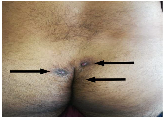
Pilonidal sinus is a common disease in adult age group of 15-40 years especially male population. The name pilonidal is derived from Latin meaning ‘nest of hairs’. It was termed jeep driver’s bottom during Second World War because many drivers were found to have this condition [1]. The incidence of Pilonidal sinus is around 26 per 100, 000 [2-3]. It received presents as a cyst, abscess or sinus tract with or without discharge [4] (Figure 1).


The condition was first described by Mayo in 1833. He suggested a congenital origin secondary to remnant of an epithelial lined tract from post coccygeal epidermal cell nests. Now the much-accepted theory suggests that congenital tracks do not contain hair and are lined by cuboidal epithelium. Karydakis pointed out three main factors for the pilonidal sinus i.e. high quantity of hair, extreme force and vulnerability to infection [5].


The treatment options range from clipping of hair, good hygiene, wide excision of area and new flap procedure [6]. Other procedures like excision and packing, excision and primary closure, marsupialization have also been suggested [7].


Regardless of procedure done recurrence rate ranged from 20%-40% [8]. Professor Limberg of Leningrad devoted his entire career to flap design. A rhomboid flap for pilonidal sinus was described by him in 1946. This is basically a technique for closing rhomboid shape defect with a transposition flap [9]. 


The flap places the suture line away from midline, giving rise to a tensionless flap of unscarred skin in middle. Thus helping in good hygiene, decreased sweat maceration, erosion and scar formation. The literature review showed that Limberg Flap was superior to primary closure and other flap procedure [10-11]. 


Figure 1: Complex Pilonidal Sinus

MATERIALS AND METHODS

The study involves 10 patients from December 2020 to November 2021. 8 were males, 2 were females. Median age was around 29 years, oldest was 41 years and youngest was 17 years.


Procedure 

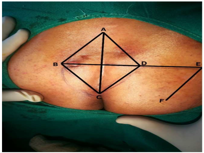
The patient was put in prone jack knife position under spinal anesthesia. The long axis of rhomboid is placed on midline marked as A-C, C being adjacent to perianal skin. Point A is placed such that all diseased tissues are included in the excision.The line B-D transects line A-C in midline and equals to 60% of length. Line D-E is equal to A-B. E-F is equal and parallel to D-C [12]. After rotation A-B is sutured to D-E and E-F is sutured A-D (Figure 2).


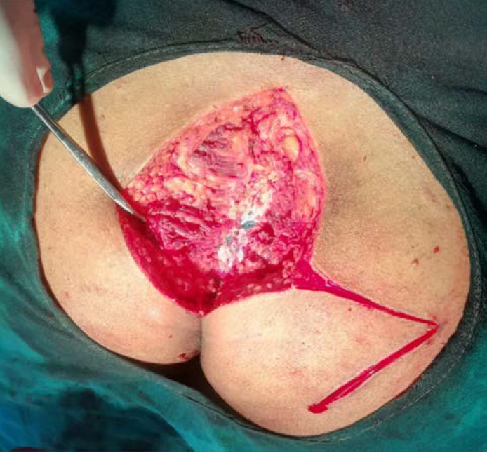
The skin and subcutaneous tissue is excised up to deep fascia (Pre sacral fascia) and rhombus shaped specimen with pilonidal sinus and its extensions are removed [12]. Then flap is raised over gluteus maximus and rotated to cover defect (Figure3). Suction drain is fixed with deep absorbable sutures and skin is closed with prolene mattress sutures [13]. This produces tension free flap of unscarred skin in middle. Antibiotics were given for 14 days (intravenously for 5 days followed by oral for 9 days) Suction drain was removed on the 5th post operative day and sutures removed on the 14th post operative day. 


Figure 2: Markings

RESULTS

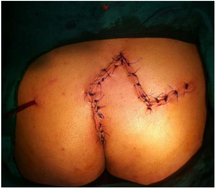
In this study 10 patients were included, median age 29 years, range 17-41 years. Out of 10 patients 6 had primary disease,4 had recurrent disease. All patients were investigated. Limberg flap surgery was done under spinal anesthesia. (Figure 4) Patient were ambulated on 2nd post operative day. Drain was removed on 5th post operative day. They were discharged on 5th post operative day with advice of not to put pressure up to 3 weeks. Sutures were removed on 14th post operative day. One patient had flap edema which resolved by 14 days and two had persistent discharge from tip which took 3 weeks to settle down. All other patient’s wounds healed well with minimal scar and post op pain. So far there has been no recurrence. Almost all patients returned to work after 4 weeks.


Figure 3: Sinus Excision and Flap Raising


Figure 4: Final Outcome

DISCUSSION

The spectrum of pilonidal sinus varies from asymptomatic to complications like abscess formation, systemic infections, squamous and verrucous carcinoma in long standing cases [14-15].


The old school practice of sinus tract excision and healing by secondary intention leads to significant discomfort and prolonged healing time (average 6 weeks). Also associated were high recurrence rates (25-40 %). Some studies have reported higher incidence of early wound disruption following excision and primary closure [16-17]. Rhomboid or Limberg flap is a well vascularized fascio-cutaneous flap that flattens the natal cleft. It leads to good primary healing and lowest recurrence rates [14]. Despite having acceptable outcome there has been reluctance among general surgeons to perform this surgery at secondary health care level. Most of the literature has been compiled and published at the level of tertiary health care institutes. This study was undertaken to check feasibility of flap surgery and compare outcomes at secondary health care level.


Previously Limberg flap was used for recurrent and complex cases only. Now it is recommended as first line management for all cases of Pilonidal sinus due to lowest complication and recurrence rates [18-19].

CONCLUSION

Limberg flap is a simple and easy to perform transposition flap technique, useful in both primary and recurrent cases of pilonidal sinus. It is having a very low complication and recurrence rate.


Conflict of Interest

None

REFERENCE
  1. Solla, J.A. and D.A. Rothenberger. “Chronic Pilonidal Disease.” Diseases of the Colon & Rectum, vol. 33, no. 9, September 1990, pp. 758–761.

  2. Humphries, A.E. and J.E. Duncan. “Evaluation and Management of Pilonidal Disease.” Surgical Clinics of North America, vol. 90, no. 1, February 2010, pp. 113–124.

  3. Søndenaa, K. et al. “Patient Characteristics and Symptoms in Chronic Pilonidal Sinus Disease.” International Journal of Colorectal Disease, vol. 10, no. 1, February 1995, pp. 39–42.

  4. Hull, T.L. and J. Wu. “Pilonidal Disease.” Surgical Clinics of North America, vol. 82, no. 6, December 2002, pp. 1169–1185.

  5. Karydakis, G.E. “Easy and Successful Treatment of Pilonidal Sinus after Explanation of Its Causative Process.” Australian and New Zealand Journal of Surgery, vol. 62, no. 5, May 1992, pp. 385–389.

  6. Chiedozi, L.C. et al. “Management of Pilonidal Sinus.” Saudi Medical Journal, vol. 23, 2002, pp. 786–788.

  7. Mohamed, H.A. et al. “Comparison between Three Therapeutic Modalities for Non-Complicated Pilonidal Sinus Disease.” The Surgeon, vol. 3, no. 2, April 2005, pp. 73–77.

  8. Berger, A. and P. Frileux. “Pilonidal Sinus.” Annales de Chirurgie, vol. 49, 1995, pp. 889–901.

  9. Wolfe, S.A. “Limberg Flap.” Plastic and Reconstructive Surgery, vol. 56, no. 2, 1975, pp. 239–240.

  10. Akca, T. and T. Colak. “Primary Closure with Limberg Flap in Treatment of Pilonidal Sinus: A Randomized Clinical Trial.” British Journal of Surgery, vol. 92, 2005, pp. 1081–1084.

  11. Azab, A.S. et al. “Radical Cure of Pilonidal Sinus by a Transposition Rhomboid Flap.” British Journal of Surgery, vol. 71, no. 2, February 1984, pp. 154–155.

  12. Farquharson, E.L. and R.F. Rintoul. Farquharson’s Textbook of Operative General Surgery. 9th ed., Hodder Arnold, 2005, pp. 457–458.

  13. Kapan, M. et al. “Sacrococcygeal Pilonidal Sinus Disease with Limberg Flap Repair.” Techniques in Coloproctology, vol. 6, 2002, pp. 388–392.

  14. Katsoulis, I.E. et al. “Outcome of Treatment of Primary and Recurrent Pilonidal Sinuses with the Limberg Flap.” The Surgeon, vol. 4, no. 1, February 2006, pp. 7–10.

  15. Werkgartner, G. “Knowledge-Based Therapy of the Pilonidal Sinus.” European Surgery, vol. 36, no. 3, 2004, pp. 170–171.

  16. Dalenbäck, J. et al. “Prospective Follow-Up after Ambulatory Plain Midline Excision of Pilonidal Sinus and Primary Suture under Local Anaesthesia.” Colorectal Disease, vol. 6, no. 6, November 2004, pp. 488–493.

  17. Abboud, B. and H. Ingea. “Recurrent Squamous-Cell Carcinoma Arising in Sacrococcygeal Pilonidal Sinus Tract.” Diseases of the Colon & Rectum, vol. 42, no. 4, April 1999, pp. 525–528.

  18. Tekin, A. “Pilonidal Sinus: Experience with Limberg Flap.” Colorectal Disease, vol. 1, 1999, pp. 29–33.

  19. Caplan, T.L. and J. Wu. “Pilonidal Disease.” Surgical Clinics of North America, vol. 82, 2002, pp. 1169–1185

Recommended Articles
Research Article
Imaging Evaluation of Ultrasonography Versus Magnetic Resonance, Imaging in Diagnosis of Carpal Tunnel Syndrome among Female Patients with Positive Nerve Conduction Study
Download PDF
Research Article
An evaluation of Profile of Medico-legal autopsies conducted at Dr. Radhakrishnan Govt. Medical College, Hamirpur, Himachal Pradesh
Download PDF
Research Article
The Efficacy of 5- Fluorouracil Cream and 25% Podophyllin Solution in Treatment of Plantar Warts in Alternative Day Regimen ; An Open Therapeutic Trial.
Download PDF
Research Article
Silent Threats: Evaluating Knowledge of Pelvic Inflammatory Disease Among Reproductive-Age Women in District Kangra
Published: 27/11/2024
Download PDF
Chat on WhatsApp
Flowbite Logo
PO Box 101, Nakuru
Kenya.
Email: office@iarconsortium.org

Editorial Office:
J.L Bhavan, Near Radison Blu Hotel,
Jalukbari, Guwahati-India
Useful Links
Order Hard Copy
Privacy policy
Terms and Conditions
Refund Policy
Shipping Policy
Others
About Us
Team Members
Contact Us
Online Payments
Join as Editor
Join as Reviewer
Subscribe to our Newsletter
+91 60029-93949
Follow us
MOST SEARCHED KEYWORDS
Copyright © iARCON International LLP . All Rights Reserved.