Introduction: Pneumonia remains one of the main conditions leading to morbidity and mortality around the world, both among adults and children. In UNHCR refugee camps, pneumonia remains one of the most prevalent diseases and one of the main causes of death in children below the age of 5 years. Objective: The study aims to show the difference in severity of CAP requiring hospitalization among Lebanese children and refugees’ children comparing 2 populations belonging to the same age group and to compare pneumonia severity between Lebanese and Syrian children. Methodology: This is an observational, retrospective study of the Lebanese and Syrian children aged between 2 months and 5 years admitted to Rafik Hariri University hospital for community acquired pneumonia from February 2015 till February 2020. The study includes 201 patients diagnosed having CAP. The data collected from the patients’ medical records includes demographic data, clinical data, physical exam, laboratory and radiology results, treatment data and vital signs. Results: Out of 5852 pediatric admission during the study period, CAP occurred in 3.4% of patients, with lower rates among Lebanese children (1.2%) compared to Syrian children (2.2%). Patients with CAP had comparable gender and age between study groups. The most prevalent clinical symptoms observed were fever (98%), cough (93%), irritability (73.1%) and decreased feeding (45.8%). Syrian children were significantly more likely to exhibit irritability and decreased feeding. Physical exam findings were more frequently observed among Syrian children, who showed significantly more illness, noisy breathing, respiratory distress, rhonchi, grunting and nasal flaring. Severe CAP was diagnosed in 57.7% and was significantly higher in Syrian patients (88.7%) compared to Lebanese patients (37.1%). Syrian children had a higher need for mechanical ventilation, IV fluids and PICU admission. Radiological findings showed that infiltration was more prevalent in Lebanese patients (82.9%) and consolidation was more prevalent in Syrian patients. Updated vaccination was less frequent among Syrian children, who required a longer period of treatment and length of hospital stay. Biomarkers of pneumonia severity (WBC and CRP) were consistently higher among Syrian children. Conclusion: This study showed that Syrian patients have significantly higher prevalence and severity of CAP. The notable differences between the two patient populations shows the clear impact of the humanitarian crisis, particularly among resource-deprived refugee and migrant populations.
Keywords
Refugees. Pediatric Intensive Care Unit (PICU)
INTRODUCTION
General Overview
Pneumonia remains one of the main conditions leading to morbidity and mortality around the world, both among adults and children [1]. Pneumonia in general remains the predominant cause of death among children below 12 years of age [2], with pneumococcal pneumonia responsible for the highest number of deaths in children 5 years or younger by 2017 [1]. Community acquired pneumonia (CAP) remains a clinical challenge, particularly among children and infants. This is due to the notable variations observed between children in regards to the clinical course of the disease, even in the case of shared etiology, which ledto the development of guidelines by the Pediatric Infectious Diseases Society (PIDS) and the Infectious.
Diseases Society of America (IDSA) [3]. Community acquired pneumonia often results in the need for hospitalization and acute medical care which incurs significant healthcare costs across the world. Studies have demonstrated significantly decreased quality of life when compared to healthy children [4]. Pneumonia symptoms such as cough induced by the sickness plays a major role in regards to quality of life, in addition to financial concerns due to the disease [5]. Other complications such as bacteremia have been reported in the literature [6-8], with respiratory-related complications, such as para pneumonic effusion and pleural empyema, affecting a high number of children with CAP [9]. In UNHCR refugee camps, pneumonia remains one of the most prevalent diseases and one of the main causes of death in children below the age of 5 (17% of child morbidity) [10]. When considering refugees and migrant populations, particularly those living in refugee camps, it is known that they are at a higher risk of disease due to the absence of basic amenities or social distancing [11]. Studies report the higher incidence of pneumonia as well as other respiratory illnesses such as asthma and chronic obstructive pulmonary disorder among refugees in camps [12], including children [13]. However, data correlating socioeconomic and lifestyle factors to the severity of CAP remain limited. Moreover, data from Lebanon regarding CAP in children is severely lacking, which fuels the need for further studies in this regard.
Review of Literature Pneumonia
Remains one of the main conditions leading to morbidity and mortality around the world, both among adults and children [1]. Pneumonia can be defined as an infection of the lung parenchyma that occurs in an acute setting. Pneumonia is not linked to a single cause and could result from an infection with bacteria, viruses or fungi [14], as will be further detailed in the ‘etiology’ section. Pneumonia is further differentiated according to the setting in which the infection is acquired, yielding the following categories: a) Community acquired pneumonia: this category refers to pneumonia due to an infection acquired outside of a hospital. b) Hospital-acquired pneumonia: this category refers to pneumonia following infection during hospitalization or within two days of discharge [15] Ventilator-associated pneumonia: this category is actually a part of the previous one, hospital-acquired pneumonia. Ventilator-associated pneumonia refers to the development of pneumonia following an infection acquired due to mechanical ventilation received during hospitalization. d) Health-care associated pneumonia: this category refers to pneumonia due to an infection acquired during receival of healthcare services in lower-acuity settings. Examples include places other than the hospital where healthcare is offered, such as nursing homes and dialysis centers [15]. As mentioned, pneumonia remains one of the leading causes of disease and deaths in the world. Among lower respiratory infections by 2017, pneumococcal pneumonia caused the highest number of deaths in children 5 years or younger by 2017 [1]. More importantly, the following two leading causes of lower-respiratory infection-related mortality were also pneumonia, namely respiratory syncytial virus pneumonia, H influenza type B pneumonia [1]. Another study from china showed that pneumonia was the predominant cause of death among children below 12 years of age [2]. This was shown based on data collected between 2006 and 2015, including 93 443 children between the ages of 1 month and 11 years [2]. More specifically, of the 510 cases of fatality recorded in this study, 36.7% were caused by pneumonia [2]. The latter dramatically overshadowed other complications such as sepsis and tumors, which accounted for 13.5% and 11.4% of deaths respectively [2]. The present review will focus on community acquired pneumonia, particularly among children.
Community Acquired Pneumonia
Community acquired pneumonia is a condition that carries a significant burden in regards to morbidity, mortality. As previously mentioned, it is characterized by the acquisition of an infection outside of a hospital setting that then leads to pneumonia. In terms of pathophysiology and clinical presentation, pneumonia can occur through the entry of pathogens into the pharynx and its consequent colonization [16]. The pathogen then enters the lower respiratory tract through the mechanism of micro-aspiration. When the host lacks proper immunological defenses, or when local immune systems are overwhelmed by a highly virulent or infectious pathogen, pneumonia can develop. In addition to micro-aspiration, pneumonia can develop due to macro-aspiration, or hematogenous spread, i.e. spreading of the pathogen through blood [16].
Symptoms
Pneumonia manifests according to different symptoms and clinical observations in children [17], such as: Fever, chills, cough, dyspnea, pleuritic chest pain, weight loss and lethargy. However, it is important to note that the age of the child as well as the causative agent influence the clinical manifestation of pneumonia. In general, pneumonia symptoms are not specific to the disease (non-specific) as they are predominantly symptoms of respiratory distress (cough, breathing difficulty) and fever [17]. When it comes to infants and young children, pneumonia might present in subtle symptoms such as decreased feeding, lethargy, irritability, inconsolable crying, etc. [17].
When the child is older, he or she might express feelings of abdominal pain, neck pain, or pleuritic chest pain [17]. In addition to the ones already cited, other physical symptoms are shown in the Table 1 above. It is important to note that tachypnea is one of the most important physical symptoms of pneumonia [18]. The definition of tachypnea adopted by the world health organization (WHO) is: 50 breaths per minute in infants 2 to 12 months of age, if 40 breaths/min in children 1 to 5 years of age and if 20 breaths/min in children 5 years of age and older. In developing countries, the WHO actually considers tachypnea to be a diagnostic factor of pneumonia when accompanied by coughing [18]. This was suggested for the diagnosis of pneumonia in low socioeconomic settings where access to chest radiography might not allow the proper diagnosis of the condition [18]. While this does not allow the distinction between radiographic pneumonia, the observation of tachypnea according to the thresholds proposed by the WHO has been correlated more highly with children who actually had pneumonia [19]. When looking at the pathogenic origins of pneumonia, some symptoms can be correlated with bacterial pneumonia, while others seem to be more prevalent in viral pneumonia [17,20]. Table 2 shows symptoms in relation with the type of pneumonia, be it bacterial or viral. Other things to note are the higher propensity of wheezing to occur in viral pneumonia, as well as infections with M. pneumonia and C. pneumonia [17,20]. M. pneumonia is further associated with extra pulmonary findings, which include myocarditis, arthritis, hemolytic anemia, as well as other conditions [17,20].
Etiology
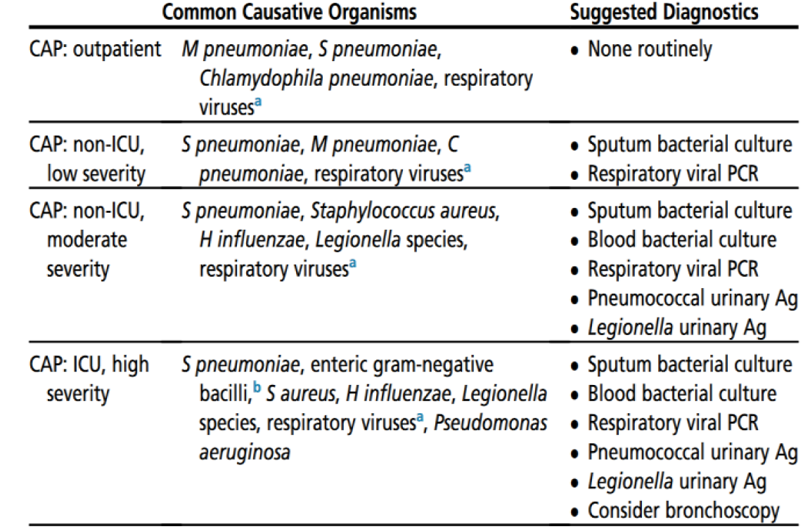
As previously mentioned, multiple pathogens have been known to cause pneumonia, be they bacteria, viruses or fungi. It was therefore logical to observe differences in the pathogenicity and cases attributed to each of these microorganisms. Moreover, in order to better manage these cases of pneumonia, clinicians attempted to understand the correlation between the causative organism and the severity of pneumonia. The most prolific bacteria to date when it comes to community acquired pneumonia remains S. Pneumonia, which is responsible for at least a quarter of community acquired pneumonia cases in the world’s adult population [21]. In general, the severity of community acquired pneumonia can be correlated with the pathogen responsible for its development, as shown in Table 3. The latter shows the probability of pneumonia and its severity in relation with the causative organism. As can be seen in the Table, when M. pneumonia, C. pneumonia and viruses (such as influenza) are detected, mild cases of pneumonia can be expected. The severity of pneumonia seems to increase when organisms such as Staphylococcus aureus, Legionella and H influenza are involved, as well as multidrug resistant pathogens [22]. Other pathogens have been known to cause pneumonia and include: Mycobacterium tuberculosis, non-Tuberculous mycobacteria, Chlamydophila species, Coxiella and Fungi (i.e. Cocci-dioides, Blastomyces, Histoplasma, Cryptococcus, Aspergillus).
Similar observations were previously observed among children, where most pneumonia cases are also caused by S. pneumonia and H. influenza type B [23]. On the other hand, severe cases of pneumonia in children can be traced back to Staphylococcus aureus and Klebsiella pneumonia [23]. According to the meta-analysis, viral pneumonia was caused by respiratory syncytial virus in at least 15% of cases, up to 40% of cases. Other viral causative agents were observed to be influenza A and B, parainfluenza, human metapneumovirus and adenovirus [23]. This data was based on evidence before the introduction of conjugate vaccines. The latter seemed to cause a shift in the pathogen profile of childhood pneumonia, showing that particularly severe cases of pneumonia are often caused by more than one pathogen [24]. The understanding of the etiology of pneumonia is thus evolving to recognize the implication of sequential or concurrent infections in the development of this condition. After the introduction of vaccines against pneumococcal pathogens (conjugate vaccine), as well as Haemophilus influenza type B conjugate vaccination, it has now become more common to see viral microorganisms as the pathogens underlying pneumonia [24].
Table 1: Physical Symptoms of Community Acquired Pneumonia among Children
Dullness to percussion (parenchymal consolidation)
Source: Retrieved from Leung [17], Barson [20] and Qin (2015) (8–10)
Table 2: Pneumonia Symptoms in Relation with the Type of Pneumonia
Symptom
Type of Pneumonia/Pathogen
High fever (> 38.5ºC)
Chills and rigors
Toxic appearance
Marked tachypnea
Localized auscultatory findings
Bacterial pneumonia
Low-grade fever
Runny nose
Myalgia
Wheezing
Diffuse and bilateral auscultatory findings
Viral pneumonia
Table 3: Organisms Commonly Causing Community Acquired Pneumonia and the Routine Diagnostics Suggested in Clinical Practice
Ag, antigen; ICU, intensive care unit; PCR, polymerase chain reaction, A: i.e. influenza, parainfluenza and respiratory syncytial virus, B: i.e. Klebsiella pneumonia, Enterococcus coli and Enterobacter spp., Source: Adopted from Lanks [22] (14)
Diagnosis and Management
Community acquired pneumonia remains a clinical challenge, particularly among children and infants. This is due to the notable variations observed between children in regards to the clinical course of the disease. In fact, pneumonia unravels differently even in children who share etiologic backgrounds and have the same type of agent responsible for the infection. As such, developing clear clinical guidelines was necessary to guide the diagnosis and management of community acquired pneumonia in children. This led to the development of guidelines by the Pediatric Infectious Diseases Society (PIDS) and the Infectious Diseases Society of America (IDSA) [3], which will be discussed in the present section. While these guidelines provide much needed suggestions for the management of children above the age of 3 months, they don’t address the handling of infants, immunocompromised children, home-based mechanical ventilation, or chronic conditions.
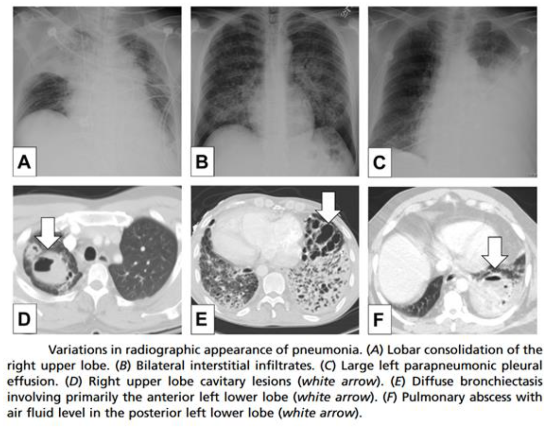
Figure 1: Variations in radiographic appearance of pneumonia
Source: Adopted from Lanks and Hsia [22]
Diagnosis
The PIDS-IDSA guidelines proposed the following for the diagnosis of children with community acquired pneumonia in an outpatient setting [3]. 1) Blood cultures are needed in children with CAP that are not fully immunized, children that are not improving clinically, or children that are deteriorating even after receival of antibiotic therapy. 2) Repeat blood cultures should only be done in children who do not show clinical improvement or in children whose pneumonia is caused by S. aureus. In other cases, repeated blood work is not necessary in order to monitor infection progression. 3) In the case of pneumonia and suspected hypoxemia, clinicians should perform pulse oximetry. 4) In an outpatient setting, community acquired pneumonia cases are generally mild and uncomplicated enough to be treated without requiring hospitalization. As a result, it is not necessary to perform routine chest radiography. Exceptions include cases of pneumonia with hypoxemia or respiratory distress, in which case post anterior and lateral chest radiography should be done. 5) When community acquired pneumonia presents with notable complications, or does not show clinical improvement or shows deterioration even after initiation of antibiotics, repeat chest radiography is needed. 6) Follow-up radiography is suggested in several situations, including clinically unstable children, deteriorating cases with respiratory distress, persistent fever despite treatment, recurrent pneumonia in the same lobe, among others. In an inpatient setting, diagnosis recommendations differ slightly in regards to blood cultures and routing chest radiography, as follows [3]. 7) Blood cultures are needed in all hospitalized children with CAP that is moderate to severe and presenting with complications. 8) All hospitalized children with CAP should undergo chest radiographs, both posteroanterior and lateral, in order to properly guide therapy choice by the characterization of parenchymal infiltrates, if existing. However, it is important to note that the radiographic presentation of pneumonia differs significantly across patients (Figure 1) [22]. While the use of CT scans could provide more sensitive information, chest radiography is more frequently and routinely done in clinical settings and provide valuable insights for the diagnosis of pneumonia by indicating parenchymal lung involvement [22]. Moreover, studies find that radiologic evidence of pneumonia cannot always be observed. This was shown in a study prospectively following 122 children with severe pneumonia, where half of the children did not exhibit radiologic manifestations of pneumonia [25]. Pneumonia was more likely to manifest radiologically when fever was detected in the affected children [25].
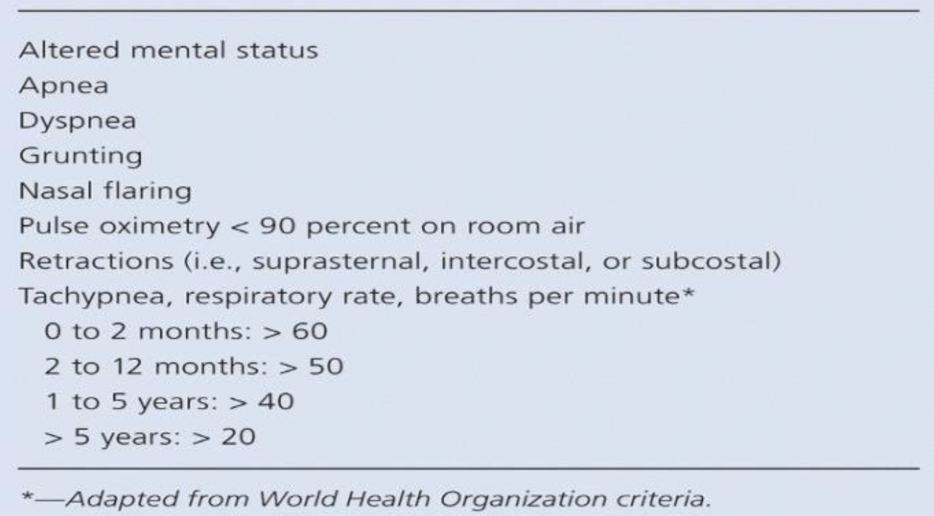
Table 4: Respiratory Distress Criteria among Children with Pneumonia, as Suggested by the PIDS-IDSA Guidelines
Source: Adopted from Bradley et al. [3]
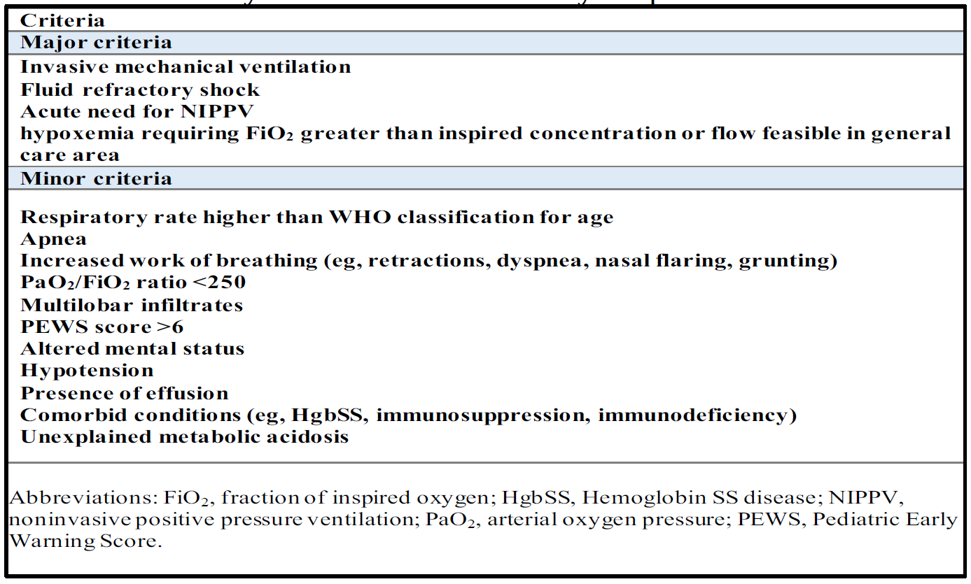
Table 5: Severity Criteria in Community Acquired Pneumonia
Source: Adopted from Bradley et al. [3]
The hospitalization of children or infants with community acquired pneumonia is often necessary when clinical presentation of the disease shows moderate to severe pneumonia, as evidenced by the observation of respiratory distress and hypoxemia (Table 4). In the event of bacterial CAP, infants 3 to 6 months of age should be hospitalized even if pneumonia is not confirmed. Other cases requiring hospitalization include inadequate home-based follow-up and care, as well as infection with a highly virulent pathogen (i.e. community-associated methicillin-resistant Staphylococcus aureus) [3].
However, one study examined the applicability of the PIDS-IDSA severity criteria (Table 5) showed that these criteria might not always be able to predict hospitalization. in fact, more than half of children who satisfied the criteria for severe community acquired pneumonia did not require hospitalization.
Management:
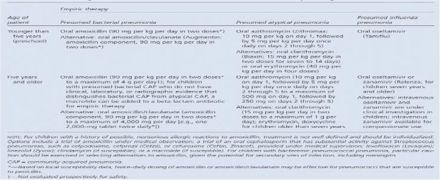
The PIDS-IDSA guidelines also proposed recommendations for the management of children with community acquired pneumonia, as shown in Table 6.
Table 6: Management of Children with Community Acquired Pneumonia
Source: Adopted from Bradley et al. [3]
Complications and Outcomes
Community acquired pneumonia often results in the need for hospitalization and acute medical care which incurs significant healthcare costs across the world. Ranging from the need for hospitalization, diagnostic tests and imaging, pain and discomfort and disease-related complications, pneumonia can result in healthcare needs that reflect negatively on both the child and caretakers. The complications and clinical outcomes of community acquired pneumonia will be explored in this section.
Quality of life and Productivity
Seeing as the treatment of pneumonia necessitates several clinic or hospital visits, as well as potential hospitalization, antibiotic treatment course and diagnostic imaging and tests, it is natural that this condition carries significant effects on the quality of life of those involved. One study examined the effect of community acquired pneumonia on the children as well as their parents. It was found that children with community acquired pneumonia suffered from significantly decreased quality of life when compared to healthy children [4]. This was consistently observed across different settings, such as a primary health clinic, a pediatric emergency department and pediatric hospital wards [4]. This reflects the significant burden of pneumonia on children, who suffer from more febrile days and more changes to their routine when compared to healthy controls. Moreover, parents are also implicated in the negative consequences of pneumonia, seeing as the treatment of their child often incurs workday losses [4]. Consistently, a study in the setting of acute respiratory infections and cough showed that these conditions significantly impair the quality of life of the children. This was reported particularly in the first few days of emergency department admission, with improvement in quality of life noticeable by the first week after admission [5]. Regardless, it seems that the cough induced by the sickness plays a major role in regards to quality of life, in addition to financial concerns [5]. This provides important insights for policy makers looking to improve both the clinical and psychological outcomes of children affected by community acquired pneumonia and their parents.
Bacteremia/Septicemia and Respiratory Complications
The clinical course of community acquired pneumonia differs according to several factors, one of which is the type of pneumonia. Literature thus seems to suggest that bacterial pneumonia is more likely to result in complications when compared to viral pneumonia [17] [26]. Clinical complications include bacteremia/septicemia, which have been reported among children admitted for community-acquired pneumonia. In one study including more than 7 thousand children, it was found that 2.5% of patients who had blood work performed upon admission to the hospital had bacteremia [6]. However, very few children had bacteremia caused by a pathogen that was resistant to penicillin. As such, evidence suggests that while bacteremia occurs in children with CAP, its prevalence might not warrant the conduction of blood tests in non-intensive care settings [6]. Another study from Thailand showed that positive blood culture could be seen in approximately 9% of children between the age of 2 months and 15 years admitted with CAP [7]. However, further examination shows that the rate of true positive blood cultures remains around 4% [7], thereby supporting the relatively low incidence of bacteremia reported in the literature [6] [8]. However, it should be noted that bacteremia could lead to notable complications (i.e. brain abscess, meningitis, osteomyelitis, septic arthritis, peri-carditis and endocarditis) [17] and should therefore be adequately and rapidly addressed. Another type of complications very common to community acquired pneumonia in children is respiratory-related complications, such as parapneumonic effusion and pleural empyema. In a study including close to two thousand children hospitalized for community acquired pneumonia, parapneumonic effusion was found to have occurred in 16.7% of children [9]. More importantly, the frequency of occurrence of parapneumonic effusion and pleural empyema due to community acquired pneumonia was found to increase over the course of 11 years, rising from 5.4% to 18.8% [9]. Another study from Italy showed that empyema was the most common complication of bacteremic pneumonia among children younger than five years of age [27]. Other complications of community acquired pneumonia include [9,17,27]: Pneumatoceles, necrotizing pneumonia, lung abscess and acute respiratory failure.
Cardiovascular Events
In a cohort of older adults, one study actually demonstrated pneumonia to be a potential risk factor for both long-term and short-term occurrence of cardiovascular events, such as myocardial infarction and arrhythmia [28]. A study from Saudi Arabia also reported this, with 16% of complications following community acquired pneumonia found to be cardiac-related complications [29]. However, heart failure was found to be significantly more likely to occur in patients with hospital-acquired pneumonia when compared to community acquired pneumonia [29]. Regardless, these events can lead to increased morbidity and mortality and should therefore be explored among pediatric populations in order to establish their potential occurrence and implications.
Predictors of severity/complications:Demographic factors
Data from Saudi Arabia showed that elderly patients were significantly more likely to exhibit complicated community acquired pneumonia [29]. However, this study failed to show any significant differences between genders in terms of CAP complications.
Biomarkers
Laboratory findings, on the other hand, seem to be consistent between children who had pneumococcal CAP and those who did not. This included C-reactive protein levels, white blood cell (WBC) counts and the percentage of neutrophils [27]. Another study showed similar results, with median WBC count, absolute neutrophil count (ANC), C-reactive protein (CRP), or procalcitonin failing to act as distinguishing factors between severe and non-severe CAP in a pediatric population [30]. That being said, adjusting the analysis to account for the child’s age as well as other factors showed that CRP and procalcitonin might have potential as predictors of the more severe outcomes of CAP in children [30].
Etiology
The severity and complications of CAP can also differ according to its etiology. Data from Italy gathered from children aged up to 5 years showed that complications most frequently occurred among cases of pneumococcal bacteremic CAP [27]. This correlation was statistically significant, along with that showing that children with pneumococcal CAP were more likely to exhibit empyema [27].
Comorbidities and Treatment
Adult patients who had diabetes mellitus and those who were receiving immunosuppressant therapy were significantly more likely to have complications during the course of their CAP [29]. This was observed in a cohort of adult patients from Saudi Arabia [29]. Among children, a study showed that CAP patients who had received ibuprofen or acetaminophen prior to hospital admission were significantly more likely to exhibit local CAP complications, such as parapneumonic effusion/pleural empyema, necrotizing pneumonia and lung abscess [9]. Similarly, children with complex chronic conditions had more than double the risk of being readmitted to the emergency department after initial admission for CAP, when compared to those who did not have these conditions [31].
Socioeconomic/Lifestyle Factors
Studies among adults suggest that lifestyle factors can predict the incidence of CAP. In fact, patients with poor dental hygiene, frequent contact with children, as well as being overweight were at significantly higher risk of CAP [32]. When considering refugees and migrant populations, this risk of CAP should be considered. It is well established that refugees, particularly those living in refugee camps, have little to no possibility of adequate hygiene, social distancing and are at a higher risk of disease due to the lack or limited availability of basic amenities such as soap and water [11]. As such, these populations are at a much higher risk of contracting infections, such as COVID-19 and pneumonia. Consistently, Turkish data demonstrated significantly higher incidence of pneumonia as well as other respiratory illnesses such as asthma and chronic obstructive pulmonary disorder among refugees in camps when compared to the general population in turkey [12]. A study from Germany showed that pediatric refugee populations were at significantly higher risk of contracting vaccine-type invasive pneumococcal disease and were thus at a higher risk of mortality when compared to German children. Moreover, higher rates of antimicrobial and antibiotic drug-resistant invasive pneumococcal disease were observed among refugee children [13]. This shows that refugee children are not only more likely to suffer from CAP, but were also at a higher risk of having a complicated drug-resistant disease course. One study looked at the predictors of CAP in a pediatric population of affected patients and healthy controls. It was found that children who were underweight, those who were in a household using unclean cooking fuel and those belonging to a low income family were significantly more likely to have CAP [33]. Children living in refugee populations in south-east Asia were examined in one study and were found to have high prevalence of pneumonia. Several factors predicted the occurrence of CAP among these refugee children and infants, including small living space (reduced volume per person), short distance between houses and shorter distance from stove to bed [34]. Data correlating socioeconomic and lifestyle factors to the severity of CAP remain limited. Moreover, data from Lebanon regarding CAP in children is severely lacking. However, the Pneumonia Etiology among Refugees and the Lebanese population (PEARL) study will be examining CAP among refugees and local populations in order to establish the etiology of CAP and guide treatment and management strategies [35].
Conclusion
CAP remains a highly prevalent condition among children, particularly those living in poor socioeconomic and living conditions (i.e. refugees). CAP can be traced to different etiologies and has differing clinical courses in patients. As such, local studies are needed in order to establish optimal treatment, management and prevention strategies for each patient population. This is crucial considering the effect of CAP on the quality of life as well as clinical outcomes of children affected by it. Moreover, more evidence is needed for the prediction of CAP severity among pediatric populations, as data on this subject remains limited across the world and particularly, in Lebanon.
Objectives
Primary Objective: The primary objective of our study is to show the difference in severity of CAP requiring hospitalization among Lebanese children and refugees’ children comparing 2 populations belonging to the same age group. The aim is to compare pneumonia severity in Lebanese and Syrian children and not between children who belong to the same population.
Secondary Objectives
The secondary goal behind this objective is to determine statistically the prevalence of community acquired pneumonia in Syrian children and in Lebanese children and to compare the prevalence between these 2 populations in order to show if the humanitarian crisis could also have an effect on the prevalence of the disease.
MATERIALS AND METHODS
Study Design and Population: This is an observational, retrospective study of the Lebanese and Syrian children aged between 2 months and 5 years admitted to Rafik Hariri University hospital for community acquired pneumonia from February 2015 till February 2020
Inclusion Criteria: Subjects included in our study met the following criteria: age between 2 months and 5 years, presenting with cough and dyspnea, diagnosed as community acquired pneumonia and onset of symptoms started within the last 14 days
Exclusion Criteria: Were excluded from our study: immunocompromised children, children confirmed having active or latent tuberculosis, patients in whom radiological findings were not suggestive of pneumonia and children whom developed pneumonia during their stay at the hospital
Sample Size: The prevalence of CAP was not well identified in the reviews and differs between the populations, whereas the incidence was highly tracked. A review was done by DeAntonio et al. [36] and evaluates the epidemiology of CAP in children aged less than 6 years in 90 developing and newly industrialized countries [36]. This review showed that the prevalence of CAP in Iranian children aged between 12 months and 5 years is 14% and in Chinese children aged between 6 months and 5 years is 12.4%. After checking the inclusion criteria, the number of patients who could be included is 201. Based on Cochran formula, this number is representative to the child diagnosed having CAP. The sample calculation was done using the following formula: N = (1.96)2 x (p q) / (B2). * p = Prevalence of CAP. * q = 1 – p. * B = 0.05% (for 95% confidence level)
Data Collection
The data was collected from the patients’ medical records at Rafic Hariri University Hospital. The first step of the data collection includes the eligibility criteria. After meeting the inclusion criteria, the patient is included in the study and received a study code. Data entry was performed directly on an electronic database and includes the following data (appendix 1):
Oxygen supplement, Mechanical ventilation, IV fluids, Antibiotics, Antiviral (oseltamivir), PICU admission, length of stay and updated vaccination.
Vital Signs
Blood pressure, heart rate, oxygen saturation and temperature. All these data were compared between Lebanese and Syrian children by comparative tables and analyzed in accordance with pneumonia severity index according to the Pediatric Infectious Diseases Society and the Infectious Diseases society of America [37-38].
Ethical Considerations
The protocol it was reviewed and granted a written study approval from the research committee in the Lebanese university. A letter was addressed by the research supervisor in order to collect the data (Appendix 3). This study was conducted in accordance with the US Code of Federal Regulation 45- CFR -46.107, 21-CFR-56.107 and Good Clinical Practice ICH Section 3 and the principles laid down by the 18th World Medical Assembly (Helsinki) and all applicable amendments. All participants had a designated code. Records will be stored and none can access the database except the researchers.
Statistical Analysis
Data was analyzed using the SPSS version 22. The statistical analysis was enrolled in order to analyze the findings between the patients as per their nationality. The tests used in the bivariate settings are the chi-square test and the Fisher exact test, in order to test the statistical difference between the nationality and the nominal variables (gender, PICU admission, vaccination). In addition, the Mann-Whitney test was used in order to test the statistical difference between the nationality and the continuous variables (age, WBC, HCT etc.) which are not normally distributed. A statistically significant correlation was set at 5% (p-value less than 0.05).
RESULTS
The study includes 201 patients diagnosed having CAP and admitted to RHUH between 2015 and 2020.
Prevalence of CAP
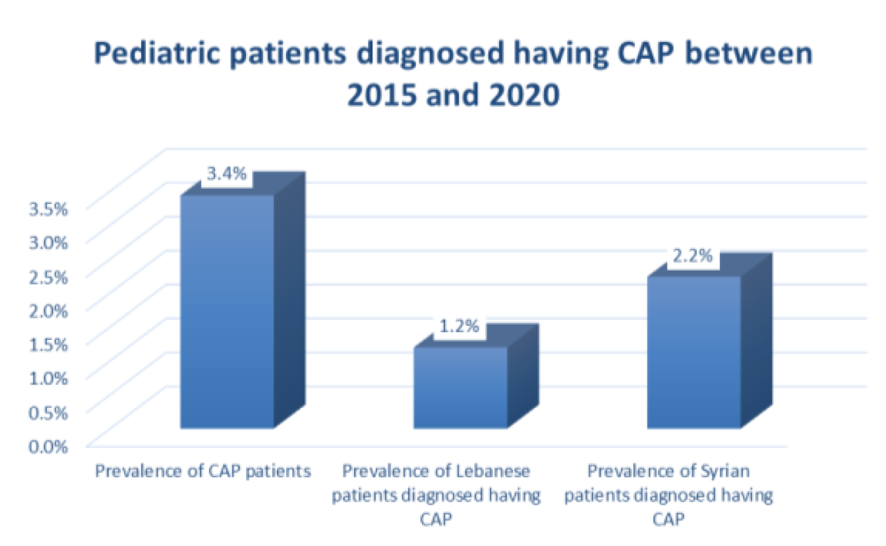
Out of 5852 pediatric admission during the study period, the prevalence CAP is 3.4% (201 patients). The prevalence of CAP in Lebanese patients is 1.2% (70 patients) and the prevalence of CAP in Syrian patients is 2.2% (131 patients) Figure 2.
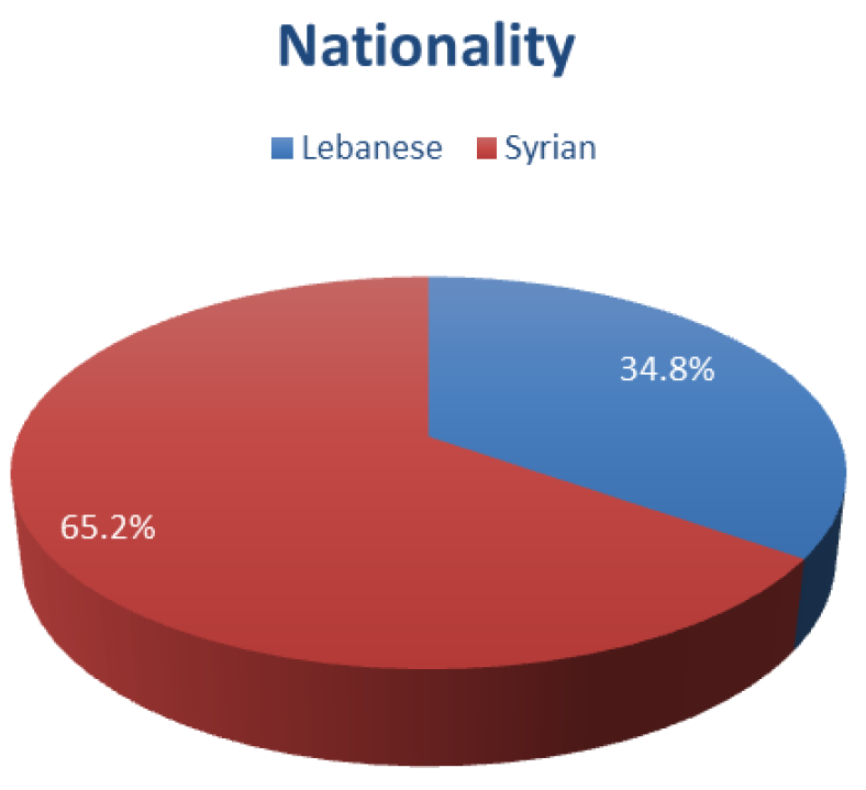
Therefore, included in the study are 201 patients distributed between 70 Lebanese patients 34.8% and 131 Syrian patients 65.2% Figure 3.
Demographic Characteristics
Gender and age do not differ between patients in function of their nationality p>0.05. The gender is equally distributed in Lebanese patients (50% boys and 50% girls) whereas Syrian patients are distributed between 54.2% of boys and 45.8% of girls. The mean age is 11.7 ±8.18 months in Lebanese patients and 11.79 ±12.29 months in Syrian patients Table 8.
Clinical Presentation
The most prevalent clinical symptoms are fever 98%, cough 93%, irritability 73.1% and decreased feeding 45.8% Table 9. The irritability was shown to be higher in Syrian patients 80.9% compared to Lebanese patients 58.6% p = 0.001. In addition, the decreased feeding was shown to be higher in Syrian patients 52.7% compared to Lebanese patients 32.9% p = 0.007.
Table 8: Demographic characteristics
Parameters
Nationality
Total
p-value
Lebanese (N = 70)
Syrian (N = 131)
Sex
Boy
35
71
106
0.570 *
50.0%
54.2%
52.7%
Girl
35
60
95
50.0%
45.8%
47.3%
Age
(Months)
Mean (SD)
11.73 (8.18)
11.79 (12.29)
11.77 (11.01)
0.219 †
Min - Max
2 - 25
2 - 72
2 - 72
* Chi-Square Test † Mann-Whitney test
Table 9: Clinical Presentation of CAP Patients
Parameters
Nationality
Total
p-value
Lebanese (N = 70)
Syrian (N = 131)
Cough
No
3
11
14
0.215 †
4.3%
8.4%
7.0%
Yes
67
120
187
95.7%
91.6%
93.0%
Fever
No
1
3
4
0.566 †
1.4%
2.3%
2.0%
Yes
69
128
197
98.6%
97.7%
98.0%
Irritability
No
29
25
54
0.001 *
41.4%
19.1%
26.9%
Yes
41
106
147
58.6%
80.9%
73.1%
Cyanosis
No
67
117
184
0.095 *
95.7%
89.3%
91.5%
Yes
3
14
17
4.3%
10.7%
8.5%
Decreased feeding
No
47
62
109
0.007 *
67.1%
47.3%
54.2%
Yes
23
69
92
32.9%
52.7%
45.8%
Vomiting
No
63
119
182
0.846 *
90.0%
90.8%
90.5%
Yes
7
12
19
10.0%
9.2%
9.5%
Seizure
No
68
124
192
0.337 †
97.1%
94.7%
95.5%
Yes
2
7
9
2.9%
5.3%
4.5%
* Chi-Square Test † Fisher's Exact Test
Figure 2: Prevalence of CAP in Pediatric Patients
Figure 3: Nationality of Patients Diagnosed Having CAP
Physical Examination Findings
The most prevalent physical exam findings are tachypnea 78.1%, illness 64.2%, nasal flaring 62.2%, crackles 55.7%, grunting 45.8% and decreased air entries 41.8% Table 10. The statistical analysis showed that Syrian patients have higher symptoms than the Lebanese patients:Illness was shown higher in Syrian patients 72.5% compared to Lebanese patients 48.6% p = 0.001.Noisy breathing was shown higher in Syrian patients 13.7% compared to Lebanese patients 0% p = 0.000.Respiratory distress was shown higher in Syrian patients 35.1% compared to Lebanese patients 17.1% p = 0.007.Rhonchi was shown higher in Syrian patients 32.1% compared to Lebanese patients 15.7% p = 0.012.Grunting was shown higher in Syrian patients 52.7% compared to Lebanese patients 32.9% p = 0.007.Nasal flaring was shown higher in Syrian patients 67.2% compared to Lebanese patients 52.9% p = 0.046.
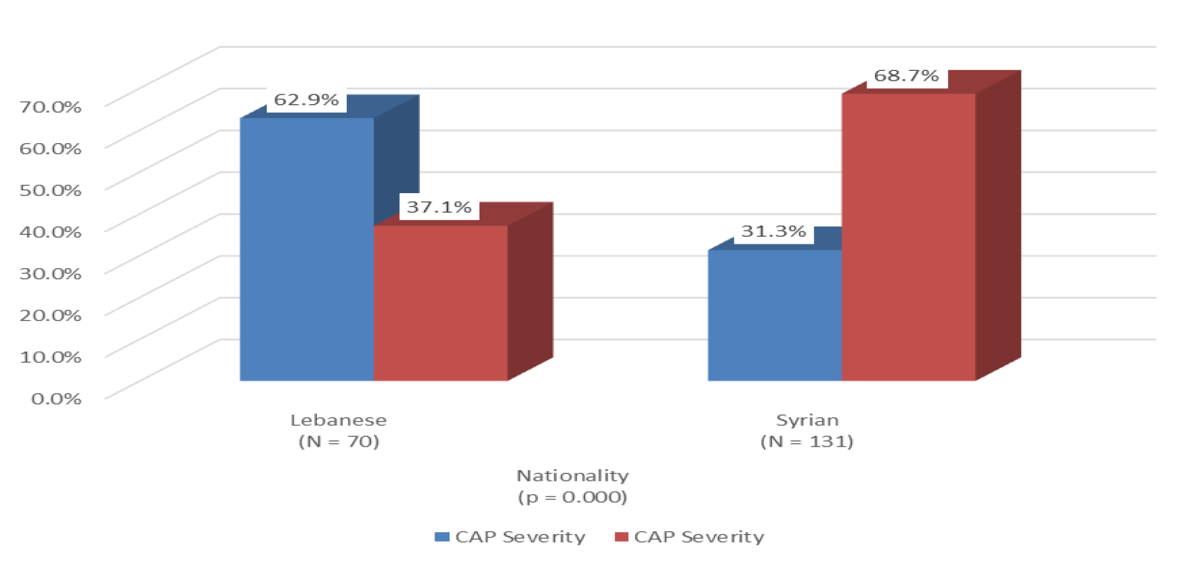
Out of 201 CAP patients, 116 patients have severe CAP 57.7% and 85% have mild CAP 42.3%. The severe CAP was shown higher in Syrian patients 88.7% compared to Lebanese patients 37.1% p = 0.000. Whereas, 62.9% of Lebanese patients have mild CAP compared to 31.3% of Syrian patients Figure 4.
Table 10: Physical Exam Findings
Parameters
Nationality
Total
p-value
Lebanese (N = 70)
Syrian (N = 131)
Illness
No
36
36
72
0.001 *
51.4%
27.5%
35.8%
Yes
34
95
129
48.6%
72.5%
64.2%
Noisy breathing
No
70
113
183
0.000 †
100.0%
86.3%
91.0%
Yes
0
18
18
0.0%
13.7%
9.0%
Respiratory distress
No
58
85
143
0.007 *
82.9%
64.9%
71.1%
Yes
12
46
58
17.1%
35.1%
28.9%
Tachypnea
No
16
28
44
0.809 *
22.9%
21.4%
21.9%
Yes
54
103
157
77.1%
78.6%
78.1%
Rash
No
67
121
188
0.275 †
95.7%
92.4%
93.5%
Yes
3
10
13
4.3%
7.6%
6.5%
Crackles
No
27
62
89
0.234 *
38.6%
47.3%
44.3%
Yes
43
69
112
61.4%
52.7%
55.7%
Wheezing
No
45
80
125
0.654 *
64.3%
61.1%
62.2%
Yes
25
51
76
35.7%
38.9%
37.8%
Rhonchi
No
59
89
148
0.012 *
84.3%
67.9%
73.6%
Yes
11
42
53
15.7%
32.1%
26.4%
Decreased air entries
No
40
77
117
0.823 *
57.1%
58.8%
58.2%
Yes
30
54
84
42.9%
41.2%
41.8%
Grunting
No
47
62
109
0.007 *
67.1%
47.3%
54.2%
Yes
23
69
92
32.9%
52.7%
45.8%
Nasal flaring
No
33
43
76
0.046 *
47.1%
32.8%
37.8%
Yes
37
88
125
52.9%
67.2%
62.2%
Cyanosis
No
67
118
185
0.127 †
95.7%
90.1%
92.0%
Yes
3
13
16
4.3%
9.9%
8.0%
* Chi-Square Test † Fisher's Exact Test
Figure 4: CAP Severity Criteria
Table 11: Distribution of Microbiological Isolates by Nationality
Parameters
Nationality
Total
p-value
Lebanese (N = 70)
Syrian (N = 131)
Moraxella Catarrhalis
No
69
131
200
0.348
98.6%
100.0%
99.5%
Yes
1
0
1
1.4%
0.0%
0.5%
EBV Igm
No
70
130
200
0.652
100.0%
99.2%
99.5%
Yes
0
1
1
0.0%
0.8%
0.5%
Blood Culture Staph Aureus
No
70
130
200
0.652
100.0%
99.2%
99.5%
Yes
0
1
1
0.0%
0.8%
0.5%
Coagulase Negative Staph
No
70
124
194
0.047
100.0%
94.7%
96.5%
Yes
0
7
7
0.0%
5.3%
3.5%
Stenotrophomonas
No
70
130
200
0.652
100.0%
99.2%
99.5%
Yes
0
1
1
0.0%
0.8%
0.5%
Heamophilus Influenza
No
70
130
200
0.652
100.0%
99.2%
99.5%
Yes
0
1
1
0.0%
0.8%
0.5%
DTA Culture
Negative
70
128
198
0.275
100.0%
97.7%
98.5%
Positive
0
3
3
0.0%
2.3%
1.5%
Sputum Culture
Negative
69
128
197
0.566
98.6%
97.7%
98.0%
Positive
1
3
4
1.4%
2.3%
2.0%
Fisher's Exact Test
Table 12: Radiological Findings
Parameters
Nationality
Total
p-value
Lebanese (N = 70)
Syrian (N = 131)
Infiltration
No
12
39
51
0.050 *
17.1%
29.8%
25.4%
Yes
58
92
150
82.9%
70.2%
74.6%
Consolidation
No
58
87
145
0.013 *
82.9%
66.4%
72.1%
Yes
12
44
56
17.1%
33.6%
27.9%
Pleural effusion
No
69
123
192
0.118 †
98.6%
93.9%
95.5%
Yes
1
8
9
1.4%
6.1%
4.5%
Thoracocentesis
No
70
125
195
0.074 †
100.0%
95.4%
97.0%
Yes
0
6
6
0.0%
4.6%
3.0%
* Chi-Square Test † Fisher's Exact Test
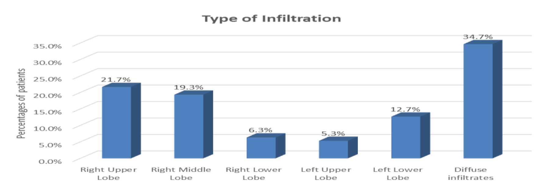
Figure 5: Type of Infiltration
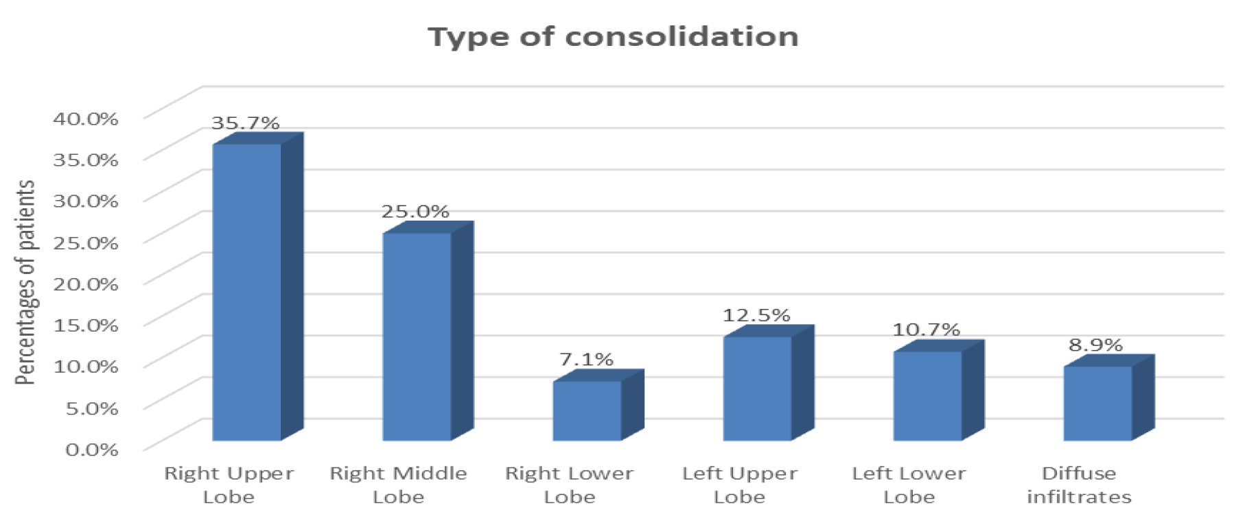
Figure 6: Type of Consolidation
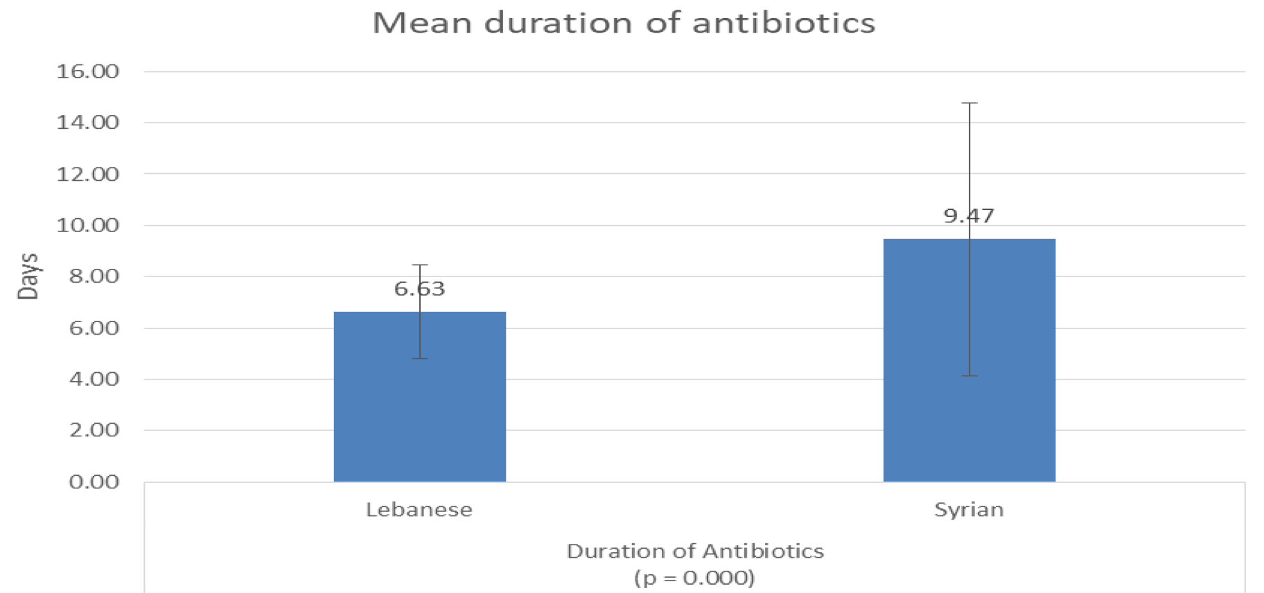
Figure 7: Duration of Antibiotics
Cultures Results
Regarding the blood culture results, 7 patients have Coagulase Negative Staph and were Syrians p = 0.047, 1 Lebanese patient had Moraxella catarrhalis, 1 Syrian patient had staph aureus, 1 Syrian patient had stenotrophomonas and 1 Syrian patient had Heamophilus Influenza. In addition, 3 patients 1.5% had positive DTA culture and 4 patients 2% had positive sputum culture Table 11.
Imaging Results
The radiological findings show that 74.6% of patients have infiltration, 27.9% have consolidation, 4.5% have pleural effusion and 6 patients 3% underwent thoracocentesis Table 12. Infiltration was shown higher in Lebanese patients 82.9% compared to Syrian patients 70.2% p = 0.05. Whereas, consolidation was shown higher in Syrian patients 33.6% compared to Lebanese patients 17.1% p = 0.013.
The most prevalent types of infiltration are diffuse infiltrates 34.7%, infiltration in the right upper lobe 21.7% and infiltration in the right middle lobe 19.3% Figure 5.
The most prevalent types of consolidation are in the right upper lobe 35.7%, consolidation in the right middle lobe 25% and consolidation in the left upper lobe 12.5% Figure 6.
Treatment
Out of 201 patients, 124 patients 61.7% need oxygen supplements. The results show that the Syrian patients need more oxygen supplements 74% compared to Lebanese patients 38.6% p = 0.000 Table 13. In addition, the quantity of oxygen given to Syrian patients is higher compared to the quantity given to Lebanese patients p = 0.05. To note that: 63% of Lebanese patients received 1 L/min nasal cannula whereas 53% of Syrian patients received a minimum of 2 L/min nasal cannula. Out of 201 patients, 27 patients underwent mechanical ventilation and all were Syrians representing 27% of the Syrian population. To note that: the mean days of mechanical ventilation is 6.1 ±5.0 days. Furthermore, the results show that 47.3% of Syrian patients needed IV fluids compared to 28.6% of Lebanese patients p = 0.010. Regarding the antibiotherapy, the majority of patients 99% received antibiotics. The type of the antibiotics differs between the two populations where 88.2% of Lebanese patients were treated by Narrow Spectrum antibiotic compared to 64.9% of Syrian patients and 11.8% of Lebanese patients were treated by Broad Spectrum antibiotic compared to 35.1% of Syrian patients p = 0.000 Table 13.
Out of 201 patients, 35 patients 17.4% were admitted to PICU. The admission to PICU was higher in the Syrian population 26%, compared to Lebanese patients 1.4% p = 0.000. To note that: the mean days of length of stay in PICU is 9.7 ±5.8 days.
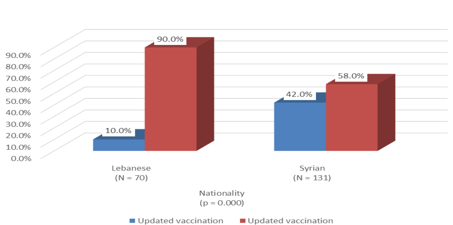
Figure 8: Updated Vaccination
Table 13: CAP Treatment Modalities
Parameters
Nationality
Total
p-value
Lebanese (N = 70)
Syrian (N = 131)
Oxygen supplement
No
43
34
77
0.000 *
61.4%
26.0%
38.3%
Yes
27
97
124
38.6%
74.0%
61.7%
Quantity of oxygen
1 l/min nasal cannula
17
45
62
0.015 *
63.0%
46.4%
50.0%
2 l/min nasal cannula
8
14
22
29.6%
14.4%
17.7%
3 l/min nasal cannula
2
8
10
7.4%
8.2%
8.1%
4 l/min nasal cannula
0
2
2
0.0%
2.1%
1.6%
5 l/min nasal cannula
0
28
28
0.0%
28.9%
22.6%
Mechanical ventilation
No
70
104
174
0.000 †
100.0%
79.4%
86.6%
Yes
0
27
27
0.0%
20.6%
13.4%
IV fluids
No
50
69
119
0.010 *
71.4%
52.7%
59.2%
Yes
20
62
82
28.6%
47.3%
40.8%
Antibiotics
No
2
0
2
0.120 †
2.9%
0.0%
1.0%
Yes
68
131
199
97.1%
100.0%
99.0%
Type of Antibiotics
Broad Spectrum
8
46
54
0.000 *
11.8%
35.1%
27.1%
Narrow Spectrum
60
85
145
88.2%
64.9%
72.9%
PICU admission
No
69
97
166
0.000 *
98.6%
74.0%
82.6%
Yes
1
34
35
1.4%
26.0%
17.4%
* Chi-Square Test † Fisher's Exact Test
Table 14: Laboratory Findings
Parameters
M
Mean
Std. Deviation
Minimum
Maximum
p-value
WBC
(10^6/L)
Lebanese
70
13522.4286
5303.91199
3600.00
26400.00
0.000
Syrian
131
18088.8092
8599.50284
4200.00
47900.00
Neutrophils
(%)
Lebanese
70
55.8314
19.63306
13.10
91.00
0.255
Syrian
131
52.6198
18.98016
17.80
95.00
Lymphocytes
(%)
Lebanese
70
32.4557
18.08639
5.00
76.30
0.145
Syrian
131
36.0855
17.22100
1.00
78.70
Monocytes
(%)
Lebanese
70
9.4986
4.11281
2.00
21.00
0.335
Syrian
131
9.4405
5.47600
1.30
47.00
Basophils
(%)
Lebanese
70
0.7657
0.91396
0.00
4.10
0.648
Syrian
131
0.9237
1.49501
0.00
6.70
Eosinophils
(%)
Lebanese
70
0.7857
0.97895
0.00
5.60
0.305
Syrian
131
1.2130
3.58502
0.00
40.00
Hemoglobin
Lebanese
70
11.3586
1.47004
7.10
14.60
0.001
Syrian
131
10.5389
1.78151
2.80
15.10
Table 14: Continue
MCV
(fl)
Lebanese
70
78.1557
9.96005
36.10
100.00
0.988
Syrian
131
78.4710
9.63960
28.30
101.00
Platelets
(10^6/L)
Lebanese
70
369828.5714
136760.33956
162000.00
737000.00
0.005
Syrian
131
442812.9771
191783.64228
12800.00
1119000.00
BUN
(mg/dl)
Lebanese
70
9.2257
4.98106
2.00
27.00
0.794
Syrian
131
10.0809
6.93944
1.30
45.00
Creatinine
(mg/dl)
Lebanese
70
0.3343
0.53565
0.20
4.70
0.186
Syrian
131
0.4718
1.98720
0.10
23.00
Sodium
(mmol/l)
Lebanese
70
137.3543
4.60418
129.00
162.00
0.716
Syrian
131
136.9176
3.90909
126.90
155.00
Potassium
(mmol/l)
Lebanese
70
4.5657
0.71645
3.40
5.90
0.000
Syrian
131
4.9489
0.71397
3.00
6.30
Chloride
(mmol/l)
Lebanese
70
101.6943
4.73693
90.20
129.00
0.546
Syrian
131
102.3260
9.57782
88.60
195.80
Bicarbonate
(mmol/l)
Lebanese
70
19.5614
3.61792
11.30
28.00
0.283
Syrian
131
19.0565
3.67654
8.30
28.60
CRP
(mg/l)
Lebanese
70
44.2929
49.68403
0.50
276.00
0.012
Syrian
131
78.2244
87.05660
0.20
418.00
HCT
(%)
Lebanese
70
34.5014
4.10081
21.70
44.00
0.001
Syrian
131
32.1053
5.23786
12.20
42.30
Mann-Whitney test
Table 15: Vital Signs Results
Parameters
M
Mean
Std. Deviation
Minimum
Maximum
p-value
ystolic BP
(mmHg)
Lebanese
70
100.97
11.007
77
120
0.148
Syrian
131
98.98
11.691
66
130
Diastolic BP
(mmHg)
Lebanese
70
58.33
9.620
40
80
0.369
Syrian
131
59.88
11.241
40
97
Heart rate
(bt/min)
Lebanese
70
135.03
13.713
103
170
0.000
Syrian
131
148.04
19.59
100
199
Temperature
(°C)
Lebanese
70
38.009
08944
36.0
40.2
0.001
Syrian
131
38.376
0.8510
36.0
40.5
Oxygen Saturation
(%)
Lebanese
70
96.057
2.6642
89.0
100.0
0.000
Syrian
131
91.344
5.5605
75.0
100.0
Mann-Whitney test
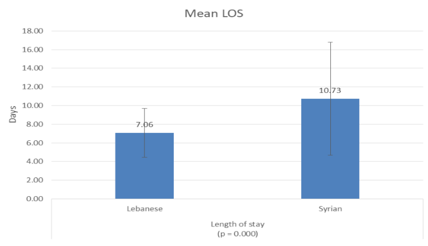
Figure 9: Length of Stay
The mean duration of antibiotic treatment differs between the two populations where Syrian patients received a mean of 9.47 ±5.3 days and the Lebanese patients received a mean of 6.6 ±1.8 days p = 0.000 Figure 7.
The results show that only 58% of Syrian patients got updated vaccination compared to 90% of Lebanese patients p = 0.000 Figure 8.
The mean length of stay differs between the two populations where Syrian patients have a mean LOS of 10.73 ±6.0 days and the Lebanese patients have a mean LOS of 7.06 ±2.6 days p = 0.000 Figure 9.
Laboratory Findings
The laboratory findings show that WBC and CRP, which are markers of the pneumonia severity, were very higher in Syrian patients compared to Lebanese patients. The mean WBC was 18088 10^6/L in Syrian patients whereas the mean WBC was 13522 10^6/L in Lebanese patients p = 0.000. The mean CRP was 78.2 mg/l in Syrian patients whereas the mean WBC was 44.3 mg/l in Lebanese patients p = 0.012 Table 14.
Vital Signs
The vital signs results showed that Syrian patients have more tachycardia p = 0.000, higher fever p = 0.001 and more desaturation p = 0.000, compared to Lebanese patients Table 15.
DISCUSSION
Two hundred and one patients diagnosed having CAP and admitted to RHUH between 2015 and 2020 were included in this study. Out of 5852 pediatric admission during the study period, CAP occurred in 3.4% of patients, with lower rates among Lebanese children (1.2%) compared to Syrian children (2.2%). Consistently, data shows that in UNHCR refugee camps, pneumonia remains one of the most prevalent diseases and one of the main causes of death of children below the age of 5 (17% of child morbidity) [10]. Moreover, significantly higher pneumonia incidence as well as other respiratory illnesses can be seen among refugees in camps when compared to the general population in turkey [12]. More specifically, children who were underweight, those who were in a household using unclean cooking fuel and those belonging to a low-income family were significantly more likely to have CAP [33]. Children in refugee populations in small living space (reduced volume per person), short distance between houses and shorter distance from stove to bed were also at a higher risk of CAP [34]. In the present study, severe CAP was diagnosed in 57.7% and was significantly higher in Syrian patients (88.7%) compared to Lebanese patients (37.1%). When considering refugees and migrant populations, it is well established that refugees, particularly those living in refugee camps, have little to no possibility of adequate hygiene, social distancing and are at a higher risk of disease due to the lack or limited availability of basic amenities such as soap and water [11]. As such, it is expected that these populations are at a much higher risk of contracting infections, such as COVID-19 and pneumonia. Consistently, pediatric refugee populations were shown to be at significantly higher risk of contracting vaccine-type invasive pneumococcal disease and were thus at a higher risk of mortality when compared to German children. Moreover, higher rates of antimicrobial and antibiotic drug-resistant invasive pneumococcal disease were observed among refugee children [13]. The most prevalent clinical symptoms observed in our study were fever (98%), cough (93%), irritability (73.1%) and decreased feeding (45.8%). Syrian children were significantly more likely to exhibit irritability and decreased feeding. Moreover, physical exam findings were more frequently observed among Syrian children, who showed significantly more illness, noisy breathing, respiratory distress, rhonchi, grunting and nasal flaring. This is consistent with the literature, which shows that pneumonia symptoms are not specific to the disease (non-specific) as they are predominately symptoms of respiratory distress (cough, breathing difficulty) and fever [17]. When it comes to infants and young children, pneumonia might present in subtle symptoms such as decreased feeding, lethargy, irritability, inconsolable crying, etc. [17], as observed in our study. Radiological findings showed that infiltration was more prevalent in Lebanese patients (82.9%) and consolidation was more prevalent in Syrian patients. It should be noted that updated vaccination was less frequent among Syrian children, who required a longer period of treatment and length of hospital stay. This is normal considering the higher severity of CAP among Syrian patients, which would translate in a longer need of healthcare. In fact, Syrian children were significantly more likely to have Coagulase Negative Staphylococcus aureus and needed more oxygen supplementation and at a higher quantity than Lebanese children. Syrian children had a higher need for mechanical ventilation, IV fluids and PICU admission. Data also shows that the severity and complications of CAP can also differ according to its etiology. Children aged up to 5 years most frequently exhibited complications in cases of pneumococcal bacteremic CAP [27], which could explain our results. Biomarkers of pneumonia severity (WBC and CRP) were consistently higher among Syrian children when compared to their Lebanese counterparts. In general, it was shown that laboratory seem to be consistent between children who had pneumococcal CAP and those who did not. This included C-reactive protein levels, white blood cell (WBC) counts and the percentage of neutrophils [27]. Another study showed similar results, with median WBC count, absolute neutrophil count (ANC), C-reactive protein (CRP), or procalcitonin failing to act as distinguishing factors between severe and non-severe CAP in a pediatric population [30]. That being said, adjusting the analysis to account for the child’s age as well as other factors showed that CRP and procalcitonin might have potential as predictors of the more severe outcomes of CAP in children [30]. This could account for the significance of biomarkers in the setting of CAP between Syrian and Lebanese patients. Regardless, data correlating socioeconomic and lifestyle factors to the severity of CAP remain limited. Moreover, data from Lebanon regarding CAP in children is severely lacking. However, the Pneumonia Etiology among Refugees and the Lebanese population (PEARL) study will be examining CAP among refugees and local populations in order to establish the etiology of CAP and guide treatment and management strategies [35].`
Impact of the Study
The results of our study will help figure out the impact of humanitarian crisis and poor social conditions on community acquired pneumonia, which remains one of the most common infectious diseases worldwide. This holds true especially in children, which highlights the need to establish their effect on pneumonia prevalence and severity in the studied population. The study offers healthcare providers a better idea regarding pneumonia severity in children especially in Syrian refugees they treat and to take special considerations in dealing with their cases and not to treat them the same as Lebanese children. Improved vaccination and ensuring better living conditions might be key to reducing the prevalence and severity of CAP among Syrian children to match those observed among Lebanese children. This would serve to improve clinical outcomes and reduce the burden of CAP on migrant and refugee populations both on the level of the children, their caretakers and the healthcare system.
Limitations
The present study had several limitations. Firstly, the study included a limited number of patients, with little more than 200 children included. Secondly, data was gathered from a single center and was prospective in nature. This prevents the generalizability of the results and their applicability on a wider scale. Thirdly, seeing as the study was retrospective, information on living conditions and income were not gathered, which does not allow the elucidation of the factors underlying the higher severity of CAP among Syrian children. Finally, the study looked at the prevalence of CAP among children only and did not include adults or siblings in order to provide a more comprehensive understanding of CAP distribution among communities.
Study Perspectives
Future studies should aim to explore CAP in a more comprehensive manner by including a high number of patients. Genders and age groups should be adequately represented, with patients included from all geographical regions. Moreover, data should be gathered prospectively in order to capture all factors potentially mediating the severity of CAP among children. Determining the influence of socioeconomic factors and living conditions is crucial especially in limited-resource setting of Syrian children and their families. Exploring CAP on the level of communities would also be important in an effort to trace the spread of this condition and formulate adequate strategies for its preventions.
CONCLUSION
The present study aimed to determine the difference in severity of CAP requiring hospitalization among Lebanese children and refugees’ children comparing 2 populations belonging to the same age group. It also aimed to examine the prevalence and characteristics of community acquired pneumonia in Syrian children and in Lebanese in an effort to investigate the possible effect of the humanitarian crisis on CAP. Two hundred and one patients diagnosed having CAP and admitted to RHUH between 2015 and 2020 were included in this study. Out of 5852 pediatric admission during the study period, CAP occurred in 3.4% of patients, with lower rates among Lebanese children (1.2%) compared to Syrian children (2.2%). patients with CAP had comparable gender and age between study groups. The most prevalent clinical symptoms observed were fever (98%), cough (93%), irritability (73.1%) and decreased feeding (45.8%). Syrian children were significantly more likely to exhibit irritability and decreased feeding. Moreover, physical exam findings were more frequently observed among Syrian children, who showed significantly more illness, noisy breathing, respiratory distress, rhonchi, grunting and nasal flaring. Severe CAP was diagnosed in 57.7% and was significantly higher in Syrian patients (88.7%) compared to Lebanese patients (37.1%). Syrian children were significantly more likely to have Coagulase Negative Staphylococcus aureus and needed more oxygen supplementation and at a higher quantity than Lebanese children. Syrian children had a higher need for mechanical ventilation, IV fluids and PICU admission. Radiological findings showed that infiltration was more prevalent in Lebanese patients (82.9%) and consolidation was more prevalent in Syrian patients. It should be noted that updated vaccination was less frequent among Syrian children, who required a longer period of treatment and length of hospital stay. Biomarkers of pneumonia severity (WBC and CRP) were consistently higher among Syrian children when compared to their Lebanese counterparts. Our results show that Syrian patients have significantly higher prevalence and severity of CAP. The notable differences between the two patient populations shows the clear impact of the humanitarian crisis, particularly among resource-deprived refugee and migrant populations. Adequate policies and programs should be formulated in order to improve the clinical outcomes of CAP among these populations. Improved vaccination campaigns could be organized along with efforts to provide basic healthcare and living needs.
REFERENCE
Roth, G.A. et al. “Global, regional and national age-sex-specific mortality for 282 causes of death in 195 countries and territories, 1980–2017: a systematic analysis for the Global Burden of Disease Study 2017.” The Lancet, vol. 392, no. 10159, 2018, pp. 1736–1788.
Zhu, Y. et al. “Causes of death in hospitalized children younger than 12 years of age in a Chinese hospital: a 10 year study.” BMC Pediatrics, vol. 18, no. 1, 2018. https://pubmed.ncbi.nlm.nih.gov/29347924/.
Bradley, J.S. et al. “The management of community-acquired pneumonia in infants and children older than 3 months of age.” Clinical Infectious Diseases, vol. 53, no. 7, 2011, pp. e25–e76.
Shoham, Y. et al. “Community-acquired pneumonia in children: Quantifying the burden on patients and their families including decrease in quality of life.” Pediatrics, vol. 115, no. 5, 2005, pp. 1213–1219.
Lovie-Toon, Y.G. et al. “Longitudinal study of quality of life among children with acute respiratory infection and cough.” Quality of Life Research, vol. 27, no. 4, 2018, pp. 891–903.
Lipsett, S.C. et al. “Predictors of bacteremia in children hospitalized with community-acquired pneumonia.” Hospital Pediatrics, vol. 9, no. 10, 2019, pp. 770–778.
Senavonge, A. et al. “Prevalence and predictors of bacteremia among children hospitalized with pneumonia.” Southeast Asian Journal of Tropical Medicine and Public Health, vol. 47, 2016, pp. 994–1000.
Neuman, M.I. et al. “Utility of blood culture among children hospitalized with community-acquired pneumonia.” Pediatrics, vol. 140, no. 3, 2017, e20171013.
Krenke, K. et al. “Clinical characteristics of 323 children with parapneumonic pleural effusion and pleural empyema due to community acquired pneumonia.” Journal of Infection and Chemotherapy, vol. 22, no. 5, 2016, pp. 292–297.
Hershey, C.L. et al. “Incidence and risk factors for malaria, pneumonia and diarrhea in children under 5 in UNHCR refugee camps.” Conflict and Health, vol. 5, no. 1, 2011, p. 24.
Kluge, H.H.P. et al. “Refugee and migrant health in the COVID-19 response.” The Lancet, vol. 395, no. 10232, 2020, pp. 1237–1239.
Kurtuluş, Ş. et al. “Chest diseases in refugees living in a tent camp and in Turkish citizens living in the district.” Turkish Thoracic Journal, vol. 19, no. 3, 2018, pp. 117–121.
Perniciaro, S. et al. “Invasive pneumococcal disease in refugee children, Germany.” Emerging Infectious Diseases, vol. 24, no. 10, 2018, pp. 1934–1936.
Mandell, L.A. et al. “Infectious Diseases Society of America/American Thoracic Society consensus guidelines on the management of community-acquired pneumonia in adults.” Clinical Infectious Diseases, vol. 44, suppl. 2, 2007, pp. S27–S72.
Kalil, A.C. et al. “Is bacteremic sepsis associated with higher mortality in transplant recipients than in nontransplant patients?” Clinical Infectious Diseases, vol. 60, no. 2, 2015, pp. 216–222.
Regunath, H. and Y. Oba. “Community-acquired pneumonia.” StatPearls, StatPearls Publishing, 2020.
Leung, A.K.C. et al. “Community-acquired pneumonia in children.” Recent Patents on Inflammation & Allergy Drug Discovery, vol. 12, no. 2, 2018, pp. 136–144.
Stuckey-Schrock, K. et al. “Community-acquired pneumonia in children.” American Family Physician, vol. 86, no. 7, 2012, pp. 661–667.
Shah, S. et al. “Lack of predictive value of tachypnea in the diagnosis of pneumonia in children.” The Pediatric Infectious Disease Journal, vol. 29, no. 5, 2010, pp. 406–409.
Barson, W.J. “Community-acquired pneumonia in children: clinical features and diagnosis.” UpToDate, 2020.
Said, M.A. et al. “Estimating the burden of pneumococcal pneumonia among adults.” PLOS One, vol. 8, no. 4, 2013, e60273.
Lanks, C.W. et al. “Community-acquired pneumonia and hospital-acquired pneumonia.” Medical Clinics of North America, vol. 103, no. 3, 2019, pp. 487–501.
Rudan, I. et al. “Epidemiology and etiology of childhood pneumonia.” Bulletin of the World Health Organization, vol. 86, 2008, pp. 408–416.
Zar, H.J. and F.P. Polack. “Childhood pneumonia: the role of viruses.” Thorax, vol. 70, 2015, pp. 811–812.
Hassen, M. et al. “Radiologic diagnosis and hospitalization among children with severe community acquired pneumonia.” BioMed Research International, 2019.
Boyd, K. “Back to the basics: community-acquired pneumonia in children.” Pediatric Annals, vol. 46, no. 7, 2017, pp. e257–e261.
Esposito, S. et al. “Bacteremic pneumococcal community-acquired pneumonia in children less than 5 years of age in Italy.” The Pediatric Infectious Disease Journal, vol. 31, no. 7, 2012, pp. 705–710.
Corrales-Medina, V.F. et al. “Association between hospitalization for pneumonia and subsequent risk of cardiovascular disease.” JAMA, vol. 313, no. 3, 2015, pp. 264–274.
Alshahwan, S.I. et al. “The prevalence of complications of pneumonia among adults admitted to a tertiary care center in Riyadh.” Annals of Saudi Medicine, vol. 39, no. 1, 2019, pp. 29–36.
Florin, T.A. et al. “Biomarkers and disease severity in children with community-acquired pneumonia.” Pediatrics, vol. 145, no. 6, 2020.
Ambroggio, L. et al. “Clinical risk factors for revisits for children with community-acquired pneumonia.” Hospital Pediatrics, vol. 8, no. 11, 2018, pp. 718–723.
Torres, A. et al. “Risk factors for community-acquired pneumonia in adults in Europe.” Thorax, vol. 68, no. 11, 2013, pp. 1057–1065.
Ngocho, J.S. et al. “Modifiable risk factors for community‐acquired pneumonia in children under 5 years of age.” Tropical Medicine & International Health, vol. 24, no. 4, 2019, pp. 484–492.
Turner, C. et al. “High rates of pneumonia in children under two years of age in a South East Asian refugee population.” PLOS One, vol. 8, no. 1, 2013, e54026.
Kesteman, T. et al. “Investigating pneumonia etiology among refugees and the Lebanese population (PEARL).” Gates Open Research, vol. 2, 2018.
DeAntonio, R. et al. “Epidemiology of community-acquired pneumonia and implications for vaccination of children.” Human Vaccines & Immunotherapeutics, vol. 12, no. 9, 2016, pp. 2422–2440.
Harris, M. et al. “British Thoracic Society guidelines for the management of community acquired pneumonia in children.” Thorax, vol. 66, suppl. 2, 2011, pp. ii1–ii23.
Alzomor, O. et al. “Management of community-acquired pneumonia in infants and children.” International Journal of Pediatrics & Adolescent Medicine, vol. 4, no. 4, 2017, pp. 153–158.
Recommended Articles
Research Article
An evaluation of Profile of Medico-legal autopsies conducted at Dr. Radhakrishnan Govt. Medical College, Hamirpur, Himachal Pradesh
Download PDF
Research Article
Imaging Evaluation of Ultrasonography Versus Magnetic Resonance,
Imaging in Diagnosis of Carpal Tunnel Syndrome among Female Patients with Positive Nerve Conduction Study
Download PDF
Research Article
The Efficacy of 5- Fluorouracil Cream and 25% Podophyllin Solution in Treatment of Plantar Warts in Alternative Day Regimen ; An Open Therapeutic Trial.
Download PDF
Research Article
Silent Threats: Evaluating Knowledge of Pelvic Inflammatory Disease Among Reproductive-Age Women in District Kangra
Pankaj sharma,
Kritika Katoch
Published: 27/11/2024
Download PDF
PO Box 101, Nakuru
Kenya.
Email: office@iarconsortium.org
Editorial Office:
J.L Bhavan, Near Radison Blu Hotel,
Jalukbari, Guwahati-India
Elias, A. M., Chokr, I., Dana, R. & Merhi, B. A. (2020). Assessing Pneumonia Severity Difference between Syrian Refugees and the Lebanese Children; a Retrospective Study over 5 Years from 2015 Till 2020. International Academic Research Journal of Internal Medicine & Public Health, 1(1), 1-16.
MLA
Elias, Anna M., et al. "Assessing Pneumonia Severity Difference between Syrian Refugees and the Lebanese Children; a Retrospective Study over 5 Years from 2015 Till 2020." International Academic Research Journal of Internal Medicine & Public Health 1.1 (2020): 1-16.
Chicago
Elias, Anna M., Imad Chokr, Rouwayda Dana and Bassem A. Merhi. "Assessing Pneumonia Severity Difference between Syrian Refugees and the Lebanese Children; a Retrospective Study over 5 Years from 2015 Till 2020." International Academic Research Journal of Internal Medicine & Public Health 1, no. 1 (2020): 1-16.
Harvard
Elias, A. M., Chokr, I., Dana, R. and Merhi, B. A. (2020) 'Assessing Pneumonia Severity Difference between Syrian Refugees and the Lebanese Children; a Retrospective Study over 5 Years from 2015 Till 2020' International Academic Research Journal of Internal Medicine & Public Health 1(1), pp. 1-16.
Vancouver
Elias AM, Chokr I, Dana R, Merhi BA. Assessing Pneumonia Severity Difference between Syrian Refugees and the Lebanese Children; a Retrospective Study over 5 Years from 2015 Till 2020. International Academic Research Journal of Internal Medicine & Public Health. 2020 Jul;1(1):1-16.


 Download PDF
 Download XML