This study investigates the levels of heavy metals in dust associated with households in Wukari, Taraba State, Nigeria. The amount of some heavy metals in the dust samples selected from Puje ward, Hospital ward and Ayvi ward were analyzed with the use of Atomic Absorption Spectrophotometer (AAS). Heavy metals determined include Pb, As, Cr, Cd, and Hg. The level of lead was highest in sample station 7 (42.550±1.91 mg/kg) from Hospital ward and lowest in sample1 (22.450±0.49 mg/kg) from Puje ward. Statistical difference exists only between the concentration of lead in sample 1 and 7. There was no statistical difference between the level of lead in sample 1 and other samples and also between sample 7 and the other samples. The level of chromium was highest in sample 1 (2.250±0.13 mg/kg) from Puje ward and lowest in sample 7 (1.300±0.19 mg/kg) and 8 (1.300±0.25 mg/kg) from Hospital ward and Ayvi ward respectively. Cadmium was highest in sample 3 (0.890±0.09 mg/kg) from Puje ward and lowest in sample 4 (0.390±0.13 mg/kg) from Hospital ward. There was a statistical difference between sample station 3 and sample stations 1, 2, 4, 6, 7, 9 and 10. Arsenic and mercury was not detected in all the dust samples. The estimated potential carcinogenic risk to the public from inhalation of dust shows the THQ30 and THQ5 for Cd to be highest in Ayvi ward than in Hospital and Puje ward, while THQ30 and THQ5 for Cr was highest in Hospital ward. The THQ30 and THQ5 for Pb was highest in Puje ward than in Hospital and Ayvi ward. The result therefore shows that Hospital ward is more predisposed to Pb toxicity, while Puje ward is more predisposed to Cr and Cd toxicity.
Wind activities in the northern part of Nigeria is extremely high round the year with special effects during the dry season. The impact of wind on soil particles has raised some issues of concern with respect to human health and indoor environment. Indoor air quality is an environmental health concern because people spend up to 90% of their time indoors in places, such as homes, offices and schools [1]. Indoor settled dusts contain various hazardous materials including heavy metals, which can affect human health. Heavy metals in indoor dusts are important indicator of pollution in agricultural and urban environments [2]. Most heavy metals are very harmful because of their non-biodegradable nature, long biological half-lives and their potential to accumulate in different body parts [3].
Dust is simply small particles in the air. Often, these particles are too small to be seen, but because they are airborne, they can be breathed in through the nose and mouth or through the skin. The size and chemical nature of the dust particles usually determine the effects they may have in the expose organisms. Very small particles can pass through the lungs into other organs of the body. Smaller particles stay in the air for much longer and can be a danger for a longer period of time. Usually the smaller particles are the most hazardous, but larger particle dust can also cause major health problems.
Dust can be caused by a huge number of processes and comes in many different forms. It is divided into two types - organic and inorganic. Inorganic dusts come from stones, chemicals and metals. Among the inorganic dusts that humans encounter are cement, coal, asbestos, metal, concrete, talc, stone, grout and sand. Organic dusts come from living things and include dust from textiles, wood, poultry, leather, grain and flour. These often also contain fungal spores and microbes. Dusts can also come from organic chemicals such as pesticides and dyes.
The two most serious health problems caused by dust are cancers of the lungs, throat and nose, and other lung conditions called Chronic Obstructive Pulmonary Disease (COPD) that includes chronic bronchitis and emphysema.
Indoor settled dusts containing heavy metals are an important source of exposure for people, especially children and other vulnerable individuals. Inhalation, dust ingestion, and dermal contact are common pathways by which toxic heavy metals can enter the body. Through breathing, airborne dust particles pose considerable health risks, such as diverse respiratory illnesses, decreased lung function, and cardiovascular diseases [4]. Heavy metals such as Cd, Ni, and Pb are common examples of metals that cause negative health impacts from breathing and have been noticed from both occupational and ambient air exposure [5]. The ingestion of dust may occur unknowingly, with food and drink, and with respect to young children via the mouthing of non-food items and repetitive hand-to-mouth activity. Otitoju et al. [6] reported that dietary exposure to heavy metals, namely cadmium (Cd), lead (Pb), zinc (Zn), arsenic (As) and copper (Cu), has been identified as a risk to human health through the consumption of contaminated food. Dermal contact is another way for exposure of heavy metals. For example, Ni is classified as a skin sensitizer and it can cause allergic contact dermatitis [7].
Heavy metals are usually non-degradable and their high levels can threaten biological life. Many of them such as Cu and Zn have some biological functions at low concentrations and cause toxic effects at higher than physiological concentrations. Heavy metals can cause anaemia, kidney failure, brain dysfunction, liver cirrhosis, cancer, and cardiovascular diseases. High exposure of Cd, Hg, and Pb may lead to the damage of bone, kidneys and the brain [8]. A number of heavy metals including Cd, Cr, Ni, Pb, and Zn are recognized as suspected carcinogens [9]. Mn may cause liver dysfunction, neurologic and neuropsychiatric illnesses. The presence of Cu irritates the mucous membranes and disturbs the digestive system. Ba is associated with muscle cramps and interferes with the heartbeat. Some heavy metals (e.g., Ba, Cr, Ni and Zn) may cause asthma [10].
In recent years, several studies have been conducted to analyze heavy metals in indoor dusts, such as household, office and classroom dusts. In Wukari city of Taraba State, Nigeria, most of the dusts associated with households are believed to be majorly due to wind effects. Wukari is located in the North-Eastern Nigeria. During winter, before and during rain fall, there is usually high wind effect which often carries these dust particles into households. These dust particles are believed to be one of the sources of heavy metals in the households of Wukari residents. Hence, the reason for this study in Wukari Taraba State Nigeria.
Due to the seasonal high wind effect in Wukari, Taraba State, Nigeria, dusts which are believed to contain accumulations of some heavy metals are been carried into households. This causes health hazards in the households and environmental pollutions. Inhalation or contact with the dust particles containing heavy metals could pose lots of health challenge to human. Therefore, investigation of the concentrations of some heavy metals in household dust in Wukari, Taraba State warrants research.
Sample Collection
Indoor dust was collected from selected sections of fifteen different households representing the three different wards in Wukari Local Government Area of Taraba State, Nigeria. The samples were collected between May – June, 2017. Using a clean plastic brush and pan, dust was collected inside the household by gradually sweeping the selected section of the household’s floor. The indoor dust was then transferred into a plastic container, brought to the laboratory, sieved and finally sun-dried.
Digestion for Heavy Metals
About 0.50g of soil sample was weighed into 125ml beaker, 100ml of distilled water was added and 0.5ml of HNO3. Finally, 5.0ml of HCl was added to the beaker. The samples were then heated on a hot plate in a well-ventilated hood until the volume has been reduced to 15-20ml. The samples were allowed to cool and precipitates were removed. After that the digested samples were analyzed using the Atomic Absorption Spectrophotometer (AAS).
Determination of Heavy Metal Concentration
The concentrated sample was taken to the UNICAM 969 Atomic Absorption Spectrophotometer (AAS) for heavy metal analysis. The sample was aspirated into an acetylene flame ignited by a hollow cathode lamp at specific wavelength peculiar to the metal lamp used for the analysis. The metals analyzed adopted the ASTM D method (Standard Test Methods).
Statistical Analysis
Statistical analysis was done with the use of Analysis of Variance (ANOVA) and further with LSD and group mean were compared for significance at p≤0.05. Results were presented as mean±standard deviation.
Estimation of target hazard quotients and target cancer risk.

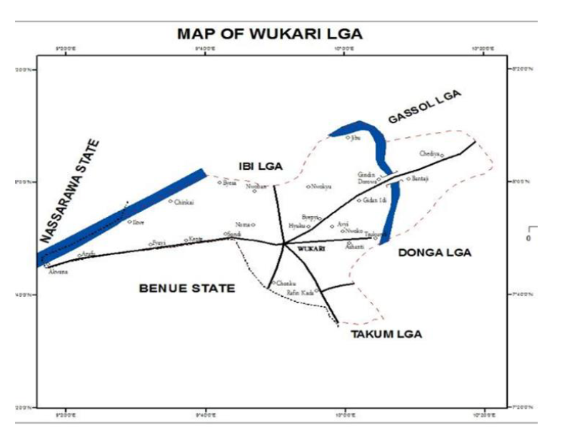
Figure 1: Map of Wukari LGA
www.kwararafauniversity.edu.ng.Retrieved 2016-12-29. 12:30am
The methodology for estimation of target hazard quotients (THQ) and target cancer risk (TR) used was provided in USEPA Region III Risk-Based Concentration Table, January–June 1996 (US EPA 1996). For carcinogenic effects (inorganic As), risk is expressed as excess probability of contracting cancer over a lifetime (70 years). For noncarcinogenic effects, risk is expressed as a target hazard quotient, the ratio between the exposure and the reference dose. The models for estimating target hazard quotients and target cancer risks (lifetime cancer risks) are:
THQ = EFr x EDtot x SFI x MCS x 10-3
RfDo x BWa x ATn
TR = EFr x EDtot x SFI x MCS x CPSo x 10-3
BWa x ATc
Where THQ: target hazard quotient; EFr: exposure frequency (350 days/year); EDtot: exposure duration, total (30 years); SFI: seafood ingestion (g/day); MCS: metal concentration in edible portion of seafood (µg/g); RfDo: reference dose, oral (mg/kg/day); BWa: body weight, adult (65 kg); ATn: averaging time, noncarcinogens (EDtot 3 365 days/year); TR: target cancer risk; CPSo: carcinogenic potency slope, oral (risk per mg/kg/day); ATc: averaging time, carcinogens (25,550 days).
The level of lead was highest in Hospital ward; ranging from 27.650±7.14 to 42.550±1.91 mg/kg. Statistical difference exists only between the concentration of lead in sample 1 and 7. There was no statistical difference between sample 1 and other samples and also between sample 7 and the other samples. The level of chromium was highest in Puje ward; ranging from 1.680±0.65 to 2.250±0.13 mg/kg. There was no statistical difference in the concentrations of Cr among all the samples. The level of cadmium was highest in Puje ward; ranging from 0.460±0.01 to 0.890±0.09 mg/kg. There was a statistical difference between sample 3 and samples 1, 2, 4, 6, 7, 9 and 10. Arsenic and mercury was not detected in all the dust samples (Table 1).
Table 1: Results of Selected Heavy Metals in Dust Samples (Mg/Kg)
| Location | Sample | Pb | As | Cr | Cd | Hg |
| Puje ward 1 | 1 | 22.450±0.49a | ND | 2.250±0.13a | 0.460±0.01a | ND |
| Puje ward 1 | 2 | 32.400±15.84a,b | ND | 1.680±0.65a | 0.540±0.11a | ND |
| Puje ward 1 | 3 | 28.900±7.21a,b | ND | 1.790±0.33a | 0.890±0.09b | ND |
| Hospital ward | 4 | 27.650±7.14a,b | ND | 1.880±0.69a | 0.390±0.13a | ND |
| Hospital ward | 5 | 34.000±0.56a,b | ND | 2.110±1.10a | 0.700±0.33a,b | ND |
| Hospital ward | 6 | 32.250±0.71a,b | ND | 1.670±0.82a | 0.420±0.01a | ND |
| Hospital ward | 7 | 42.550±1.91b | ND | 1.300±0.19a | 0.420±0.04a | ND |
| Ayvi ward | 8 | 29.300±4.10a,b | ND | 1.300±0.25a | 0.600±0.16a,b | ND |
| Ayvi ward | 9 | 26.750±6.72a,b | ND | 1.670±0.82a | 0.420±0.01a | ND |
| Ayvi ward | 10 | 36.050±5.87a,b | ND | 1.740±0.66a | 0.440±0.08a | ND |
Values are mean ± standard deviation of duplicate determinations. Mean in the same column having different letters of the alphabet are statistically significant (p<0.05). ND = Not detected
Table 2 shows the results of selected heavy metals in dust samples (mg/kg) based on locations. The average level of lead concentration was highest in Hospital ward (34.113±6.41 mg/kg). The average levels of chromium and cadmium concentrations were highest in Puje ward (1.905±0.43 and 0.628±0.21 mg/kg respectively).
Table 2: Results of Selected Heavy Metals in Dust Samples (Mg/Kg) Based on Locations
| Location | Pb | As | Cr | Cd | Hg |
| Puje ward 1 | 27.971±9.00 | ND | 1.905±0.43 | 0.628±0.21 | ND |
| Hospital ward | 34.113±6.41 | ND | 1.740±0.66 | 0.480±0.19 | ND |
| Ayvi ward | 30.700±6.14 | ND | 1.572±0.53 | 0.483±0.12 | ND |
Values are mean ± standard deviation of location (ward) determinations. ND = Not detected.
Table 3 show the estimation of potential health risks of selected heavy metals in dust samples based on locations.
Metal concentrations detected in dust from Wukari were estimated to investigate potential carcinogenic and non-carcinogenic risk to the public from inhalation of dust. The table shows target hazard quotients (THQ) and target cancer risk (TR) caused by inhalation of dust. The THQ30 and THQ5 for Cd was highest in Ayvi ward than in Hospital and Puje ward, while THQ30 and THQ5 for Cr was highest in Hospital ward. The THQ30 and THQ5 for Pb was highest in Hospital ward than in Puje and Ayvi ward.
The TR5 for Cd was highest in Puje ward, while TR30 was highest in Ayvi ward. The TR5 and TR30 for Cr was highest in Puje ward. The TR5 for Pb was highest in Ayvi ward, while TR30 was highest in Hospital ward.
Table 3: Estimation of Potential Health Risks of Selected Heavy Metals in Dust Samples Based on Locations
| Location | Heavy metals | THQ5 | THQ30 | TR5 | TR30 |
| Puje ward 1 | Pb | 4.1 x 10⁻⁹ | 4.1 x 10⁻⁹ | 8.8 x 10⁻¹³ | 5.3 x 10⁻¹² |
| Cr | 1.3 x 10⁻⁹ | 1.3 x 10⁻⁹ | 6.0 x 10⁻¹⁴ | 3.6 x 10⁻¹³ | |
| Cd | 6.6 x 10⁻⁹ | 6.6 x 10⁻⁹ | 1.9 x 10⁻¹⁴ | 1.2 x 10⁻¹² | |
| Hospital ward | Pb | 9.9 x 10⁻⁹ | 9.9 x 10⁻⁹ | 1.1 x 10⁻¹² | 6.4 x 10⁻¹² |
| Cr | 9.8 x 10⁻⁹ | 9.8 x 10⁻⁹ | 5.4 x 10⁻¹⁴ | 3.2 x 10⁻¹³ | |
| Cd | 6.9 x 10⁻⁹ | 6.9 x 10⁻⁹ | 1.4 x 10⁻¹⁴ | 8.5 x 10⁻¹⁴ | |
| Ayvi ward | Pb | 9.2 x 10⁻⁹ | 9.2 x 10⁻⁹ | 9.8 x 10⁻¹³ | 5.9 x 10⁻¹² |
| Cr | 6.4 x 10⁻⁹ | 6.4 x 10⁻⁹ | 5.1 x 10⁻¹⁴ | 3.0 x 10⁻¹³ | |
| Cd | 9.6 x 10⁻⁹ | 9.6 x 10⁻⁹ | 1.5 x 10⁻¹⁴ | 9.3 x 10⁻¹⁴ |
Legend: THQ5= Target hazard quotient for 5 years, THQ30= Target hazard quotient for 30 years, TR5= Target cancer risk for 5 years, TR30= Target cancer risk for 30 years.
In this study, the presence and concentrations of heavy metals in dust associated with households in Wukari, Taraba State, Nigeria was investigated. Three different wards were selected for the study. They include Puje ward, Hospital ward and Ayvi ward. The concentrations of the selected heavy metals in the dust samples from the three wards were analyzed with the use of Atomic Absorption Spectrophotometer (AAS). Heavy metals determined include Pb, As, Cr, Cd, and Hg.
The average level of lead concentration was highest in Hospital ward (34.113±6.41 mg/kg) and lowest in from Puje ward (27.971±9.00 mg/kg). Lead (Pb) is a highly toxic metal whose widespread use may cause extensive environmental contamination and health problems in many parts of the world, especially, Wukari populace of Taraba State, Nigeria. As a result of the wide applications and usage, exposure of humans to lead through inhalation of dust particles is unavoidable. Lead poisoning is a widely studied occupational and environmental hazard. The result of this study shows that lead concentration in dust samples among the three wards examined was highest in Hospital ward. This shows that people living in hospital ward will be predisposed to lead toxicity and may suffer health hazards resulting from lead poisoning or toxicity.
The sources of lead exposure may include mainly industrial processes, food and smoking, drinking water, domestic sources, gasoline, paints and dust particles [11]. Lead is an extremely toxic heavy metal that does not play any major biological function, but disturbs various physiological processes and unlike other metals. A high lead concentration fastens the production of reactive oxygen species (ROS), causing lipid membrane damage that ultimately leads to damage of some vital biological processes and could suppresses the overall growth. This means that people living in Puje ward may suffer less harm that may result from lead exposure than those living in Ayvi ward and Hospital ward of Wukari Local Government Area of Taraba State. Many researchers have shown that oxidative stress in living cells is caused by the imbalance between the production of free radicals and the generation of antioxidants to detoxify the reactive intermediates or to repair the resulting damage. Under the influence of lead, the level of the reactive oxygen species (ROS) increases and the level of antioxidants decrease. Lead can substitute calcium even in picomolar concentration affecting protein kinase C, which regulates neural excitation and memory storage [12].
Arsenic and mercury were not detected in all the dust samples. Humans may encounter arsenic by natural means, industrial source, or from unintended sources. Drinking water may get contaminated by use of arsenical pesticides, natural mineral deposits or inappropriate disposal of arsenical chemicals. Accidental consumption of arsenic by children may result in cases of acute poisoning [13]. Arsenic is a protoplastic poison since it causes malfunctioning of cell respiration, cell enzymes and mitosis. Since arsenic was not detected in the three different wards, it means that people living in these wards will not suffer from arsenic toxicity. Mercury is often considered as the most toxic heavy metal in the environment. Mercury poisoning is referred to as acrodynia or pink disease. Exposure to metallic mercury vapours at higher levels for shorter periods of time can lead to different diseased conditions such as lung damage, vomiting, diarrhoea, nausea, skin rashes, increased heart rate or blood pressure. Since mercury was not also detected in the three different wards, it means that people living in these wards will not suffer from mercury toxicity. Since arsenic and mercury were not detected in all the dust samples, it implies that human and environmental activities that cause accumulations of these metals are less practiced or experienced.
The average level of cadmium concentration was highest in Puje ward (0.628±0.21 mg/kg) and lowest in Hospital ward (0.480±0.19 mg/kg). This shows people living in Puje ward are prone to exhibit the toxicity that may exist due to Cd toxicity. Cadmium interacts with essential nutrients through which it causes its toxicity effects. Premature birth and reduced birth weights are the issues that arise if cadmium exposure is high during human pregnancy. Inhabitants of Hospital ward and Ayvi ward are less prone to these toxic effects than inhabitants of Puje ward.
The average level of chromium was highest in Puje ward (1.905±0.43 mg/kg) and lowest in Ayvi ward (1.572±0.53 mg/kg). Chromium is extensively used in industries such as metallurgy, electroplating, production of paints and pigments, tanning, wood preservation, chemical production and pulp and paper production. These industries play a major role in chromium pollution with an adverse effect on biological and ecological species [14]. A wide range of industrial and agricultural practices increases the toxic level in the environment causing concern about the pollution caused by chromium. Exposure to chromium compounds can result in the formation of ulcers, which will persist for months and heal very slowly. Ulcers on the nasal septum are very common in case of chromate workers. Exposure to higher amounts of chromium compounds in humans can lead to the inhibition of erythrocyte glutathione reductase, which in turn lowers the capacity to reduce methemoglobin to haemoglobin [15]. Exposure to a higher amount of chromium could be more possible in Puje wards than in Ayvi ward and Hospital ward.
The results of this study have shown the concentrations of the selected heavy metals in dust samples from Hospital ward, Puje ward and Ayvi ward. The result therefore shows that Hospital ward is more predisposed to Pb toxicity, while Puje ward is more predisposed to Cr and Cd toxicity. Therefore, adequate precautionary measures should be taken by people living in these wards to avoid excessive inhalation of these metals through dust particles in the households. This will help prevent the toxicity that may arise as a result of heavy metal toxicity.
Tran D.T. et al. “Elemental characterization and source identification of size resolved atmospheric particles in French classrooms.” Atmospheric Environment, vol. 54, 2012, pp. 250-259.
Lu X. et al. “Contamination assessment of copper, lead, zinc, manganese and nickel in street dust of Baoji NW, China.” Journal of Hazardous Materials, vol. 161, 2009, pp. 1058-1062.
Otitoju O. et al. “Quantification of heavy metals in some locally produced rice (Oryza sativa) from the northern region of Nigeria.” Journal of Environment and Earth Science, vol. 4, no. 4, 2014, pp. 67-71.
Turner A. and Hefzi B. “Levels and bioaccessibilities of metals in dusts from an arid environment.” Water Air Soil Pollution, vol. 210, 2010, pp. 483-491.
Vincent J.H. “Health-related aerosol measurement: a review of existing sampling criteria and proposals for new ones.” Journal of Environmental Monitoring, vol. 7, 2005, pp. 1037-1053.
Otitoju O. et al. “Heavy metal contamination of green leafy vegetable gardens in Itam Road construction site in Uyo, Nigeria.” Research Journal of Environmental and Earth Sciences, vol. 4, no. 4, 2012, pp. 371-375.
Mazinanian N. et al. “Nickel release and surface characteristics of fine powders of nickel metal and nickel oxide in media of relevance for inhalation and dermal contact.” Regulatory Toxicology and Pharmacology, vol. 65, 2013, pp. 135-146.
Barregard L. “Health effects of inorganic mercury.” Toxicology Letters, vol. 164, 2006, p. S11.
Cook A.G. et al. “Health effects of natural dust: role of trace elements and compounds.” Biological Trace Element Research, vol. 103, 2005, pp. 1-15.
Khaparde V.V. et al. “Influence of burning of fireworks on particle size distribution of PM10 and associated barium at Nagpur.” Environmental Monitoring and Assessment, vol. 184, 2012, pp. 903-911.
Thurmer K. et al. “Autocatalytic oxidation of lead crystallite surfaces.” Science, vol. 297, no. 5589, 2002, pp. 2033-2035.
Flora S.J.S. et al. “Heavy metal induced oxidative stress and its possible reversal by chelation therapy.” Indian Journal of Medical Research, vol. 128, 2012, pp. 501-523.
Mazumder G. “Chronic arsenic toxicity and human health.” Indian Journal of Medical Research, vol. 128, no. 4, 2008, pp. 436-447.
Ghani A. “Effect of chromium toxicity on growth, chlorophyll and some mineral nutrients of Brassica juncea L.” Egyptian Academic Journal of Biological Sciences, vol. 2, no. 1, 2011, pp. 9-15.
Schlatter C. and Kissling U. “Acute fatal bichromate poisoning.” Beitrage zur Gerichtlichen Medizin, vol. 30, 1973, pp. 382-388.