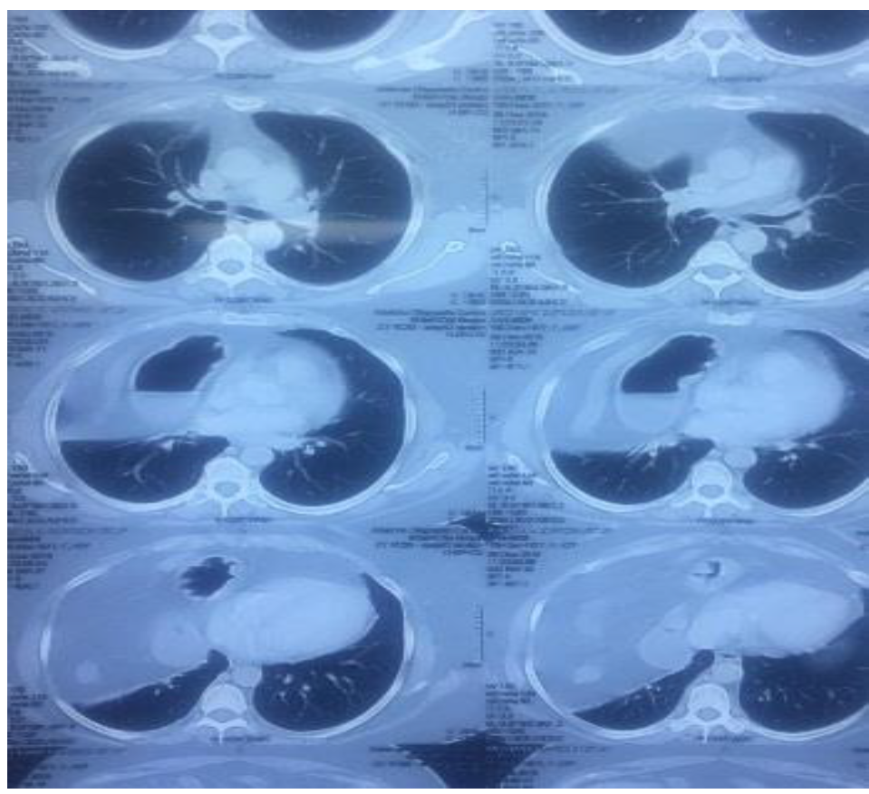
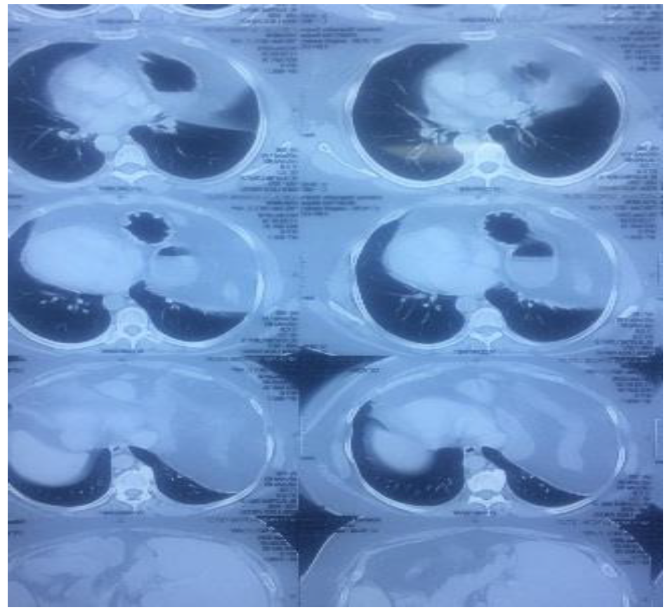
Diaphragm is a musculoaponeurotic barrier dividing thoracic cavity from that of abdominal. Traumatic diaphragmatic rupture is a rare clinic pathological entity and a missed diagnosis. Diaphragmatic injury may result in the development of delayed herniation and strangulation of abdominal organs into the thoracic cavity. Spontaneous (effort or delayed) rupture of diaphragm is rarely seen with the incidence of approx. 1% without obvious blunt trauma of abdomen. This poses a great difficulty to the surgeons and radiologists to diagnose. Very easily the diagnosis is missed or confused with pneumothorax or hydro pneumothorax. The Spontaneous rupture of Right sided diaphragm is very rare in comparison to left sided rupture. Diaphragmatic hernia without trauma in adults is a very rare entity. The reported cases of Right sided diaphragmatic hernia in literature so far is only less than 20. We report 2 cases of Right sided spontaneous delayed diaphragmatic hernia, who presented in our out patients clinic with the chief complaint of breathlessness on exertion. In both the cases, the defect was repaired with primary closure and reinforced with marlex mesh.
Keywords
Ruptured Diaphragmatic Hernia
Spontaneous Diaphragmatic Hernia
Stranuous Athletic Exercise
Blunt Abdominal Injuries
Motor Vehicle Accidents and Effort Diaphragmatic Rupture
INTRODUCTION
In 1848, Vincent Alexander Bochdalek first described the nonfusion of Posterolateral Right or Left foramina of diaphragm leading to mainly left sided Bochdalek hernia [1-2].
The diaphragm is dome shaped Musculotendinous structure, which is located between Thorax and Abdomen and separating the thoracic contents from that of abdominal cavity. It consists of central tendon with Right and Left leaflets composed of striated muscles. Three large openings for Oesophagus, Aorta and IVC disrupt the continuity of diaphragm. The diaphragm plays an important role in respiratory function too [3-4].
Though the traumatic diaphragmatic rupture is a rare clinic pathological condition [5-6], yet it may occur due to the location of diaphragm that it exposes to the injuries either by closed (blunt) or penetrating trauma through the thoracoabdominal transition area on either side [3-4].
Right sided diaphragmatic rupture is less common than that of left side, which is due to the protective anatomic lie of liver which acts as buffer, therefore it becomes difficult challenge to diagnose Right diaphragmatic injury, because the clinical signs are often nonspecific [5-6]. A spontaneous ruptured (acquired) diaphragmatic hernia without any apparent h/o trauma or any violent activity which leads to Valsalva maneuver causing a sudden surge in abdominothoracic pressure gradient, is an even more rare presentation and difficult to diagnose.
Approximately less than 20 cases of right sided ruptured diaphragm without apparent trauma have been reported in the literature so far [7]. Here we are reporting 2 more such cases of right sided spontaneous (delayed/efforts) diaphragmatic hernia.
Literature Search
Incidence: Petrone et al. [3] reported traumatic diaphragmatic hernia 75% on Left side and only 25% on Right side, whereas other authors have reported more than 80% Left sided diaphragmatic rupture [8-9]. However, an increase in incidence on Right sided diaphragmatic rupture being reported due to an increased improvement in imaging techniques [3].
Traumatic diaphragmatic hernias frequently caused by pretreating abdominal injuries are 10-19% whereas blunt thoraco abdominal trauma causes only 5% [10].
Mechanism of injuries: The diaphragm separates the negative pressure thorax from that of positive pressure abdomen. The rupture of diaphragm occurs when intra-abdominal pressure suddenly rises above the tensile strength of diaphragm. The commonest cause of spontaneous acquired diaphragmatic disorders is by trauma, which may either be blunt or penetrating. The blunt trauma produces larger radial tears of 5-15 cm of the diaphragm. Blunt diaphragm injury occurs more frequently on Lt side due to the congenitally weak area in the diaphragm.
Diaphragmatic rupture [11] may be:
Indirect or tension type due to distortion of bony thorax
Direct or impact type resulting from blunt force on the bony thorax
The most common mechanism of spontaneous rupture of diaphragm involves sudden and forceful Valsalva maneuver that can lead to an in-coordination between the expiratory muscles causing rupture [12]. This Valsalva maneuver causes a sudden increase in abdomino thoracic pressure gradient, which is believed to be the primary underlying mechanism responsible for diaphragmatic rupture. Relatively less pressure is required to disrupt the diaphragm in spontaneous (effort) rupture in comparison to blunt traumatic diaphragm rupture.
Mechanism of rupture may be due to:
Shearing of a stretched membrane/diaphragm thought to be an important factor in pathogenesis of the diaphragmatic hernia [13]
Avulsion of diaphragm from its points of attachments
Sudden transmission of force through viscera acting as viscous fluid
The spontaneous closure of the diaphragm tear is unlikely because of abdomino thoracic pressure gradient and the progression to enlargement of the defect which lead to thoracic herniation of intra-abdominal viscera.
The factors increasing intra-abdominal pressure and abdominothoracic pressure gradient can be pregnancy (parturition) [14], child birth (vaginal delivery) [15] blunt abdominal trauma, sneezing, violent vomiting and coughing [16-17], excessive retching, stranuous physical exercise and dancing [18] or athletic events e.g. Heavy weight lifting [19-20], straining at stool (i.e. Valsalva maneuver for constipation) [21], asthma and pertusis and even large meals can increase the risk of herniation. Road traffic accident is another most common cause [22].
Hernial Contents
The hernial contents depend on the type and site of injury and the side of the diaphragm ruptured. Mostly the diaphragm rupture occurs on Left side than right side. Hence the hernial contents may be Liver, stomach, colon, spleen, pancreas, greater omentum and large bowel etc. Aydin Kurt et al. [23] reported these viscera as the hernial contents in non-traumatic right diaphragmatic hernia for the first time.
Diagnosis
Diagnosis is based on clinical presentation. A spontaneous rupture implies an absence of trauma, but there is always the possibilities that a diaphragmatic defect arose from some forgotten trauma in the past [20]. Hence acquired diaphragmatic hernias are usually traumatic and blunt thoraco abdominal trauma accounts for 75-81% of all the diaphragm trauma cases [24]. Depending on the side of the diaphragmatic rupture, size of the defect and the hernial contents, the symptoms may be related to pulmonary, gastro intestinal or a combination [25].
The symptoms in adult spontaneous diaphragmatic hernias are usually or predominantly chronic, vague and Intermittent in nature but can present acutely with constant and special symptoms [25]. The symptoms relating to GIT, the patient can have recurrent abdominal pain, back pain, abdominal distension, post prandial fullness, nausea and vomiting, whereas the respiratory symptoms in the adults are less common yet the patient may present to the outpatient clinics with c/o shortness of breath, breathlessness on exertion (Dyspnoea), Cyanosis, Chest pain, Palpitation or referred pain to shoulder.
Imaging X-ray Chest
There is no single investigation that provides a reliable diagnosis of traumatic diaphragmatic hernia with bowel contents or coiled NG tube in the chest at initial presentation. Though, the diagnosis of diaphragmatic hernia may be obvious, still the routine Chest X-ray is non diagnostic in up to 40% of cases. This percentage may further be reduced in intubated patients with positive pressure ventilation, which prevents herniation of abdominal contents into the chest. Further, it could be difficult to differentiate from diaphragmatic abdominal hernia especially a “hernial sac”. Right sided diaphragmatic injuries, unless resulting in large defects, may particularly be difficult to identify on X-ray chest. The chest X-ray with NG tube insertion for upper GI Barium or Abdominal CT scan or Abdominal MRI has been found to be the best diagnostic tool. This may also show unilateral elevation of diaphragmatic dome, sub diaphragmatic densities and displacement of abdominal organs. Chest radiograph in diaphragmatic hernia may reveal nonspecific findings, like an arch like shadow of raised hemi diaphragm, extraneous densities above the diaphragm and mediastinal shifts to contralateral side and disc like atelectasis adjacent to raised diaphragm [9].
Ultrasound
Ultrasound is now frequently used in early evaluation of trauma patients for free fluid in abdomen, chest or pericardium but an experienced operator may be able to visualize an injury to the diaphragm. A negative study does not exclude the diagnosis.
C.T. Scanning
CT scanning may be useful in detecting diaphragmatic injury and to rule out different diagnosis like pulmonary embolism, pneumonia, spontaneous pneumothorax etc. However, most of the patients with penetrating injury still don’t receive a correct preoperative diagnosis. The findings consistent with diaphragmatic injury such as “waist like constriction of abdominal viscera i.e. collar sign”, intrathoracic herniation of abdominal viscera and diaphragmatic discontinuity may be recorded. The helical CT, especially the coronal and sagittal reformatted images is useful in the diagnosis of acute diaphragmatic rupture after blunt trauma. Helical CT can detect 78% of Left sided and 50% of right sided injuries [26].
MRI
In most of the cases, a standard chest X-ray or CT may reveal the diagnosis, yet in some cases more subtle signs require careful analysis of CT images and MRI in some specific situations. MRI with breath hold permits good visualization of diaphragmatic abnormalities, but the technique cannot be performed in an emergency situation [27].
Laparoscopy and Thoracoscopy
Some authors have advised to induce pneumoperitoneum being a safe and accurate diagnostic tool for differentiating diaphragmatic hernia from a paralyzed or eventuated diaphragm. In diaphragmatic hernia, the injected air will enter from peritoneum into the pleural cavity [28]. Thoracoscopy may be used to visualize the diaphragm when the diagnosis of diaphragmatic rupture is considered and in case laparotomy is not required to manage other injuries. Laparoscopy has a sensitivity of about 88% and a sensitivity of nearly 100% in evaluating for diaphragmatic Injury [29]. N. D’souza et al. further emphasized that diagnostic laparoscopy offers a diagnostic and therapeutic tool to prevent progression of occult traumatic injury to chronic diaphragmatic hernias [30]. Right sided traumatic diaphragmatic injury is a risk factor for morbidity and mortality.
Case-1
This 42 years old male patient got admitted in our hospital with h/o of breathlessness on exertion since last 4yrs and hypertension 2yrs, for which he was on regular treatment with Tab Olmezest H80 mg OD. During Preanesthetic checkup, he disclosed a h/o fall 4yrs ago and hitting his right side of chest on a sofa couch and after that he used to feel discomfort on exertion. It was also revealed that the he had pneumonia at the age of 4yrs and had right 10th rib partial resection with ICD insertion for 4-5 months, but he did not respond the treatment and hence he switched over to some homeopathic medication and got recovered completely. In later life, the patient remained a marathon runner (12km) consecutive for 3yrs without any problem. He was a chronic alcoholic and used to ingest 350ml spirit every day, which he left since approx. 5 months ago. He was a tobacco, gutka chewer and chain smoker. He used to get breathless off and on while dancing and exertional work for which he used to take rest in between.
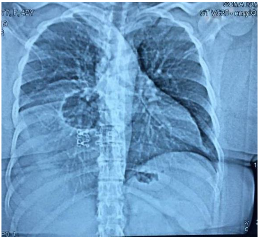
Earlier, the patient got admitted at some other hospital for his fever of 102-103oF. His fever subsided with the treatment given but breathlessness still persisted, for which he underwent flexible bronchoscopy there, which revealed normal vocal cards, trachea and carina but there was an evidence of extrinsic compression over right middle and lower lobe. Hence C.T. Chest was advised, which revealed right sided diaphragmatic hernia.
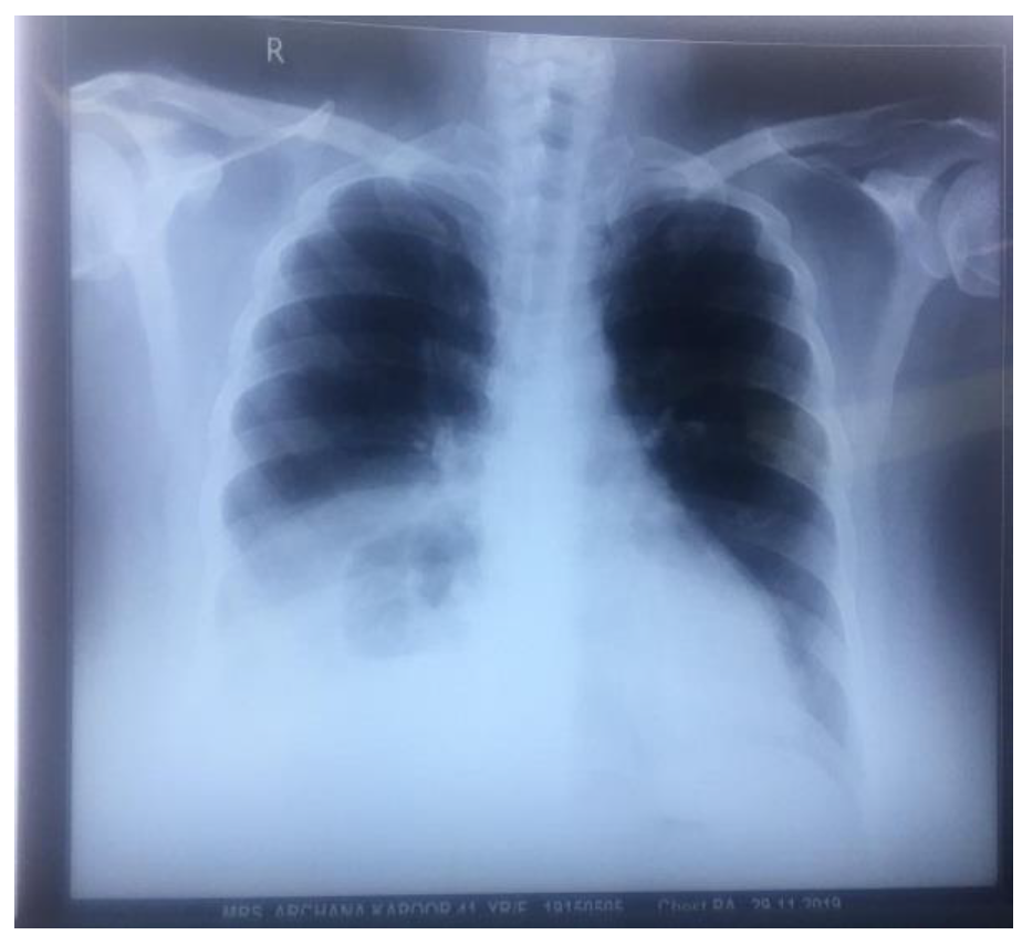
Now the patient was planned for progressive pneumoperitoneum to confirm the diaphragmatic rupture. Preoperative his CBC, KFT, LFT, Blood sugar, serum electrolytes ABG, EKG and 2D ECHO all were found within normal limits. X-ray Chest (PA view) revealed right middle and lower zone opacification.
Pneumoperitoneum was performed ↓GA with spontaneous respiration by slow injection of 2 lit of gas over 1/2hr period. The post pneumoperitoneum X-ray chest did not reveal any evidence of pneumothorax. A day after, the patient was posted for right diaphragmatic hernia repair
Case-2
This 42yrs. old female patient was referred to our pre-anesthesia clinic from outpatient department of our hospital. She presented in OPD with c/o cough with expectoration since last 8 months, hypertension 6 months, hypothyroidism 5 yrs and breathlessness on exertion since last 5 months and the patient was on regular treatment.
During pre-anesthetic checkup she revealed that she had forceful projectile vomiting about 3½ yrs. ago with epigastric pain radiating to her back. Since then she was able to climb only one flight of stairs. Her past surgical history revealed the excision of her right breast fibroadenoma approx. about 11yrs ago under GA, which was uneventful.
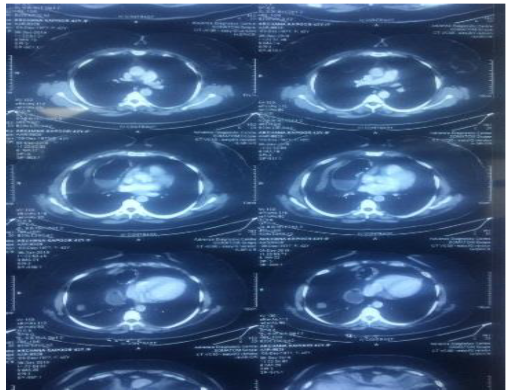
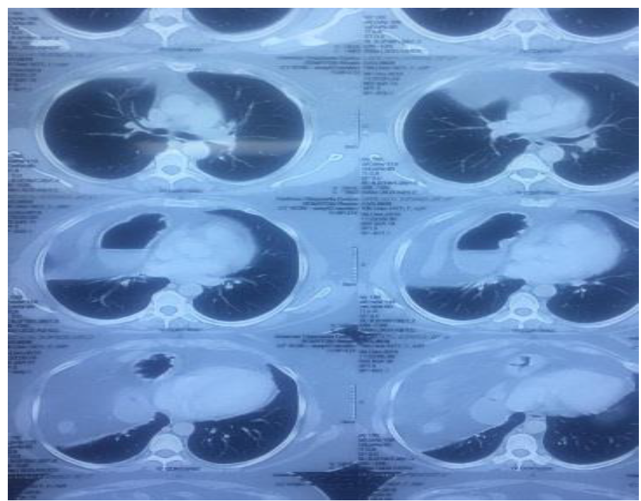
On examination, her BP was 160/90mmg, HR 106/mt and Spo2 97% on room air. Chest auscultation revealed bilateral decreased breath sounds on the bases anteriorly and heart sounds appeared clinically normal with no murmur. Her CBC, blood sugar, KFT, urine examination, ECG all were within normal limits. 2D ECHO revealed LVEF 55% with normal study. X-ray chest PA view showed elevated right dome of diaphragm with gas shadows and area of fluid and air level. This raised the suspicion of abdominal viscera in right thorax. Hence the patient was advised CT chest, which revealed the herniation of fat, colon and duodenal loop in right thorax and confirmed the diagnosis of Rt. Sided Spontaneous Ruptured Diaphragmatic Hernia. The patient was recounselled and discussed about the pathology and was advised to get her hernia repaired at the earliest. She was also advised to continue her antihypertensive and thyroxin treatment till the time of surgery.
The patient took almost 3½ month to decide to undergo surgery. At the time of admission her BP was well controlled and there was no fresh complaint. Hence the patient was posted for her diaphragmatic hernia repair on the next morning.
Figure 1: X-ray Chest PA View - Elevated Right Dome of Diaphragm with Gas Shadows and Area of Fluid and Air Level
Figure 2: CT Chest
Figure 3: Axial Thoracic CT Scan
Figure 4: Axial HRCT Thorax Showing Pulmonary and Mediastinal Structures
Figure 6: Axial CT Scan Images of the Thorax Demonstrating Lung and Mediastinal Structures
Anesthetic Management
The surgeon took case 1 for progressive pneumoperitoneum under GA as described in case report, which turned out to be negative for pneumothorax, while in case 2, the surgeon deferred the procedure and decided to take the patient for surgical correction on CT finding.
In case 1, the diaphragmatic defect was repaired through thoracoabdominal approach and for which tracheal intubation was done with left sided medium size Robertshaw DLT. In case 2, the surgical repair was done through epigastric and right subcostal incision, for which single lumen endotracheal intubation was performed. NIBP, ECG, pulse oximetry, ECCO2, urine output, temperature and wide bore IV cannula were instituted before induction of General anesthesia.
During the surgical procedure, in case 1, the diaphragmatic rupture was identified in the posterior half of the diaphragm whereas in case 2, the diaphragmatic rupture was identified in anterior half of the diaphragm.
The hernial contents found in case 1 were omentum, right sided colon and part of right lobe of liver and in case 2 were omentum, Tr colon and stomach were reduced into the abdominal cavity and the diaphragmatic defect was repaired by primary closure with silk and marlex mesh reinforcement was done. ICT was inserted and kept in situ.
The total duration of surgical correction of right sided diaphragmatic hernia lasted for 3-3.30hrs in both the cases.
Anesthetic technique in both the cases more or less was similar as induction of Gen. Anesthesia was initiated with pre oxygenation with 100% for 5 min, Inj. Midazolam, fentanyl propofol/thiopental, O2 and N2O, sevoflurane and intubation was facilitated with suxamethonium. The endo tracheal tube was properly secured in situ and ointment was applied to the both eyes and closed with tape. Anesthesia was maintained with O2, N2O, Isoflurane and vacuroneum, fentanyl, dexmededitomidine and IPPV with circle absorber. At the end of surgery before the skin closure, the wound was infiltrated with Bupivacaine 0.125% 20-30ml for post of analgesia. The residual muscle relaxant effect was neutralized with injection Glycopyrrolate and Neostigmine. Both the patients were extubated on the table and shifted to ICU for post-operative monitoring and observation for 48hrs. In ICU, inj. dexmedetomidine infusion was started @ 0.3 – 0.4 mcg/kg/hr, injection diclofenac and paracetamol 1000mg every 6 hrs. For postoperative pain management. O2 was supplemented with Venturi mask @ 6 lit/min. Both the patient. Made satisfactory recovery in ICU and after 48hrs the patients were shifted to room.
DISCUSSION
Bochdalek hernia is usually most common congenital deformity due to the failure of closure of Pleuroperitoneal ducts during 4th and 12th weeks of gestation with an incidence 1:4000 live births [31-32] ranging from 1:2200-1: 12,500 live births [25,5]. The majority of these cases are presented during neonatal life with respiratory distress but rarely due to posterolateral congenital diaphragmatic defects. Small Bockdalek’s hernias remain asymptomatic and undiagnosed until childhood [33-34].
It can however be acquired in adulthood due to the reopening of coronary ligaments as a result of trauma. In adults, the clinical presentation of Bochdalek hernia are rare and usually asymptomatic and incidentally detected on x-ray chest and CT scan. The incidence is only about 1% of all the diaphragmatic hernias. It may be left or right hemidiaphragmatic [13,16] (right sided being rare approx. in 10-20% only) or bilateral diaphragmatic hernia [6,35] Nakadi et al. [35] reported a case of bilateral diaphragmatic rupture in 23 yrs. old female. There are fewer than 100 cases of Bochdalek hernia reported in adults. The right sided Bochdalek hernia in adult is an extremely rare entity with less than 20 of such cases have been reported in the literature earlier [36-37]. Further, only 12 articles describing adult right sided Bochdalek hernia containing colon requiring surgery have been reported in the literature. These two more cases of right sided spontaneous delayed diaphragmatic hernia have been reported in the literature.
The symptomatic adult Bochdalek hernias are usually left sided, possibly due to caudate lobe of liver compressing right pleuroparitoneal canal and thus prevent right sided herniation [38,7]. This may also be due to the fact that right hemi diaphragm being fully developed and formed embryologically before the left side [39,25]. Interestingly, right sided Bochdalek hernias are rarely symptomatic as the defect is usually larger than left sided defect [26].
Frank Edwin and Mark in 2009 [40] suggested/proposed that the use of term spontaneous to describe diaphragmatic hernias is a misnomer and diverts the clinician’s attention from clinical recognition of antecedent (Precipitating) factors that may raise the clinician index of suspicion. Hence, this entity should be best described as “Effort Diaphragmatic Rupture”. These two reported cases of right sided diaphragmatic hernia are in consistence with this definition. In delayed diaphragmatic hernia cases, the time interval between the trauma and presentation ranges from 2 weeks to 40 years [37]. Both the cases (I and II) presented in our outpatient department with the chief c/o breathlessness on exertion 4 yrs. and 3.9 yrs. after their initial (original) episode of blunt trauma hitting right side of his chest and forceful projectile vomiting respectively. Grimes in 1974 [38-39] classified the diaphragmatic rupture depending on the presentation time as:
Acute when the presentation and diagnosis is made at the time of injury
Delayed when the presentation and diagnosis is made after an interval of time since the original injury
Chronic when the presentation and the diagnosis is made with symptoms of visceral incarceration due to the associated herniation of abdominal contents into thorax
According to this classification, both these cases may be classified into “delayed” type of acquired (spontaneous) diaphragmatic hernia.
The diaphragmatic injuries may either be Penetrating due to direct trauma or blunt due to indirect trauma, causing the diaphragmatic hernias. The direct penetrating injuries to the diaphragm is more common in about 2/3 of cases, whereas the rest 1/3 injuries may be due to blunt trauma e.g. motor vehicle crashes/accidents in young men [41]. D’souza and Clarke [29] reported the mean age of 30yrs and the majority (92.4%) of the patients are male in traumatic diaphragmatic injuries.
The diaphragmatic injury is usually accompanied by other injuries, which indicate that more severe trauma might have occurred. The injury to the left hemi diaphragm occurs three times more frequent than the injuries to right hemi diaphragm following blunt trauma. The case 1 had blunt trauma on right side of his chest by hitting a sofa couch.
The penetrating injuries tend to be smaller in size measuring less than 2 cm and as a result of this, these injuries are more likely to be occult and frequently result in delayed diagnosis. The blunt trauma more commonly causes left diaphragmatic hernia possibly due to the buffering effect of the liver on the right hemi diaphragm, which distributes the pressure evenly over the diaphragmatic surface [12]. However, the relative paucity of Rt. sided injuries may also have been associated with under diagnosis [26]. The impact of injuries occurring from lateral sides of the chest will cause the distortion of the chest wall and shears the diaphragm (as occurred in case 1), whereas a direct front injury will lead to increase in intra-abdominal pressure [12].
The spontaneous rupture of diaphragm leading to spontaneous acquired diaphragmatic hernia without any apparent h/o trauma is further more rare entity to occur e.g. parturition [14], violent projectile vomiting [16], stranuous physical exercise or dancing [18] etc. Both the reported cases had no apparent h/o blunt trauma. Case 1 had fallen on sofa couch while the case 2 had forceful projectile vomiting and later both the cases developed breathlessness on exertion. In addition case 2 used to have radiating pain in the back as well.
The symptoms in adults due to the injury/trauma depend on the site and type of injury causing the diaphragmatic defect, time of presentation after the injury or initial episode and the herniated abdominal viscera e.g. stomach, liver, omentum, bowel or colon etc. The case 1 had omentum, right Colon, right lobe of liver whereas the case 2 had omentum, transverse colon and stomach as the hernial contents. The diagnosis is delayed from months to years after the initial injury or episode. The symptoms are generally less severe, vague and intermittent in nature but can present acutely with constant and specific symptoms relating to GIT. Although respiratory symptoms are less common, yet may be due to the reduction in chest cavity volume/capacity. The respiratory symptoms may include shortness of breath, dyspnoea, orthopnoea, chest pain or referred pain to shoulder or in the back.
Right sided Bochdalek hernia patients may present to an emergency department with c/o dyspnoea or acute abdominal pain (may be due to acute intestinal obstruction) to be investigated with X-ray chest and CT scanning [42,35]. To diagnose spontaneous (effort) delayed rupture of diaphragm poses a great difficulty to the physician and radiologist as well. It may be difficult to interpret on X-ray chest because it can easily be missed and confused with pneumothorax or pleural effusion in adults resulting from an increase in abdominothoracic pressure gradient during any stranuous physical activities.
A delay in early diagnosis and treatment of diaphragmatic hernia cases may cause an increased morbidity and mortality in these patients. Salacin S et al. reported a case of delayed Bochdalek hernia in adult leading to death was first diagnosed at Medico legal autopsy [1]. Bisgaard et al. [18] reported an adult case with 4 months’ delay in diagnosis of spontaneous rupture of left hemidiaphragm as a result of physical exercise. De Alwisk et al. reported a case of sudden death in an adult no traumatic diaphragmatic hernia, which developed due to undiagnosed congenital diaphragmatic hernia (CDH), though very rare occurrence [43]. C.J. Barclay-Buchanan et al. [21] reported 69 yrs. old patient who presented to an emergency department with dyspnea after Valsalva maneuver for chronic constipation. On investigation, CT scan showed a large anteromedial diaphragmatic rupture with herniation of stomach, small intestine and colon into right hemi thorax and the patient died within 24 hours. Both the cases presented were managed successfully and had uneventful recovery in post-operative period and during their stay in the hospital and were discharged in satisfactory conditions.
CONCLUSION
The signs and symptoms in an adulthood Rt. sided diaphragmatic hernia are vague and nonspecific. Awareness of this rare entity and recognition of various precipitating factors like stranuous physical activities or any thoracic abdominal trauma leading to surgical intervention or any violent effort conditions leading to an increase in thoracoabdominal pressure gradient are the first step towards the diagnosis in patients with effort or non-traumatic diaphragmatic rupture. Therefore, such patients require a careful and thorough H/o past such ailment during preoperative assessment which may help in differentiating some diagnosis and conclusion.
The diagnosis in such patients is not easy in an emergency department and also poses a great challenge to physician and radiologist as well, especially when the tear is small or non-traumatic. The diagnosis is often delayed. Hence, a very high index of suspicion of diaphragmatic rupture to be kept in mind in such patients. The importance of plain X-ray chest with N.G. tube in situ may highlight and alert the possibility of diaphragmatic rupture. Further, an early acquiring CT scan or MRI is warranted when dealing with unexplained symptoms in spontaneous diaphragmatic rupture to reduce the morbidity and mortality.
Conflicts of Interest
There is no conflicts of interests
Acknowledgment
The authors are highly grateful and appreciated Mrs. Neeru Sehgal for her sincere assistance and untiring support in typing this manuscript to generate this article in presentable shape.
REFERENCE
Salacin S. et al. “Bochdalek hernia in adulthood: a review and autopsy case report.” J Forensic Sci, vol. 39, no. –, 1994, pp. 1112–1116.
Haller J.A. “Professor Bochdalek and his hernia: then and now.” Prog Paediatr Surg, vol. 20, no. –, 1986, pp. 252–255.
Petrone P. et al. “Diaphragmatic injuries and post traumatic diaphragmatic hernias.” Current Problems in Surgery, vol. 54, no. 1, 2017, pp. 11–32.
Thiam O. et al. “Traumatic diaphragmatic injuries: epidemiological, diagnostic and therapeutic aspects.” Springer Plus, vol. 5, no. 1, 2016, article no. 1614.
Yamaguchi M. et al. “Thoracoscopic treatment of Bochdalek hernia in adult: case report of a case.” Ann Thorac Cardiovasc Surg, vol. 8, no. –, 2002, pp. 106–108.
Mullins M.E. et al. “Prevalence of incidental Bochdalek's hernia in a large adult population.” AJR Am J Roentgenol, vol. 177, no. –, 2001, pp. 363–366.
Kumar A. et al. “Caecal perforation with faecal peritonitis: unusual presentation of Bochdalek hernia in an adult.” World J Emerg Surg, vol. 4, no. –, 2009, p. 16.
Kaur R. et al. “Blunt traumatic diaphragmatic hernia: pictorial review of CT sign.” Indian J Radiol Imaging, vol. 25, no. 3, 2015, pp. –.1010-1013.
Glenn F. et al. “Young man with pain in the chest and pulmonary infiltrate in the left lower lobe.” JAMA, vol. 251, no. 3, 1984, pp. 387–388.
Mariadason J.G. et al. “Management of stab wounds to thoracio abdominal region: a clinical approach.” Ann Surg, vol. 207, no. –, 1988, pp. 335–340.
Scaglione M. et al. “Use of helical CT in the assessment of traumatic rupture of the diaphragm.” Eur J Radiol, vol. 29, no. –, 1999, pp. 49–54.
Furák J. et al. “The role of minimal access thoracic surgery in the management of traumatic diaphragmatic rupture.” Interact Cardiovasc Thorac Surg, vol. 3, no. –, 2004, pp. 536–540.
Simpson J.H. et al. “Rupture of the diaphragm due to blunt trauma.” Thorax, vol. 23, no. –, 1968, pp. 545–549.
Fikri M. et al. “A giant left-sided Bochdalek hernia in an adult with a thoracic kidney and colon.” BMJ Case Rep, vol. –, no. –, 2014, p. bcr2013201596.
Thoman D.S. et al. “Congenital posterolateral diaphragmatic hernia in an adult.” Am Surg, vol. 68, no. –, 2002, pp. 746–749.
Dal Agnol M.M. et al. “Bochdalek hernia in an adult: case report of laparoscopic repair.” Arq Bras Cir Dig, vol. 26, no. 1, 2013, pp. 72–74.
Al-Shehri M.Y. et al. “Bochdalek hernia presenting as tension pneumothorax in an adult.” Ann Thorac Med, vol. 5, no. –, 2010, pp. 244–246.
Aghajanzadeh M. et al. “Congenital diaphragmatic hernia in an adult.” Med J Malaysia, vol. 65, no. –, 2010, pp. 146–147.
Brown S.R. et al. “Congenital posterolateral diaphragmatic hernia (Bochdalek's) presenting in adulthood: case report and review of the literature.” J Emerg Med, vol. 14, no. –, 1996, pp. 305–309.
Clifton M.S. et al. “Congenital diaphragmatic hernia: therapeutic interventions and strategies.” Pediatr Clin North Am, vol. 59, no. 5, 2012, pp. 971–985.
Parlakgümüş A. et al. “The Bochdalek hernia in adult: a rare cause of intraabdominal organ herniation.” Ulus Travma Acil Cerrahi Derg, vol. 15, no. –, 2009, pp. 86–88.
Birmole B. et al. “A rare case of Bochdalek hernia with pulmonary hypoplasia in an adult.” Lung India, vol. 26, no. –, 2009, pp. 44–46.
Nitecki S. et al. “Late presentation of Bochdalek hernia: our experience and review of the literature.” Hernia, vol. 6, no. –, 2002, pp. 205–209.
Brown S. et al. “Congenital diaphragmatic hernia presenting in an adult.” Thorax, vol. 48, no. –, 1993, pp. 941–942.
Adewale L. et al. “Bochdalek hernia in adulthood: a rare presentation.” J Med Cases, vol. 3, no. 5, 2012, pp. 318–320.
Sharma R.K. et al. “Congenital diaphragmatic hernia presenting as tension pneumothorax in an adult.” Indian J Surg, vol. 75, no. 1, 2013, pp. 469–471.
Fujii Y. et al. “An adult case of Bochdalek hernia successfully treated by laparoscopic surgery.” Int J Surg Case Rep, vol. 5, no. 12, 2014, pp. 1281–1283.
Loff S. et al. “Surgical approach to Bochdalek hernia: laparotomy or thoracotomy?” J Pediatr Surg, vol. 31, no. –, 1996, pp. 538–541.
Thomas T.V. et al. “Bochdalek hernia in an elderly female: a rare presentation.” J Clin Diagn Res, vol. 7, no. 5, 2013, pp. 921–922.
Eren S. et al. “Imaging of congenital diaphragmatic hernias in adults: a pictorial review.” Clin Imaging, vol. 32, no. 5, 2008, pp. 330–336.
Al-Salem A.H. et al. “Congenital diaphragmatic hernia: outcome analysis of 237 cases.” Indian J Pediatr, vol. 76, no. 7, 2009, pp. 707–711.
Özdemir A. et al. “A rare cause of acute abdomen: Bochdalek hernia.” Ulus Travma Acil Cerrahi Derg, vol. 15, no. 6, 2009, pp. 625–627.
Garg P.K. et al. “Bochdalek hernia in adults: a rare cause of visceral strangulation.” BMJ Case Rep, vol. –, no. –, 2012, p. bcr2012006862.
Killeen K.L. et al. “Imaging of traumatic diaphragmatic injuries.” Radiographics, vol. 19, no. –, 1999, pp. 1034–1050.
Slesser A.A.P. et al. “Congenital diaphragmatic hernias presenting in adulthood.” BMJ Case Rep, vol. –, no. –, 2012, p. bcr2012006488.
Tokunaga Y. et al. “A case of laparoscopic repair of adult Bochdalek hernia.” Surg Laparosc Endosc Percutan Tech, vol. 18, no. 6, 2008, pp. 641–643.
Rowe D.H. et al. “Thoracoscopic repair of a Bochdalek hernia in an adult.” Ann Thorac Surg, vol. 73, no. –, 2002, pp. 1646–1647.
Ng B.H. et al. “Adult Bochdalek hernia presenting as haemothorax.” Med J Malaysia, vol. 67, no. –, 2012, pp. 105–106.
Rao D.S. et al. “Bochdalek hernia with gastric perforation in an adult.” Indian J Surg, vol. 72, no. 2, 2010, pp. 167–169.
Rattan K.N. et al. “Bochdalek hernia with intestinal gangrene: a rare presentation in an adult.” J Emerg Trauma Shock, vol. 3, no. –, 2010, pp. 408–409.
Chattopadhyay S. et al. “Bochdalek hernia with intrathoracic kidney in an adult.” Saudi J Kidney Dis Transpl, vol. 23, no. 6, 2012, pp. 1229–1231.
Kocaoglu M. et al. “An adult case of Bochdalek hernia with retrocardiac stomach and severe dyspnea.” Diagn Interv Radiol, vol. 17, no. –, 2011, pp. 77–79.
Saito T. et al. “An adult case of Bochdalek hernia complicated with gastric volvulus.” Ann Thorac Cardiovasc Surg, vol. 13, no. –, 2007, pp. 201–203.
Nam S.H. et al. “Laparoscopic repair of Bochdalek hernia in an adult.” Asian J Surg, vol. 27, no. –, 2004, pp. 68–70.
Recommended Articles
Research Article
Evidence Guided, Experience Based and Patient Compatible 3-Step Protocol for Managing Moderate to Severe Category of COVID19 Infections
Farah Husain,
...
Kirti N Saxena
Published: 05/01/2021
Download PDF
Research Article
A Descriptive Cross-Sectional Study to Evaluate the Knowledge about Symptoms and Risk Factors for Heart Attack (Myocardial Infarction) Among the General Population of New Delhi
Aastha Sehgal,
...
Avneet Kaur
Published: 06/05/2023
Download PDF
Research Article
Knowledge About Risk and Preventive Factors for Osteoarthritis Knee among Ortho OPD Patients in Regional Hospital, Keylong in District Lahaul and Spiti of Himachal Pradesh
Ajay Thakur,
Jaspal Chaudhary
Published: 20/06/2022
Download PDF
Research Article
A Randomized Controlled Trial Comparing the Effect of Oral Preemptive Pregabalin Compared to IV Lignocaine in Attenuating Hemodynamic Response to Laryngoscopy
P. Aashik,
...
B.N. Reddy
Published: 17/05/2023
Download PDF
PO Box 101, Nakuru
Kenya.
Email: office@iarconsortium.org
Editorial Office:
J.L Bhavan, Near Radison Blu Hotel,
Jalukbari, Guwahati-India
Garg, R., Tangari, K. K. & Kumari, C. (2021). Right Sided Spontaneous (Delayed) Ruptured Diaphragmatic Hernia: Cases Report and Review. IAR Journal of Anaesthesiology and Critical Care, 2(1), 1-7.
MLA
Garg, Roshan, K. K. Tangari and Chitralekha Kumari. "Right Sided Spontaneous (Delayed) Ruptured Diaphragmatic Hernia: Cases Report and Review." IAR Journal of Anaesthesiology and Critical Care 2.1 (2021): 1-7.
Chicago
Garg, Roshan, K. K. Tangari and Chitralekha Kumari. "Right Sided Spontaneous (Delayed) Ruptured Diaphragmatic Hernia: Cases Report and Review." IAR Journal of Anaesthesiology and Critical Care 2, no. 1 (2021): 1-7.
Harvard
Garg, R., Tangari, K. K. and Kumari, C. (2021) 'Right Sided Spontaneous (Delayed) Ruptured Diaphragmatic Hernia: Cases Report and Review' IAR Journal of Anaesthesiology and Critical Care 2(1), pp. 1-7.
Vancouver
Garg R, Tangari KK, Kumari C. Right Sided Spontaneous (Delayed) Ruptured Diaphragmatic Hernia: Cases Report and Review. IAR Journal of Anaesthesiology and Critical Care. 2021 Jan;2(1):1-7.


 Download PDF
 Download XML